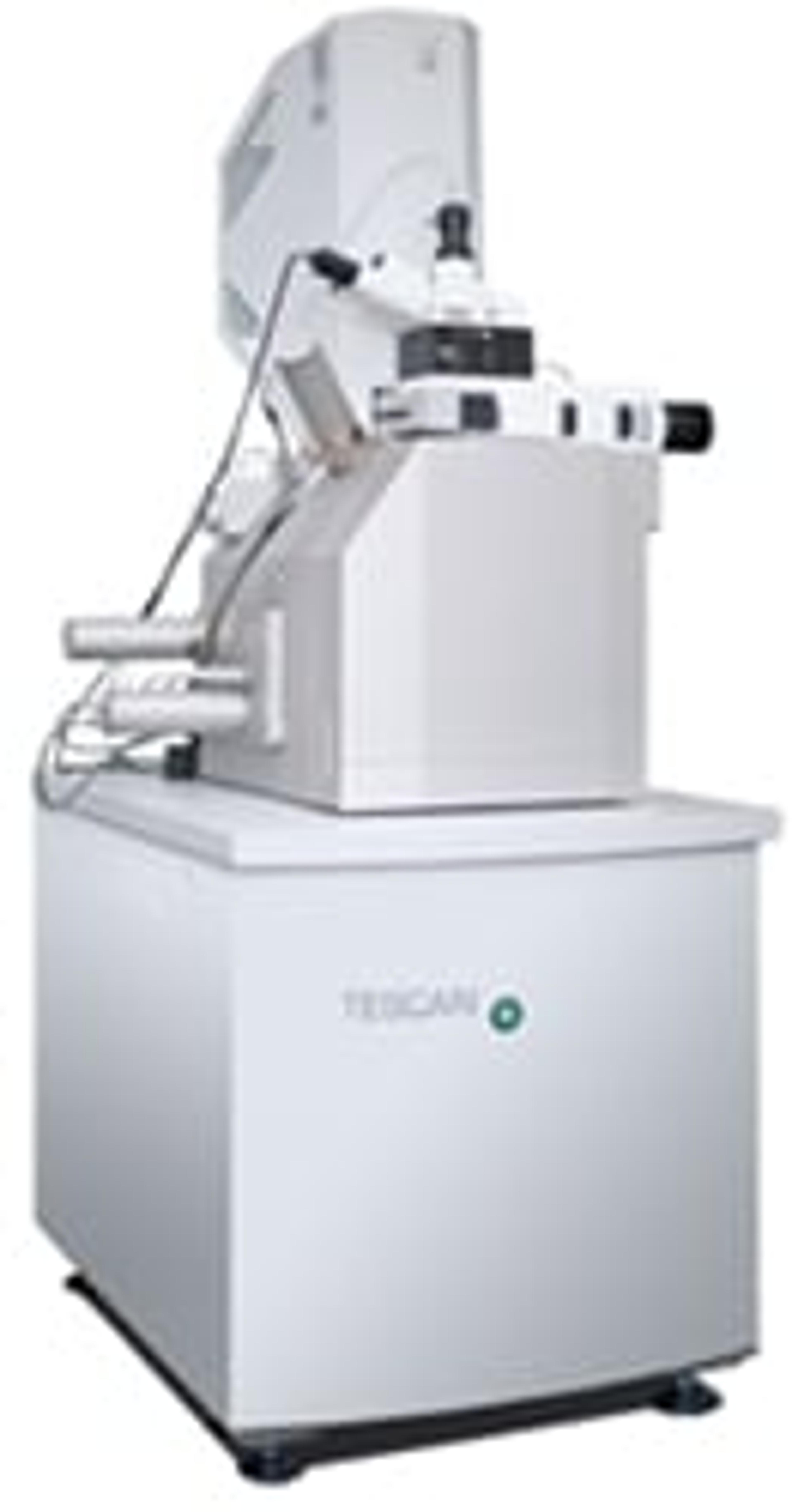
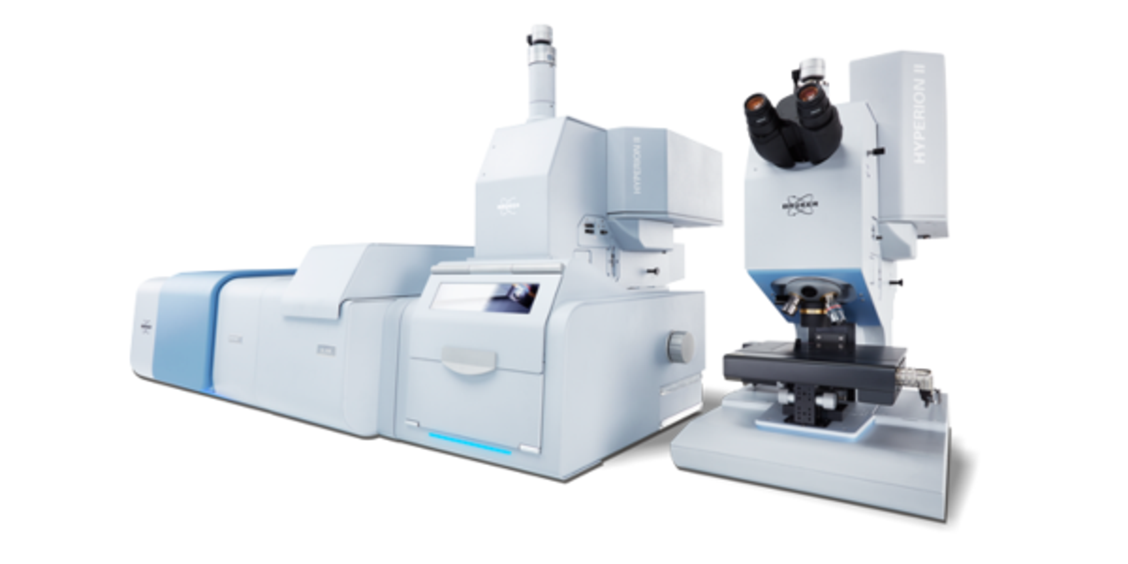
RISE Microscopy - Correlative Raman Imaging and Scanning Electron Microscopy (Raman-SEM)
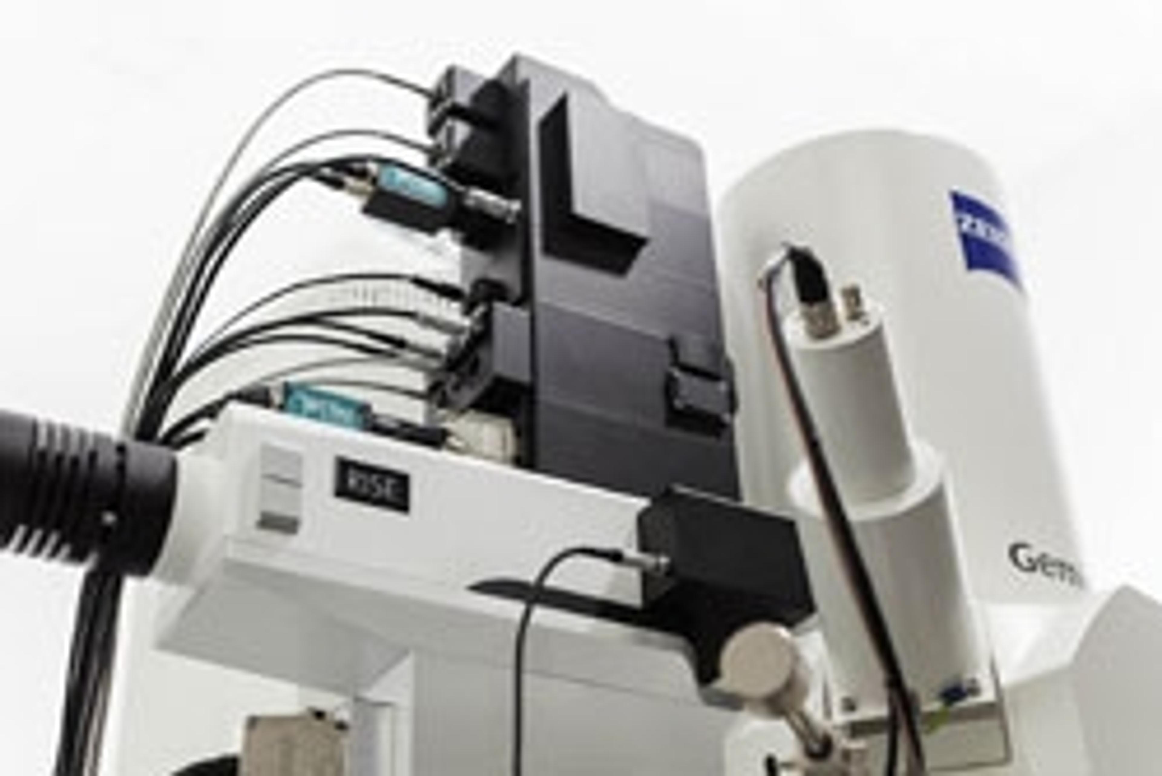
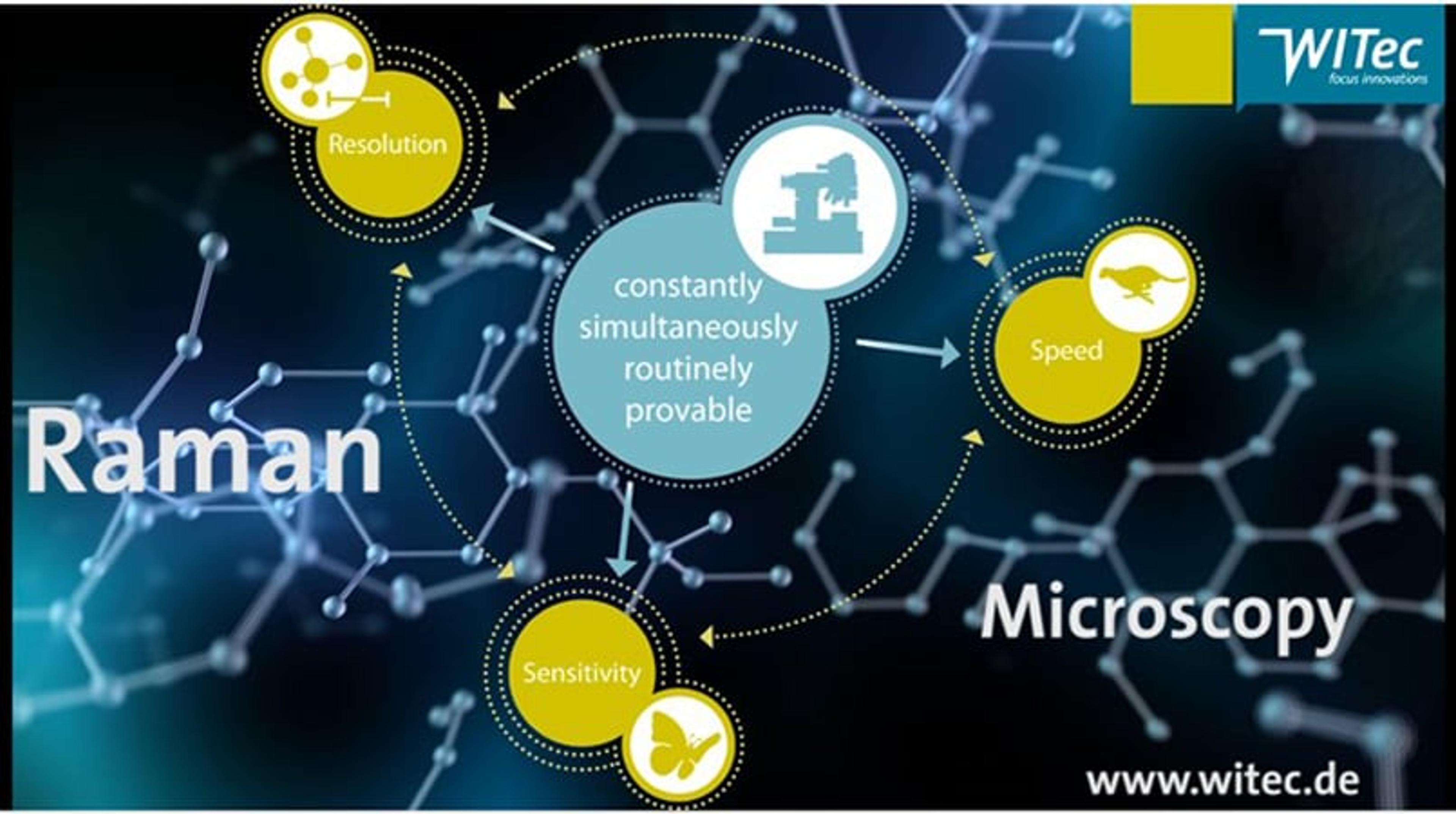
RISE Microscopy is a novel correlative microscopy technique that combines SEM and confocal Raman Imaging and links ultra-structural surface properties to molecular compound information. RISE Microscopy combines an SEM and a confocal Raman microscope. The confocal Raman microscope is integrated into the vacuum chamber of the electron microscope. Non-destructive Raman and SEM measurements are consecutively performed at two diff…


The supplier does not provide quotations for this product through SelectScience. You can search for similar products in our Product Directory.
Amazing results with clarity on data
Analyze nanoparticles at a single particle level
Ease of operation and analysis.
Review Date: 2 Apr 2022 | Oxford Instruments Raman
RISE Microscopy is a novel correlative microscopy technique that combines SEM and confocal Raman Imaging and links ultra-structural surface properties to molecular compound information.
RISE Microscopy combines an SEM and a confocal Raman microscope. The confocal Raman microscope is integrated into the vacuum chamber of the electron microscope. Non-destructive Raman and SEM measurements are consecutively performed at two different positions inside this chamber using an automatic transfer stage. Calibration of the sample position ensures scanning of the same sample area in both SEM and Raman modes.
The integrated RISE software carries out the required parameter adjustments and instrument alignments. The acquired results can then be correlated and the Raman and SEM images overlaid. RISE Microscopy pairs ease-of-use with exceptional analyzing benefits and is therefore suited for a wide variety of applications such as nanotechnology, materials science, and life science.
Features:
- Quick and convenient switching between Raman and SEM measurement
- Automated sample transfer from one measuring position to the other
- Integrated software interface for user-friendly measurement control
- Correlation of the measurement results and image overlay
- No compromise in SEM and Raman imaging capabilities
Applications:
- All fields of application that require a comprehensive and detailed sample analysis, e.g. materials science, pharmaceutical science and industry, geo science, nanotechnology, life science etc…

Looking into batteries with RISE microscopy
Understanding structure-composition-property-performance relationships is fundamental in developing better batteries. RISE (Raman imaging and scanning electron) microscopy is an extremely useful technology for investigating these features. Non-destructive Raman imaging microscopy can visualize structural and chemical information acquired from the battery’s internal components such as their molecular composition, grain fractures, the formation of solid electrolyte interphase (SEI) layers and degradation processes at the electrodes. In the application note, WITec documents changes of new and used electrodes with correlative RISE microscopy.
Correlative Raman imaging characterizes crystal properties of 2D materials
In this poster, WITec present correlative Raman imaging as a versatile tool for investigating three prominent examples of 2D materials, namely graphene and the transition metal dichalcogenides (TMDs) molybdenum disulfide (MoS2) and tungsten disulfide (WS2).
3D Raman imaging: Best practice examples from various applications
Confocal Raman imaging is a microscopy technique used for the identification and imaging of chemical and molecular compounds. This non-destructive, non-invasive and label-free method is used in various fields of application such as nanotechnology, materials and surface research, geological and environmental sciences and life sciences and pharmaceutics.
Confocal Raman Imaging, Correlative Raman-SEM, and Atomic Force Microscopy: Geo-Science Applications
Raman spectroscopy has long been applied in geoscience, for example for the identification and characterization of minerals, or in the observation of mineral phase transitions in high- and ultra-high pressure/ temperature experiments. By means of confocal Raman imaging (CRI), such characteristics can be evaluated from large-area scans on the centimeter scale to the detailed investigations with sub-micron resolution. In this study, WITec confocal Raman microscopes of the alpha300 and alpha500 series were used in order to carry out high-sensitivity measurements on a sample of diorite.
Correlative Confocal Raman Microscopy for 2D Materials Investigation
This application note presents a variety of application of the WITec alpha300 Confocal Raman Microscope Series in material testing and investigation. It covers examples such as analysis of graphene, analysis and imaging of transition metal dichalcogenides and photoluminescence imaging of layers and defects of WS2 crystals. The alpha300 series can carry out advanced confocal Raman imaging with multiple correlative microscopy technique options, including AFM, SNOM, SEM (RISE), fluorescence and photoluminescence.
Comprehensive Analyses of Graphene
Flexible and adaptive analyzing methods can support the effective investigation of graphene and accelerate the progress in graphene research and product development. This application note discusses the imaging of nano-carbon samples with combined Raman-AFM, SNOM, Nearfield-Raman, and Raman-SEM (RISE). It details the principles behind multiple investigations into graphene including; imaging of multilayered graphene, chirality investigations and nearfield-Raman imaging.
On the RISE: Correlative Confocal Raman and Scanning Electron Microscopy
Correlative imaging techniques are gaining more importance in many fields of applications as more characteristics of a specimen can be analyzed with one instrument. This application note describes the use of RISE (Raman Imaging and Scanning Electron) microscopy for a variety of applications including imaging of; geological samples, non-conductive organic materials, and hamster brain tissue. It also describes the ability of rise to analyze polymorphs and correlate structure with chemical phases.
Imaging of Nano-Carbon Samples with Combined Raman-AFM, SNOM, Nearfield-Raman, and Raman-SEM
In this application note, an overview of the WITec imaging techniques is given through various application examples from graphene research. A list of publications from graphene studies based on data obtained with a WITec microscope is presented.
RISE Microscopy - Correlative Ultra-Structural SEM and Molecular Raman Imaging
RISE Microscopy is a novel correlative microscopy technique that combines SEM and confocal Raman Imaging. Through RISE Microscopy ultra-structural surface properties can be linked to molecular compound information. This unique combination opens up new dimensions for more comprehensive sample characterization. In this application note, WITec’s RISE Microscope, was used to investigate a geological diorite sample.
Correlative confocal Raman imaging microscopy
In this video, WITec GmbH offers insight into the range of potential applications of correlative Raman imaging microscopy. This promises to combine the sensitivity and resolution of 3D chemical characterization with other techniques.
RISE Microscopy at Analytica 2014
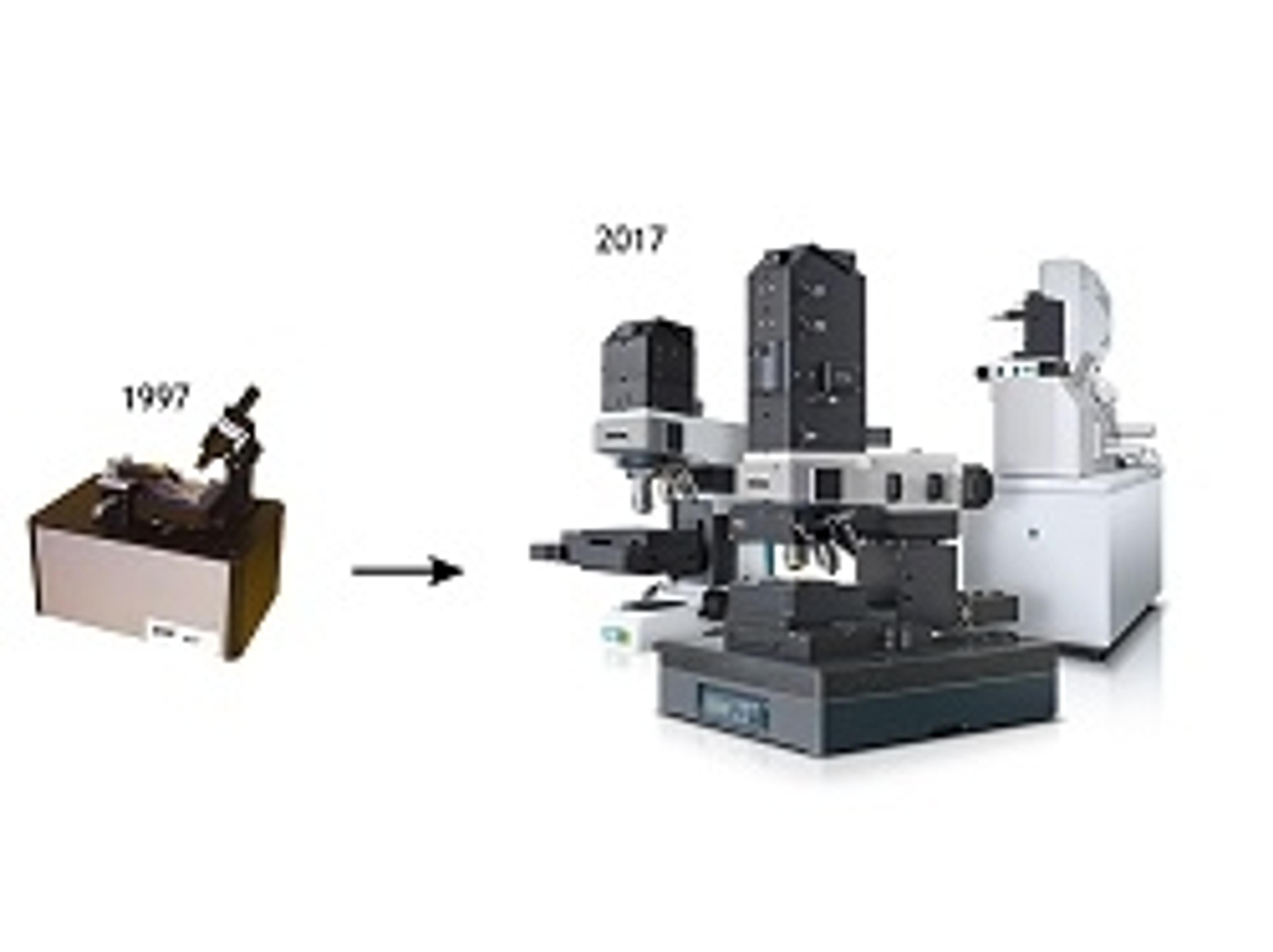
RISE Microscopy was launched at Analytica 2014 in Munich, Germany. Get an impression of the world's first fully integrated Raman Imaging & Scanning Electron Microscope and WITec's activities at the show.

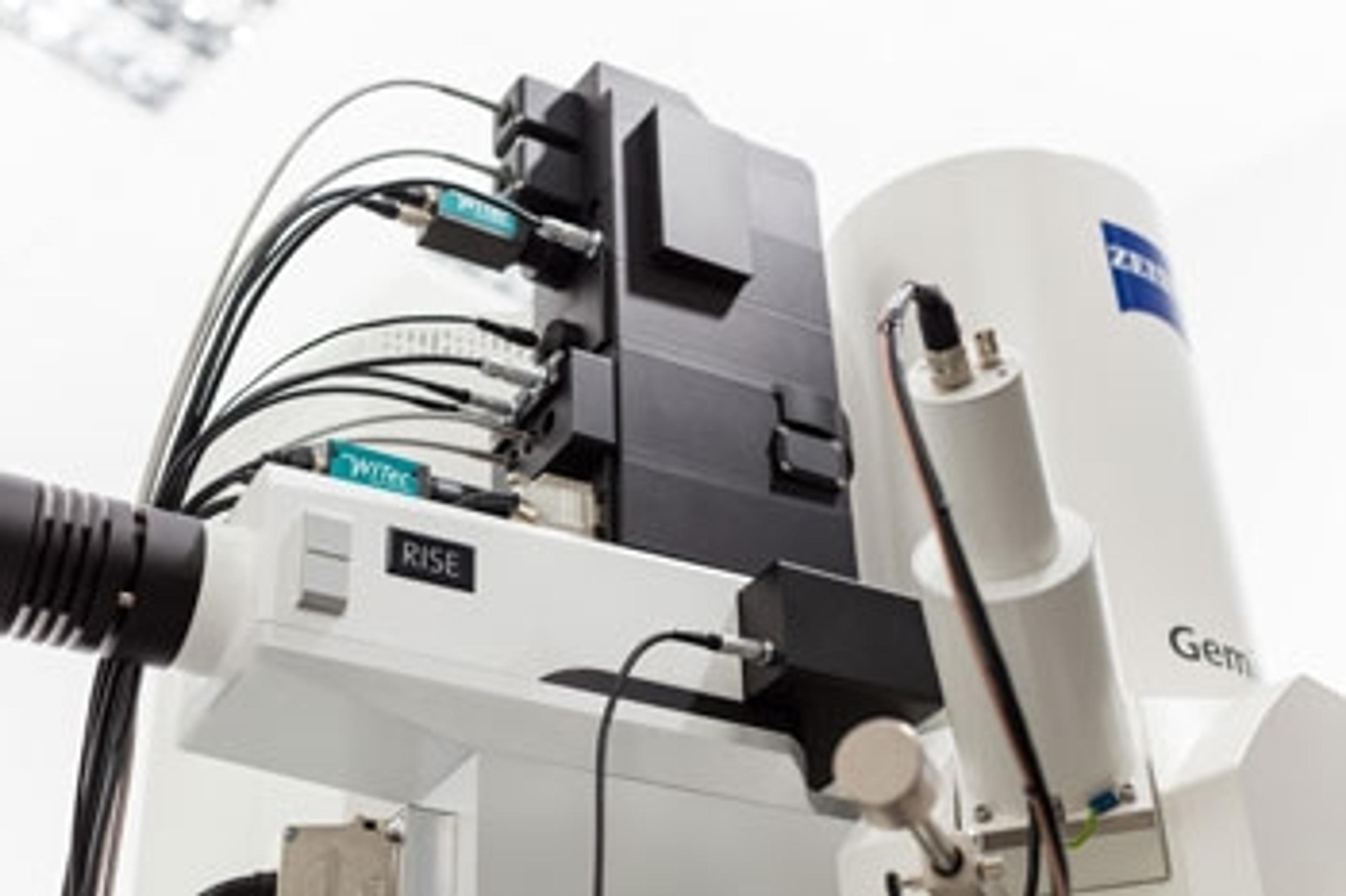
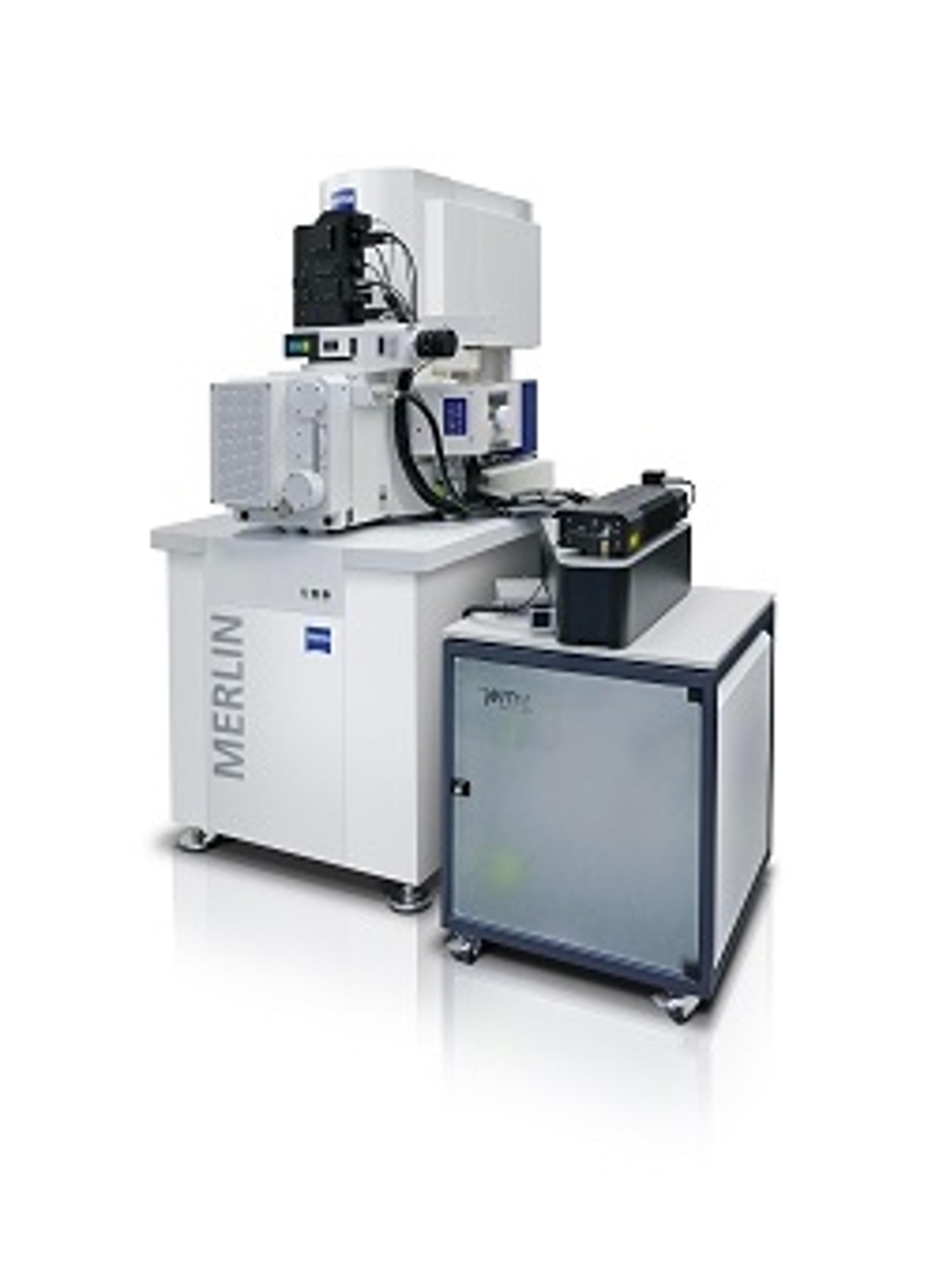
WITec’s RISE Microscopy now available with ZEISS Sigma 300 Scanning Electron Microscope
Improving ease-of-use and accelerating the experimental workflow