ResourceMaterials

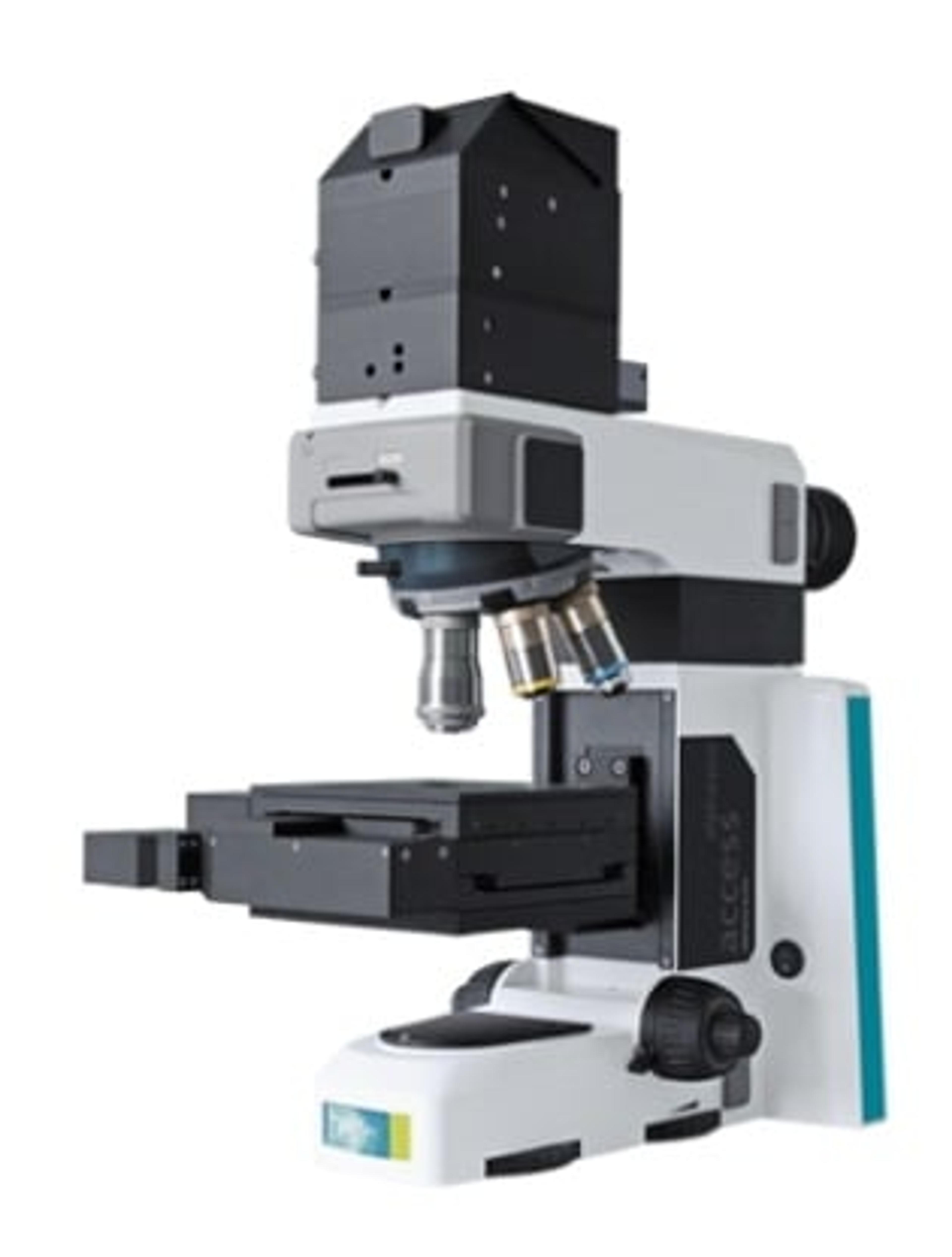
Correlative Confocal Raman Microscopy for 2D Materials Investigation

9 May 2017

This application note presents a variety of application of the WITec alpha300 Confocal Raman Microscope Series in material testing and investigation. It covers examples such as analysis of graphene, analysis and imaging of transition metal dichalcogenides and photoluminescence imaging of layers and defects of WS2 crystals. The alpha300 series can carry out advanced confocal Raman imaging with multiple correlative microscopy technique options, including AFM, SNOM, SEM (RISE), fluorescence and photoluminescence.

Scanning Probe Microscope Series WITec alpha300

Oxford Instruments Raman

Scanning Probe Microscope Series WITec alpha300 The WITec microscopy series features scanning probe as well as high resolution optical and Raman microscopy techniques in either a single instrument or combined system configurations for the highest flexibility throughout a wide range of microscopy applications. For example, it is possible to start with Confocal Raman Microscopy and upgrade later to Atomic Force Microscopy or vice versa. With such a combined instrument, chemical information can be directly linked to structural AFM information from the same sample area using only one instrument. For high-resolution optical information, the system can even be equipped with SNOM capabilities. All of these methods enable nondestructive sample analysis on the nanometer scale while requiring only minimal sample preparation, if any. This extensive modularity and ease of use facilitates a more comprehensive understanding of the sample. Typical applications are found in all fields of surface and materials science, geosciences, life science, pharmaceutical research, thin films and coating analysis as well as nanophotonics and nanotechnology. WITec’s technological leadership and the high-quality design of the alpha300 series pushes the frontiers of high-resolution Raman- and SPM-imaging abilities even further. This will aid our customers as they endeavor to become leaders in their varied fields.

(1)

RISE Microscopy - Correlative Raman Imaging and Scanning Electron Microscopy (Raman-SEM)

Oxford Instruments Raman

RISE Microscopy is a novel correlative microscopy technique that combines SEM and confocal Raman Imaging and links ultra-structural surface properties to molecular compound information. RISE Microscopy combines an SEM and a confocal Raman microscope. The confocal Raman microscope is integrated into the vacuum chamber of the electron microscope. Non-destructive Raman and SEM measurements are consecutively performed at two different positions inside this chamber using an automatic transfer stage. Calibration of the sample position ensures scanning of the same sample area in both SEM and Raman modes.The integrated RISE software carries out the required parameter adjustments and instrument alignments. The acquired results can then be correlated and the Raman and SEM images overlaid. RISE Microscopy pairs ease-of-use with exceptional analyzing benefits and is therefore suited for a wide variety of applications such as nanotechnology, materials science, and life science.Features: Quick and convenient switching between Raman and SEM measurement Automated sample transfer from one measuring position to the other Integrated software interface for user-friendly measurement control Correlation of the measurement results and image overlay No compromise in SEM and Raman imaging capabilities Applications: All fields of application that require a comprehensive and detailed sample analysis, e.g. materials science, pharmaceutical science and industry, geo science, nanotechnology, life science etc…

(1)

Links

Tags