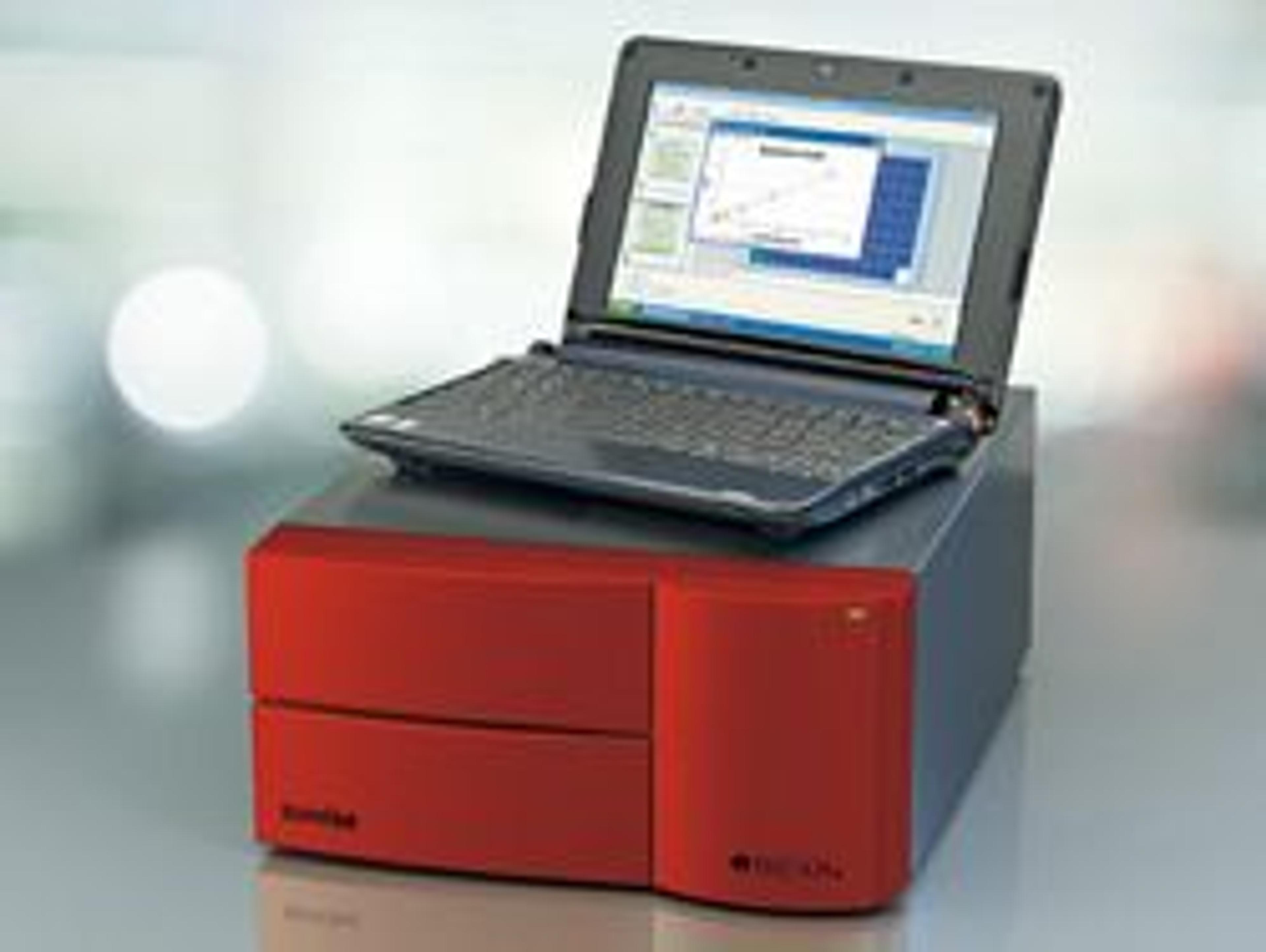
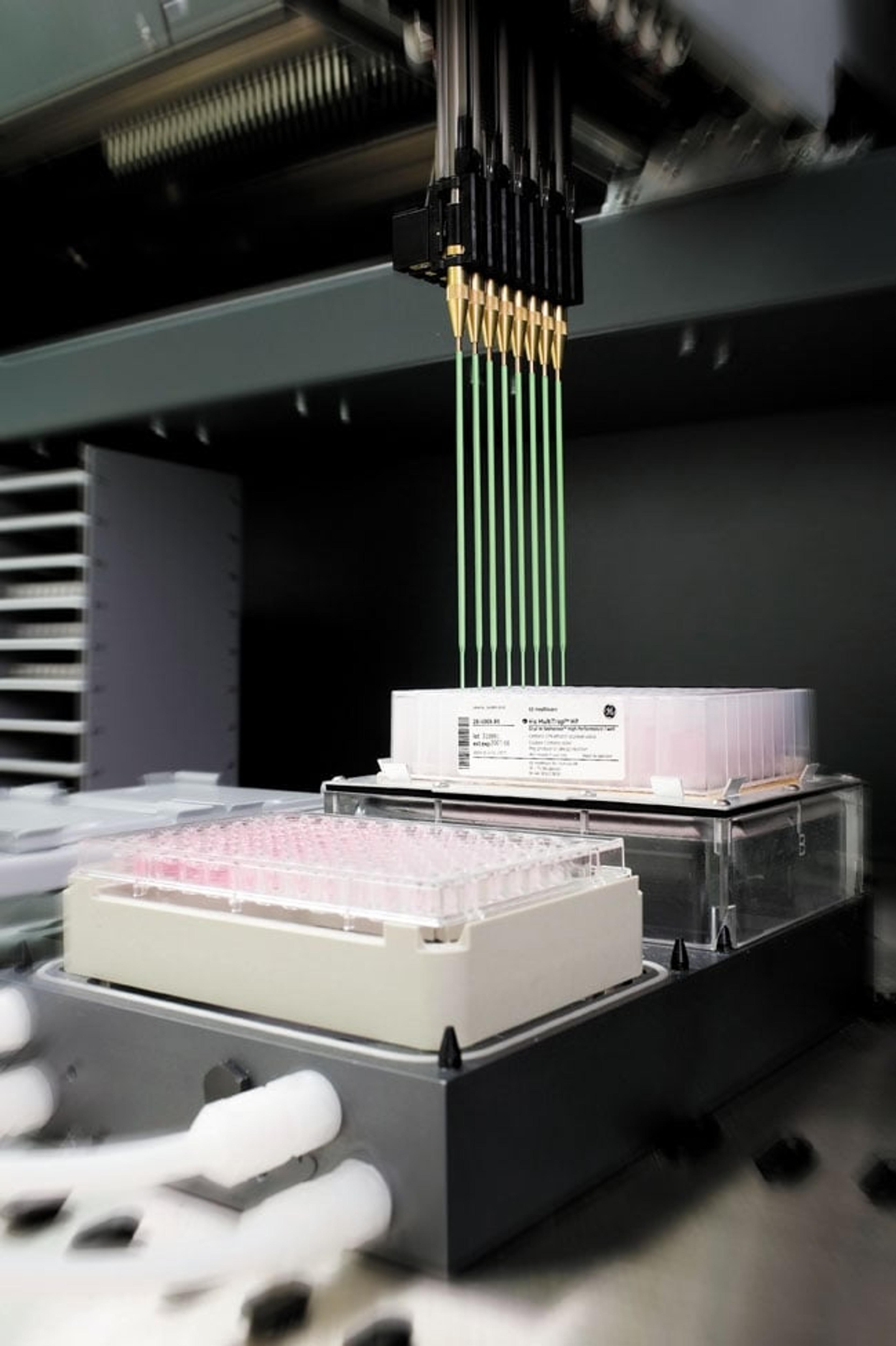
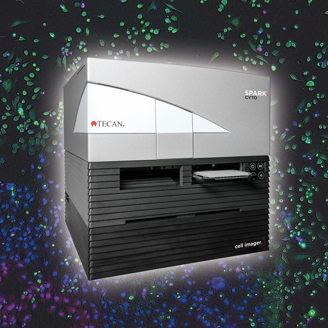
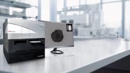
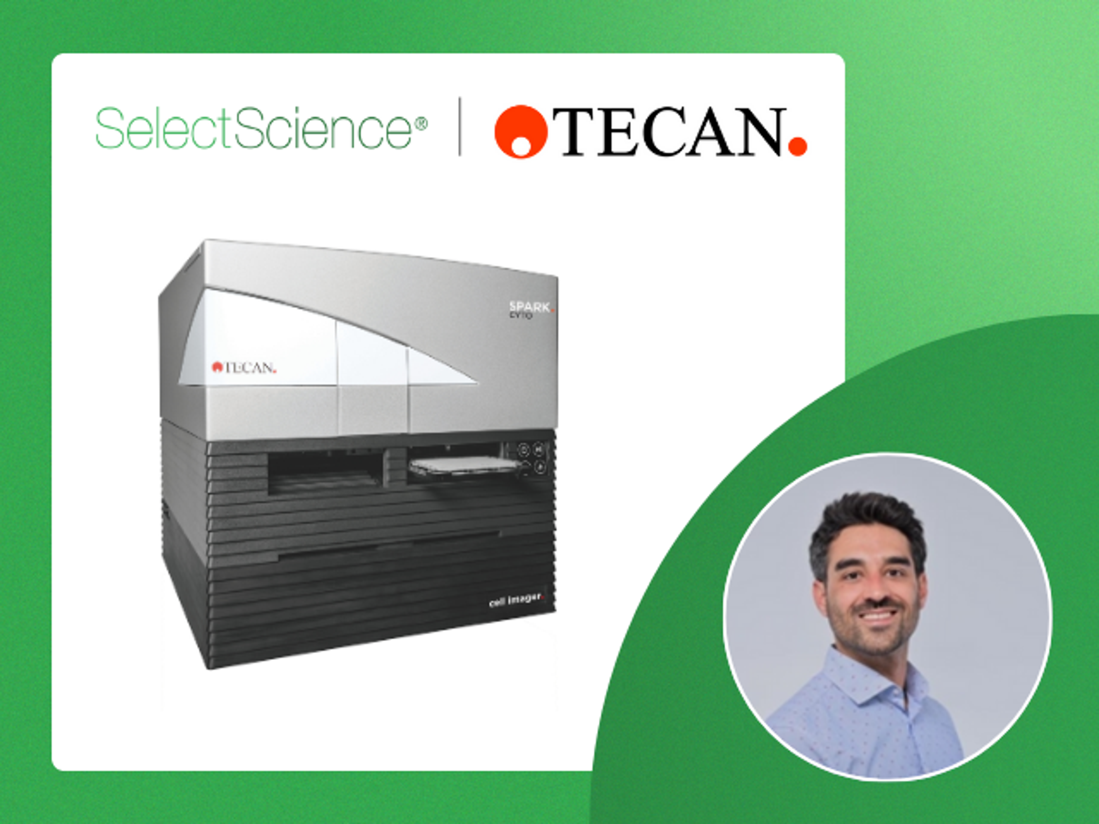
Spark® Cyto
Spark Cyto is the first live cell plate reader to detect biological, chemical or physical events – capturing the maximum amount of data possible from each plate well, at the same time and under the same conditions.


Receive your quote directly from the manufacturer.
Perfect.
Tissue samples
Great experience.
Review Date: 19 Jul 2021 | Tecan
Utterly recommended!
Proliferation, ELISA
Great equipment! I use it very week for 96 well plate based assays.
Review Date: 10 Jul 2021 | Tecan
Tecan's Spark Cyto is very good.
Cellular fluorescence imaging
Tecan's Spark Cyto is very good. It can automatically perform bright-field and fluorescence imaging of the cells in the well plate, and statistically output the results as needed. This greatly saves our time. At the same time, its fluorescence imaging signal is very strong and clear. It is much clearer than the general inverted fluorescence microscope. In addition, it can set temperature and carbon dioxide, and perform real-time detection of cells, making real-time tracking possible.
Review Date: 9 Mar 2021 | Tecan
Great one, I like it!
Clinical lab
This product is the first one that I liked and it has good value for money. It is also easy to use.
Review Date: 9 Feb 2021 | Tecan
Great user friendly instrument, would highly recommend it!
Drug development
We have found that being able to do so many different applications within one product is great for our lab. We can do cell culture wound healing for drug development, nucleic acid analysis, cell counting, luminescence assays, colorimetric assays, etc. It has been extremely user friendly and has increased the efficiency and reliability of our experiments.
Review Date: 10 Feb 2020 | Tecan
The Spark Cyto can be an excellent support in any cell biology laboratory.
Phenotypic screening, cell-based assay development, target identification...
Using the Spark Cyto for long term proliferation experiments I was impressed by its capabilities for automated cell incubation and analysis: we observed that the cells grow very homogeneously and with a similar doubling time compared to a dedicated cell incubation approach. The Spark Cyto is equipped with excellent optics, an ultra-sensitive camera and I particularly appreciate the presence of a 2x objective which allows perfect whole-well imaging at cellular resolution in a 96-well plate. The imaging capability of this instrument, both in bright field and in multiple fluorescence channels, allows reproducible analysis in real time from live cell cultures. The flexibility and ease of use of this instrument make it particularly suitable for both cell-based assay development and for medium-high throughput phenotypic screening. The Spark Cyto represents, in my opinion, an excellent support for daily activities in any cell biology laboratory.
Review Date: 2 Oct 2019 | Tecan
Can be recommended to labs with many students.
Cell survival assays, ELISA
Using the Spark Cyto for cell imaging, enzymatic tests and immunoassays, we were impressed by how intuitive and easy to use the software is. Our diploma and PhD students instantly learned how to work with Spark Cyto and obtain high quality results.
Review Date: 17 Jul 2019 | Tecan
Great system with broad detection portfolio. Ideal for assay development and screening!
HTS for drugs (70.000 compounds) and gene function (RNAi & CRISPR-Cas)
This detection system nicely bridges the gap between a plate reader and imaging systems like automated microscopes. It combines multi parameter screening (all kinds of fluorescence detection) with the features of image based screening (cytometric measurements) in one machine. Coupling kinetic measurements over long time periods to injection of reagents at the most effective time points within the reader. Ideal system to be used in assay optimisation and in screening. Nice graphical interface for assay set up and a lot of nice analysis features like confluence or viability measurements.
Review Date: 4 Jul 2019 | Tecan
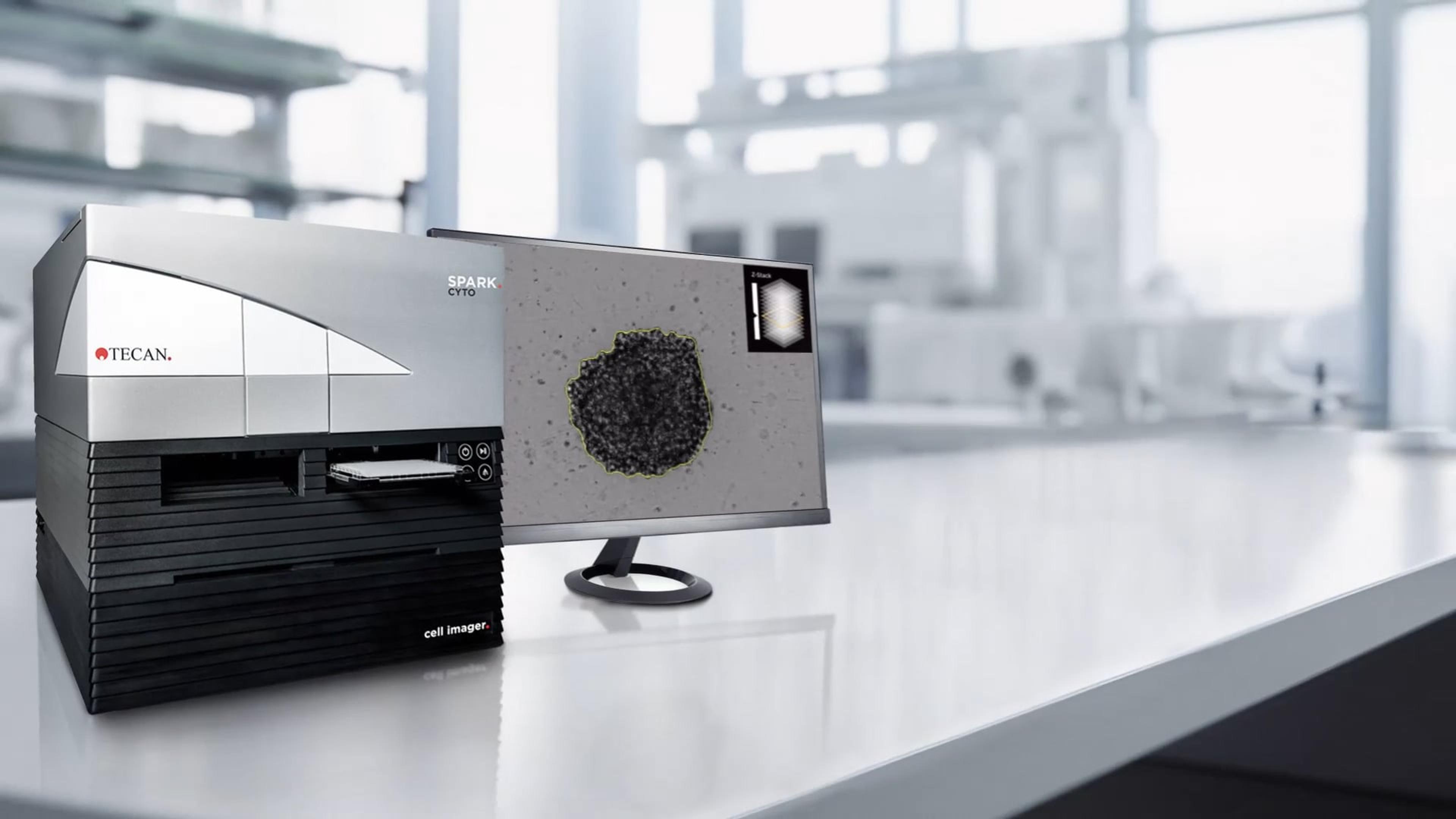
Spark Cyto builds on the foundation of the original Spark platform, combining the flexibility of a high-end multimode plate reader with whole well imaging and comprehensive environmental control to allow multiparametric measurements for cell-based applications.
This dynamic instrument uses real time data acquisition and analysis to deliver meaningful insights faster than ever before, ensuring that no key event is ever missed. It combines live cell imaging with industry-leading detection technologies, allowing qualitative and quantitative information to be integrated into unique multiparameter data sets. Top-of-the-range camera components and patent-pending LED autofocus technology offer three magnification levels and four acquisition channels (for fluorescence and bright field imaging), allowing the investigation of entire cell populations, and enabling the whole well area of a 96- or 384-well microplate to be recorded with just one image, without tiling or distortion.
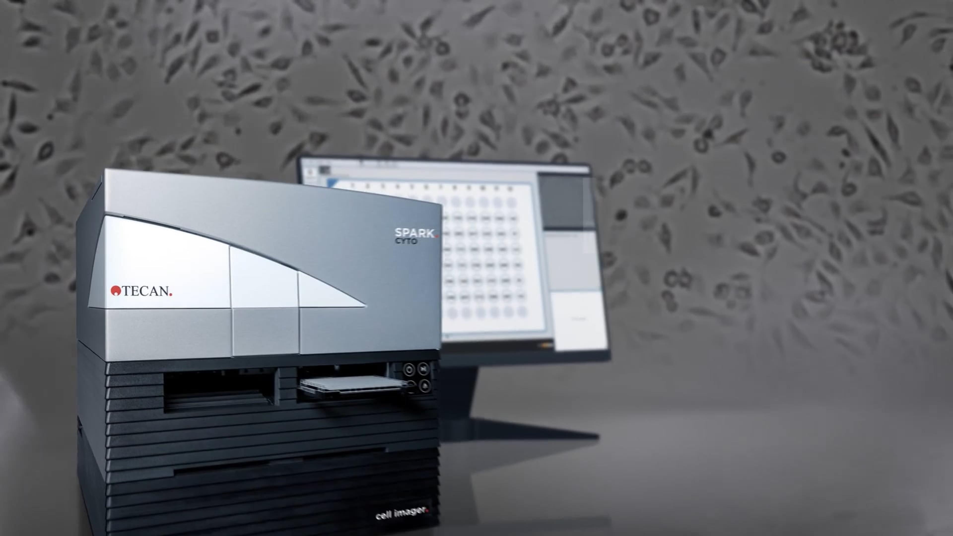
Spark Cyto is designed to handle a broad range of common cytometry applications, offering a new level of experimental control without compromising on ease of use and convenience. Powerful SparkControl™ and Image Analyzer™ software give the operator complete control of all the parameters associated with their experiment and Real Time Experimental Control (REC™) to enable kinetic experiments to be performed automatically – to deliver multiplexed live cell data from a single instrument, unlocking new possibilities for cell-based research and giving increased confidence in the statistical relevance of the results.
Spark Cyto is available in five specialized configurations with different detection modalities, each fully equipped for real-time live cell imaging cytometry.
Please note that Spark Cyto is for research use only. Not for use in diagnostic procedures.
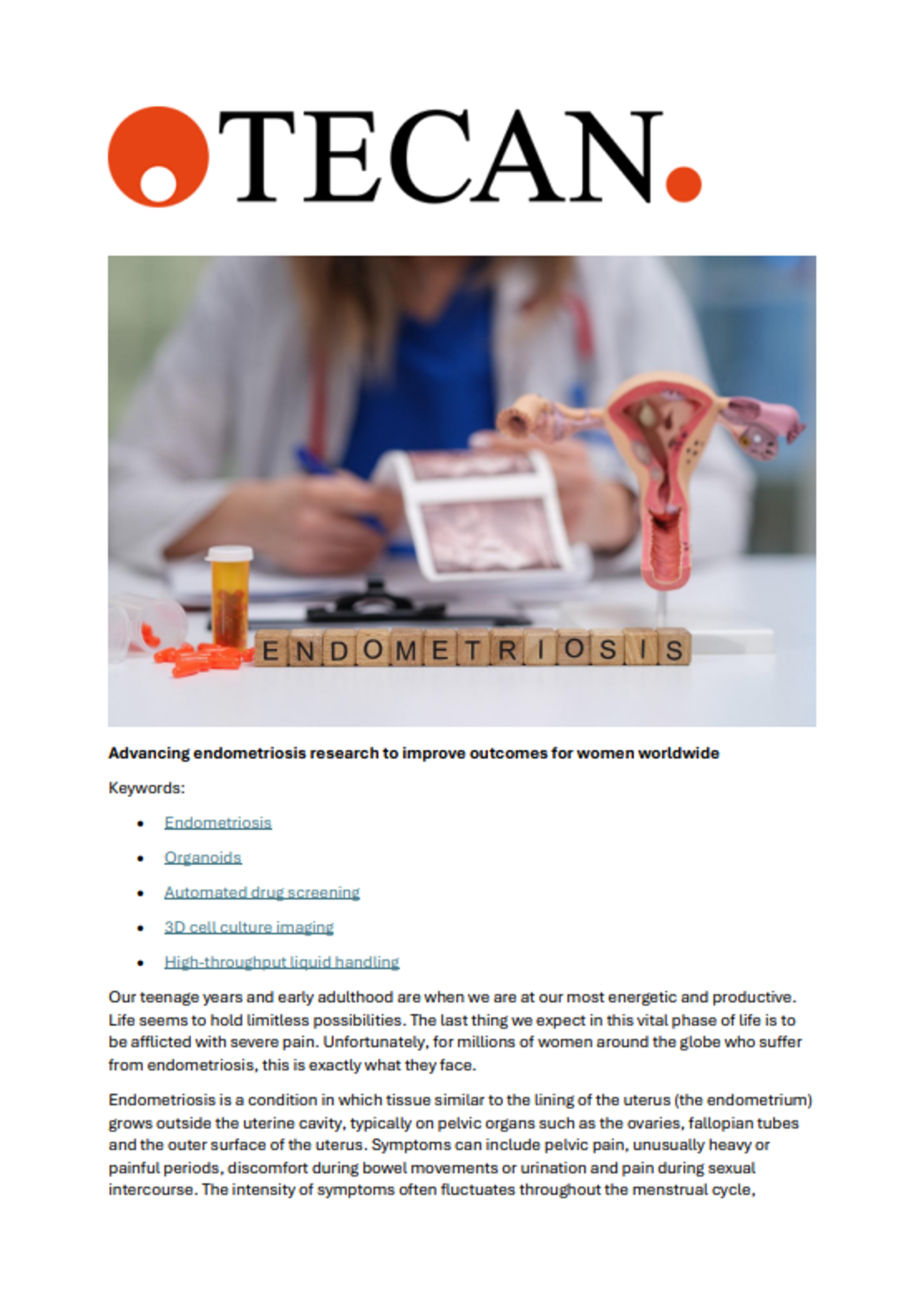
Advancing endometriosis research to improve outcomes for women worldwide
Organoid models have the potential to transform and deepen our knowledge of diseases in many fields of biomedical research and accelerate solutions to patients. Discover how a collaboration between GynQura (an innovative project within the BioInnovation Institute’s BioStudio program in Copenhagen) and Tecan is harnessing patient-derived organoid models to identify and develop novel, targeted treatments for endometriosis.
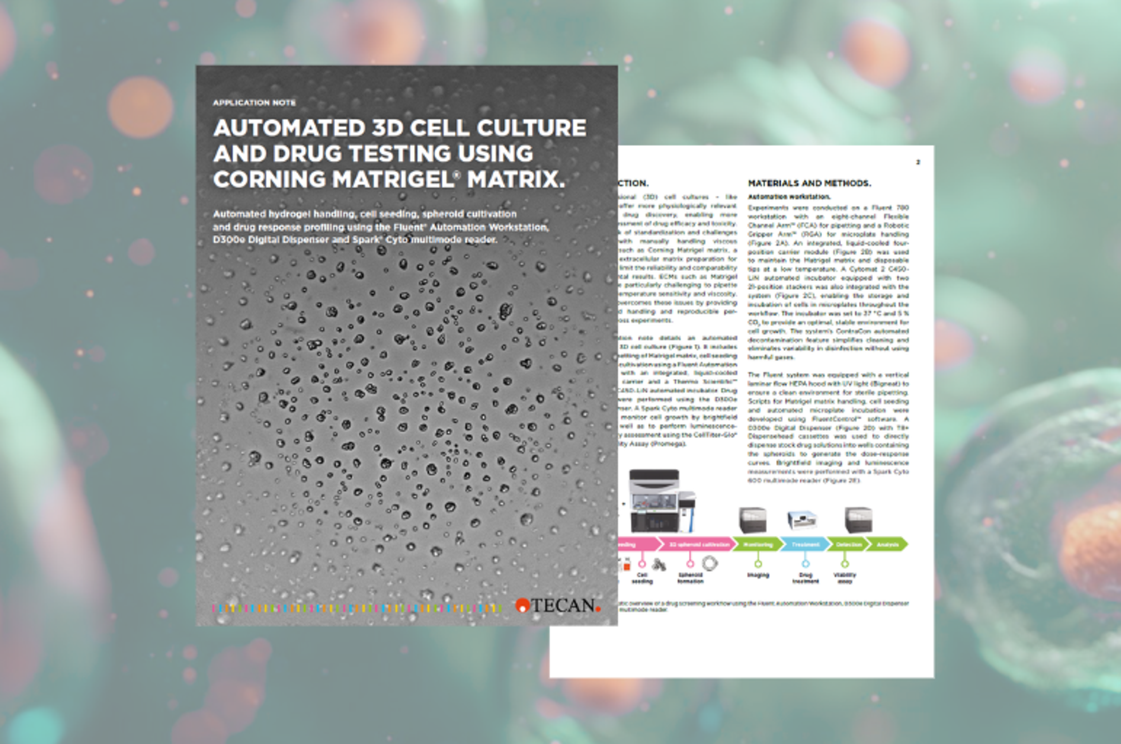
Automated 3D cell culture and drug testing using Corning Matrigel Matrix
This application note details an automated workflow for 3D cell culture. It includes controlled pipetting of Matrigel® matrix, cell seeding and spheroid cultivation using a Fluent Automation Workstation with an integrated, liquid-cooled four-position carrier and a Thermo Scientific™ Cytomat™ 2 C450-LiN automated incubator. Drug treatments were performed using the D300e Digital Dispenser. A Spark® Cyto multimode reader was used to monitor cell growth by brightfield imaging, as well as to perform luminescence-based viability assessment using the CellTiter-Glo® 3D Cell Viability Assay (Promega).
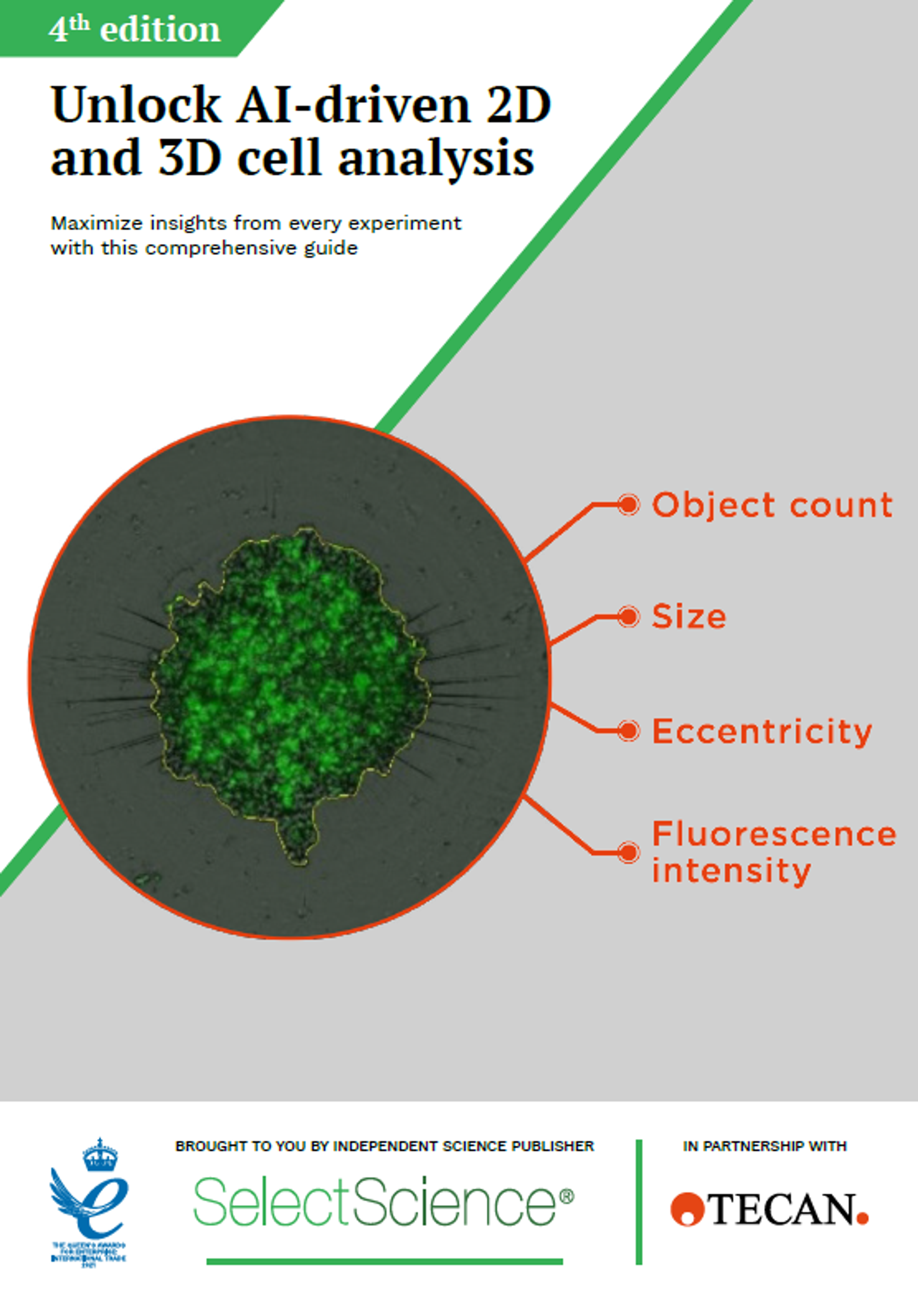
Unlock AI-driven 2D and 3D cell analysis
This application eBook presents an in-depth analysis of how advances in cell-based technology – such as label-free live-cell imaging in 2D and 3D with artificial intelligence and deep-learning-driven insights – are bringing higher levels of automation, reproducibility, and flexibility to cell-based research.
Download your free copy of this fourth edition eBook to find out about:
- Simplified cell-based assay workflows
- In vitro drug screening on cancer cells
- Real-time monitoring of spheroid growth
- Label-free multiple spheroid analysis
- Automated spheroid production for screening applications
- Assessing drug response in patient-derived colon organoids
- 3D liver tissue reconstruction
- Simplify cell line development
- Cell model development for SARS-CoV-2 infection studies
- Multicolour apoptosis detection

Assessing drug response in patient-derived colon organoids
Explore how the Spark Cyto multimode plate reader, equipped with a deep learning-based 3Dai algorithm, enhances automated imaging and patient-derived organoids (PDOs) analysis for efficient drug screening and cytotoxicity evaluation, streamlining personalized medicine and drug development.
Real-time growth monitoring of spheroids in round bottom microplates
Three-dimensional (3D) cell culture systems have emerged as promising techniques in the field of cell-based assays, with approaches such as multicellular 3D spheroids (dense cell aggregates mostly comprised of cell lines) gaining widespread attention. Different methods of spheroid generation have been explored, such as magnetic levitation, hanging drop methods, and the use of ultra-low attachment (ULA) round bottom microplates for scaffold-free generation of 3D aggregates independent of solid carriers. Tecan describes the generation of spheroids in Corning 96-well, black, round bottom, ultra-low attachment microplates, and monitoring of spheroid growth in the Spark® Cyto multimode reader using brightfield (BF) and fluorescence (FL) imaging. Discover how the Spark Cyto provides ease of use, multiplexing capabilities, and the possibility of executing an entire workflow on one instrument.
Please note that Spark Cyto is for research use only. Not for use in diagnostic procedures.
In vitro drug screening on cancer cells using the Spark Cyto imaging cytometer
Cell-based in vitro assays represent essential tools for evaluating cytostatic and cytotoxic effects in drug screening projects. Explore the advantages of combining the bright field (BF) and fluorescence-based object identification algorithms of the Spark® Cyto reader with a commercially available highly sensitive viability and toxicity assay. Tecan also discusses relevant considerations when establishing such a cell-based assay.
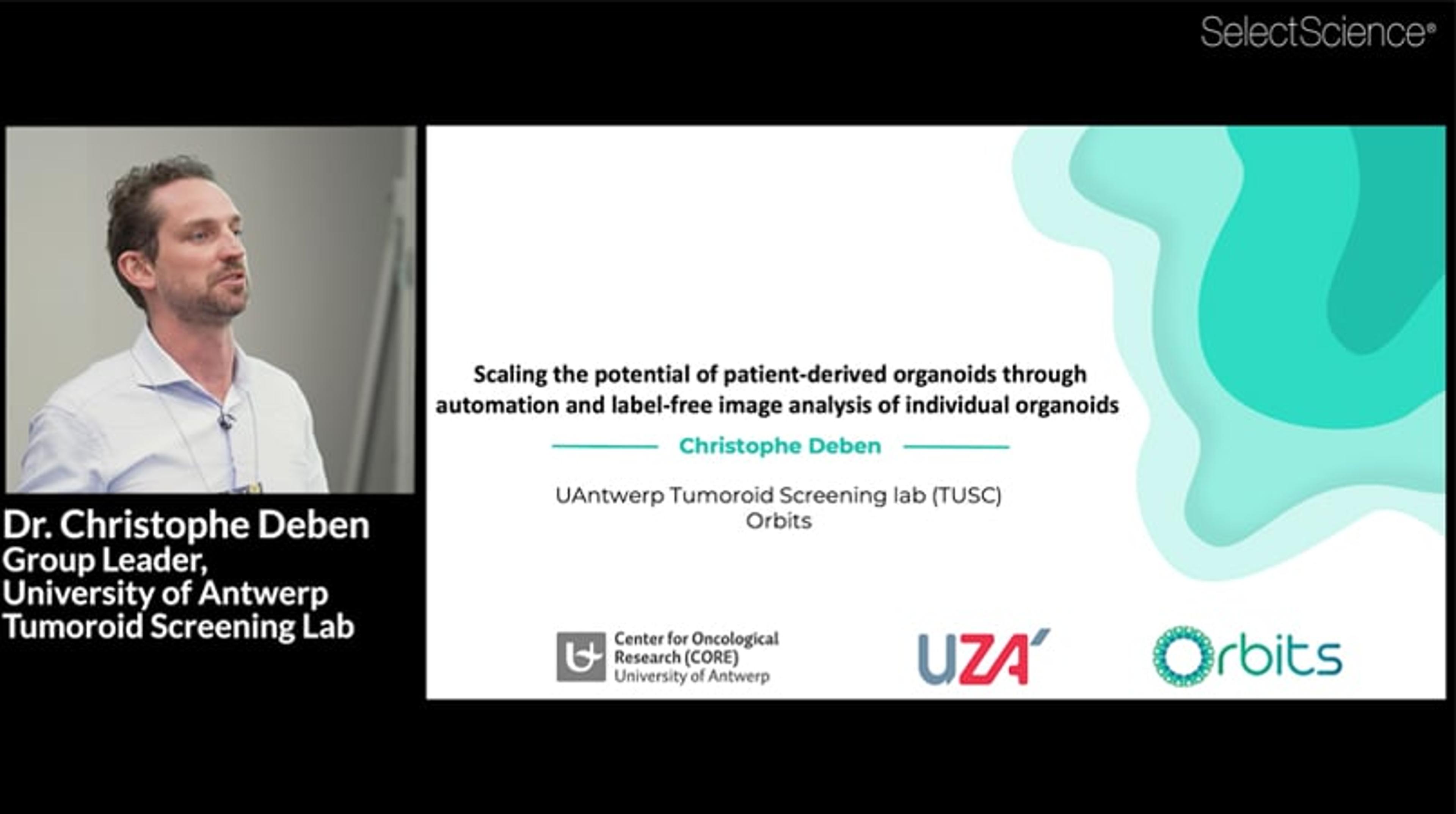
Scaling the potential of patient derived organoids through automation and label free image analysis of individual organoids
3D patient-derived tumor organoids hold great potential for many basic and translational applications. However, traditional approaches to organoid image analysis and downstream data processing are often challenging and labor-intensive. Download this presentation to discover how Dr. Christophe Deben, Group Leader of the University of Antwerp’s Tumoroid Screening Lab, has developed a label-free brightfield image analysis software (OrBITS) to monitor organoid growth rates and track movements.
Dr. Deben also shares a semi-automated workflow for cell seeding drug treatment using the Tecan D300e digital drug dispenser and live-cell imaging using the Tecan Spark® Cyto multimode plate reader.
Real-time cell imaging and analysis to provide a more complete picture of cell death
This application note seeks to provide a practical demonstration of how real-time assays and a plate reader with bright field and fluorescence imaging functionality can work in unison to reveal cell and compound-specific features of an apoptotic response.
Top tips for addressing the “reproducibility crisis” in cell biology research
The dangers of irreproducibility are becoming increasingly well-recognized. However, irreproducible results can be caused by a number of different factors, from the methods and instrumentation used to the level of expertise within the lab. The current “reproducibility crisis” is therefore a daunting task for scientists to start to address. This resource outlines expert tips for overcoming the top three challenges highlighted by those working in cell biology.
In this infographic, we outline how to overcome the top three challenges with cell biology, as highlighted by scientists:
Complex analytical workflows
Combining multiple methods
Laborious experimental practices
For further information, visit our content hub full of free resources exploring the one-stop solution designed to mitigate the risk of irreproducibility.
Multiple methods for normalizing cellular fluorescent signals to the cell number per well
Quantitative cell-based experiments are crucial for the analysis of cellular response to stimulation. This technical note describes how to perform quantitative cell-based analyses using a multimode plate reader with whole-well imaging capabilities.

Mini organs, big breakthroughs – an Accelerating Science Forum
Organoids are self-assembling 'mini organs' at the core of a new era, driving big breakthroughs in personalized medicine and next-generation therapies. These industrialized 3D in vitro models are transforming our ability to study disease biology and test and develop drugs with unprecedented accuracy, mirroring human response in ways traditional models cannot.
Join our on demand Accelerating Science Forum – held in partnership with Tecan – where world-leading experts will explore the practical blueprint for capitalizing on this breakthrough technology. As part of our Accelerating Science Feature, we will move beyond the scientific complexity to focus on how to successfully transition these powerful 3D models from the lab bench into industrialized, high-throughput screening environments to truly deliver the next generation of drug discovery.
Attend this online roundtable discussion to:
- Gain actionable insights on how to move organoid models into high-throughput screening
- Explore the critical path to achieving true functional maturity in multi-organ systems
- Hear expert predictions on the industry landscape, key applications, and pace of clinical adoption for organoids over the next five years
Certificate of attendance:
When you view the on-demand webinar, you can request a certificate of attendance by emailing editor@selectscience.net.
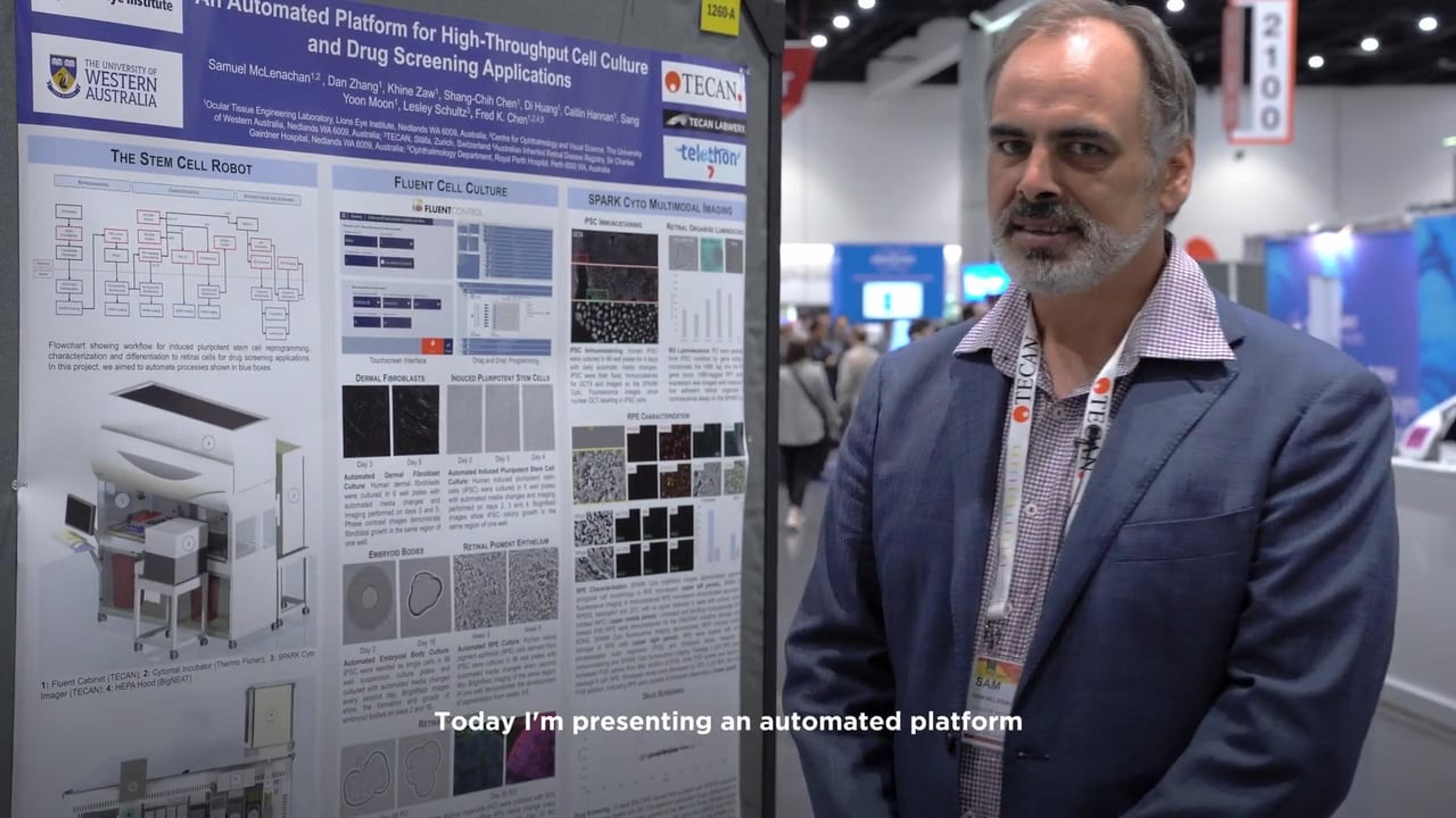
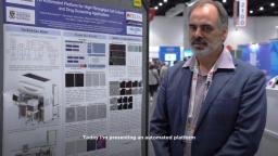
Can't wait? Watch this poster presentation from the Lions Eye Institute in Perth about automating the culture of various cell types, including iPSCs, dermal fibroblasts, and retinal organoids.
Spark Cyto is for research use only. Not for use in diagnostic procedures.
TechTalk: Automated organoid analysis with Spark® Cyto imaging plate reader
Friday, March 6 at 15:00 GMT | 16:00 CET | 10:00 EST | 07:00 PST
Organoids offer unprecedented insights, yet many laboratories are held back by the complexity and high cost of the required infrastructure. Typical workflows often demand sophisticated protocols and a high level of expertise that is simply not available in every lab.
In this 20-minute SelectScience TechTalk, we will introduce the Spark® Cyto, an imaging microplate reader designed to break down these barriers. Discover how this compact instrument, with integrated real-time environmental control, easily automates the monitoring and analysis of 3D structures such as organoids, enabling high-quality fluorescence and label-free imaging without a steep learning curve.
We will also demonstrate effortless assay multiplexing by integrating high-quality imaging data with quantitative metabolic readouts, such as ATP-based luminescence.
Key learning objectives:
- Understand how to perform live-cell imaging of 3D cultures, including organoids, with real-time environmental control
- Learn how to combine high-quality imaging with quantitative metabolic assays (e.g. ATP-based luminescence) for seamless multiplexed analysis
- Discover how AI-assisted image analysis simplifies data interpretation and reduces manual workload
- Explore strategies for automating 3D cell culture workflows to improve reproducibility, efficiency, and scalability
Who should attend?
- Researchers working with 3D cell cultures
- Anybody interested in automating cell-based assays
Certificate of attendance
If you attend the live TechTalk, you will automatically receive a certificate of attendance, including a learning outcomes summary, for continuing education purposes.
If you view the on-demand TechTalk, you can request a certificate of attendance by emailing editor@selectscience.net.
Spark is for research use only. Not for use in diagnostic procedures.
TechTalk details
- Cost: Free to attend
- Location: Online
- Duration: 20 minutes
Registration is required to secure your place. If you register but can’t attend live, you will receive a link to the on‑demand recording once it becomes available
Scaling 3D cell experiments with confidence with the new Spark® Cyto 3Dai analysis tools coming soon and start to end workflow automation
Complex 3D cell models like spheroid and organoid systems hold great potential for many applications, from drug development to personalized medicine. However, lack of reproducibility poses a particular challenge where automation and quality control is key to increase throughput while achieving consistent and reliable results.
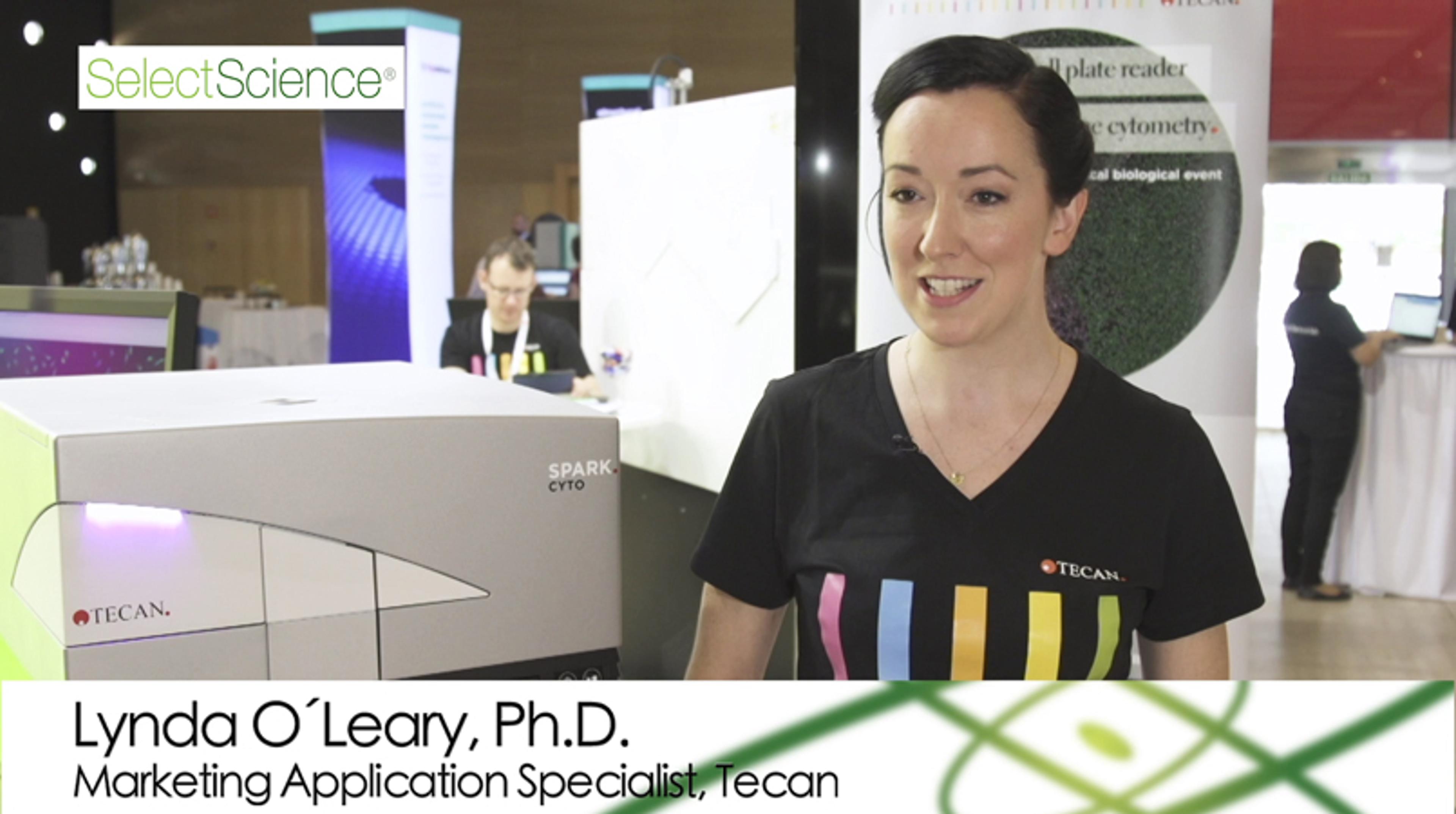
In this webinar, join Roman Petrovsky, Product Manager Cell Imaging and Lynda O’Leary, Manager Marketing Application Detection, Tecan, as they present novel automated tools for cell culture, quality control, and AI-based analysis to level up your cell research from start to end out of one hand.
Key learning objectives
- Learn how to simplify and automate spheroid and organoid experiments addressing reproducibility, quality control and analysis challenges
- Explore how to generate high-quality spheroids and organoids at scale
- Discover how multiplex analysis of spheroids and organoids with AI provides insights faster
Who should attend?
- Academic scientists interested in the techniques and applications of 3D cell models for research, engineering, and drug development.
- Industry scientists interested in identifying promising platforms for drug discovery and targets for novel therapies in 3D cell models.
Certificate of attendance
All webinar participants can request a certificate of attendance, including a learning outcomes summary, for continuing education purposes.
Spark Cyto is for research use only. Not for use in diagnostic procedures.
Lions Eye Institute showcases robotics for retinal organoid development
Discover how the Lions Eye Institute in Perth is revolutionizing ocular research with an automated platform for high-throughput cell culture and drug screening. Senior Scientist Sam McClennan explains how the Stem Cell Robot System integrates liquid handling, imaging, and incubation technologies to streamline the process of reprogramming patient fibroblasts into stem cells and differentiating them into retinal organoids and pigment epithelial cells. Learn how this cutting-edge system enables large-scale drug screening and advanced imaging for retinal disease research.
3D cell biology powered by AI
Discover the power of 3D cell biology powered by AI. The Spark Cyto is a label-free cell counting tool crucial for cell biology. It offers accurate, non-invasive quantification without cytotoxic effects from staining. The user-friendly software features a live viewer for monitoring growth and an intuitive method editor for easy measurement setup.
Advancing cell counting with Tecan Spark Cyto
Tecan introduces Spark Cyto, a label-free cell counting tool crucial for cell biology. It offers accurate, non-invasive quantification without cytotoxic effects from staining. The user-friendly software features a live viewer for monitoring growth and an intuitive method editor for easy measurement setup.
Scaling the potential of patient-derived organoids through automation and label-free image analysis of individual organoids
3D patient-derived tumor organoids hold great potential for many basic and translational applications. However, traditional approaches to organoid image analysis and downstream data processing are often challenging and labor-intensive.
In this presentation, Dr. Christophe Deben, Group Leader of the University of Antwerp’s Tumoroid Screening Lab, demonstrates a semi-automated workflow for cell seeding drug treatment using the Tecan D300e digital drug dispenser and live-cell imaging using the Tecan Spark® Cyto* multimode plate reader. In addition, Deben illustrates the development and integration of label-free brightfield image analysis software (OrBITS) to monitor organoid growth rates and track movements.
This talk was presented, in partnership with Tecan, at SLAS2023.
*Spark® Cyto is for research use only. Not for use in diagnostic procedures.
Unlock New Possibilities for Your Cell-Based Assays: New Plate Reader with Live Cell Imaging and Real Time Cytometry
Hear how the Tecan Spark® Cyto multi-mode plate reader with fluorescence imaging and cytometry capabilities can help you to gain more meaningful insights from your cell-based research. Lynda O’Leary, Marketing Application Scientist, Tecan, shares how the Spark Cyto can help you to save time and samples and standardize procedures for long-term live-cell kinetic experiments procedures to gain a better understanding of the complex processes within cell biology.
Video filmed at SLAS Europe 2019 – visit the SelectScience special feature for more videos from the event.
Meeting the Needs of Live Cell Analysis for High-Throughput Screening
Dr. Jens Peter von Kries is Head of the Screening Unit at the Leibniz-Forschungsinstitut für Molekulare Pharmakologie (FMP). Learn how the unit’s chemical biology platform supports high-throughput experiments for academic projects from around the world and hear how the new Tecan Spark® Cyto plate reader will help meet the needs of the most demanding screening projects, through multiparameter fluorescence measurements and the ability to perform real-time analyses of long-term live-cell assays.
Video filmed at SLAS Europe 2019 – visit the SelectScience special feature for more videos from the event.

Mini organs, big breakthroughs: Scaling organoids for next-generation drug discovery
Experts explore the technologies and innovations transforming organoids into scalable platforms for drug discovery

Automation advances stem cell culture and differentiation for retinal disease research
Hear how the Ocular Tissue Engineering Laboratory is addressing retinal cell culture challenges via an automated stem cell robot platform

Organoids revolutionize cancer treatment
Find out how research at HUB Organoids is paving the way for more effective cancer therapeutics
The role of cell-based assays in the modern lab, beyond the petri dish
This special feature explores the latest research, applications, and advances in cell-based assay technology
3D Cell Culture: Tumor-specific cancer treatments, organoid quality, drug screening and more
Keep up to date with the latest research and discover the tools and techniques advancing 3D cell culture and its applications
Tumor-specific 3D cell culture models for accurate testing of anticancer agents
Watch this on-demand webinar to discover how tissue-specific 3D culture platforms can be used throughout the drug development workflow

How to streamline development of a cytotoxicity assay for high-content compound screening
Watch this on demand webinar to learn how to speed up your assay development process

Predicting drug responses using pancreatic cancer organoids and multimodal plate imaging: Your questions answered
Watch this on-demand webinar to discover how pancreatic cancer-derived organoids can be used as a predictive tool for personalized medicine

Pancreatic cancer-derived organoids and multimodal plate imaging to predict drug responses
Register for this webinar to discover how pancreatic cancer-derived organoids can be used as a predictive tool for personalized medicine