The dangers of irreproducibility are becoming increasingly
well-recognized, and there has been a recent and growing concern surrounding the validity of certain studies and
practices as a result.
Baker, M. 1,500 scientists lift the lid on
reproducibility. Nature 533, 452–454 (2016).
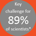
A key challenge for researchers hoping to address this “reproducibility crisis” is that there is no one cause of irreproducibility. Non-standardized methods and protocols, differing techniques and instrumentation used, a lack of standardized quality control, and complex workflows, along with laborious tasks prone to human error, are all critical factors influencing reproducibility.
In the resource below, we outline the top three challenges that cell biology labs currently face, provide top tips from expert cell biologists around the world, and explore the one-stop solution designed to mitigate the risk of irreproducibility.

A key challenge for those managing a lab or workflow is the varying levels of expertise and existing gaps in training for certain techniques or technologies.
The complex analysis tools that are required to provide relevant and robust insights often need expert knowledge to not only operate but also to analyze the subsequent data. For some labs, it can be a challenge to keep up with the expertise needed to optimize the use of these analytical tools.
To address the challenges associated with missing or novice know-how, check out these top tips from cell biology experts:
See the below resources for further assistance on how you can best address varying levels of expertise.
How can you revolutionize your cell assays without the expertise of a core lab?
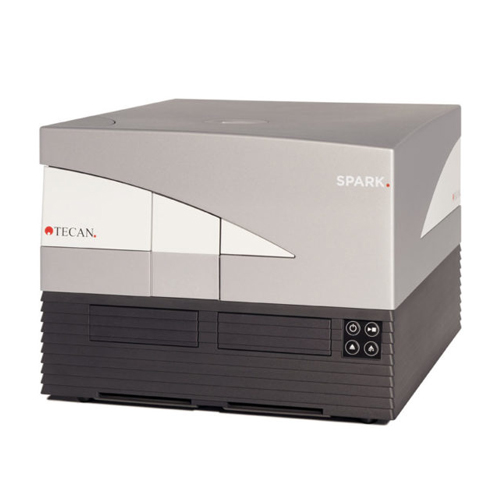
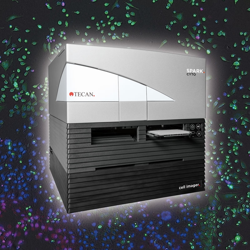
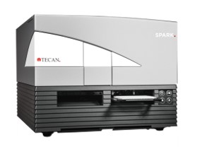
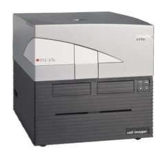
The Spark and Spark Cyto multimode readers with cell imaging are designed to simplify complex methods - from whole-well to single-cell and 3D models - in one easy-to-use system, without the need for dedicated experts.
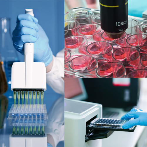
Issues with reproducibility often arise when labs try to combine various approaches - whether to consolidate data, implement a new line of investigation, or in preparation of collaboration with other labs.
The use of multiple different techniques and instrumentation can mean that the analysis of cells takes place at different time points and under different conditions –for example, analyzing cell viability on a microplate reader, counting cells under a microscope, and quantifying signals with a flow cytometer. This variation in approaches can lead to inconsistent data if not appropriately controlled.
To help consolidate your methodology and reduce the risk of data inconsistency, adhere to the following top tips:
For more guidance on how to ensure standardized, optimized, and well-planned workflows and achieve robust experimental practices in your cell biology research, take a look at the free resources below.
Ready to multiplex your experiments in a single instrument?
The Spark and Spark Cyto multimode readers with cell imaging are designed to standardize and automate a plethora of cell assays, while offering multiplexing capabilities and providing full experimental control for better results with fewer replicates.
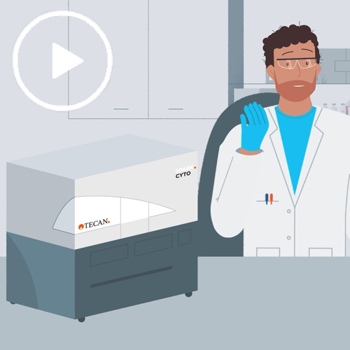
Watch video
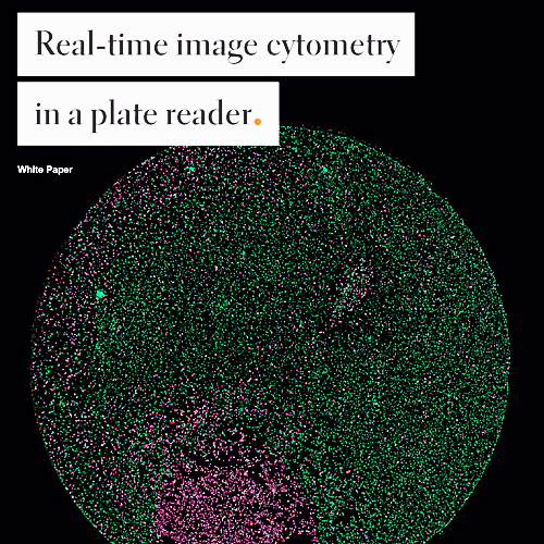
Free download
Free download
Expert webinar

A key pain point for laboratories around the world is the laborious and manual nature of certain tasks within a workflow.
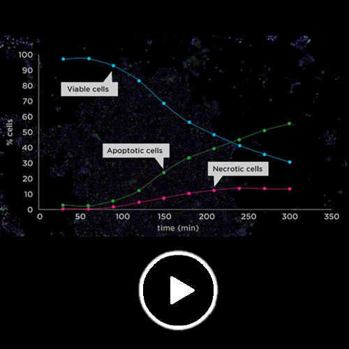
The need to measure multiple different cell parameters, at different time points, and under different conditions, increases the risk of variability in results. As a result, scientists often try to overcome this by conducting live-cell analysis, which involves taking measurements at different time points from the same cells under the same conditions. However, this is a very laborious approach, often requiring lab attendances over weekends or at night.
To overcome this laborious approach, cell biology experts recommend the following:
Explore the resources below to find out more about how automation can relieve your live-cell imaging workload.
Ready for live-cell assays without spending weekends in the lab?
Never miss a critical biological event with the Spark and Spark Cyto multimode readers with cell imaging that are designed to fully automate your live-cell assays, with complete environmental control for temperature, gas, and humidity to enable long-term live-cell assays.
Free download
Free download
Free download
Expert webinar

The Spark and Spark Cyto platforms from Tecan are designed for the seamless integration and combination of various functions, as outlined in the figure below. Hover over the features to find out more.
Cell incubation &
live-cell analysis
Temperature and gas
control, and evaporation protection
Smart automation
Integrated plate stacker extension, automated lid lifting, and seamless integration in
robotic solutions simplify and automate experiments
Low-volume DNA/RNA
quantification
Using just 2ul sample detecting down to 1 ng/ul
UV/vis spectroscopy
Plate
and cuvette-based spectroscopy offering ultra-fast absorbance scans and single wavelength assays (ELISA)
Imaging & microscopy
(Spark Cyto only) integrated automated brightfield and multichannel fluorescence
microscopy
Benchtop single-cell cytometry
(Spark Cyto only) For cell proliferation, apoptosis, cytotoxicity, and many more
studies
Fluorescence & luminescence assays
Enabling high-throughput assays such as fluorescence polarization (FP), TR-FRET, and
ALPHA screening
Benchtop cell counting
(Spark only) Accurate cell count and viability assessment for up to eight samples/run
in less than 30 seconds/sample
Please note that the Spark and Spark Cyto are for research use only
The Spark and Spark Cyto platforms from Tecan are designed for the seamless integration and combination of various functions, including:
Please note that the Spark and Spark Cyto are for research use only

*The advice highlighted in this resource is based on the results of a survey conducted
by SelectScience® in partnership with Tecan®.
**Certain images and/or photos on this page are the copyrighted property of 123RF.com,
its contributors or its licensed partners and are being used with permission under the relevant license. These
images and/or photos may not be copied or downloaded without permission from 123RF.com. Other images courtesy
of Tecan®.