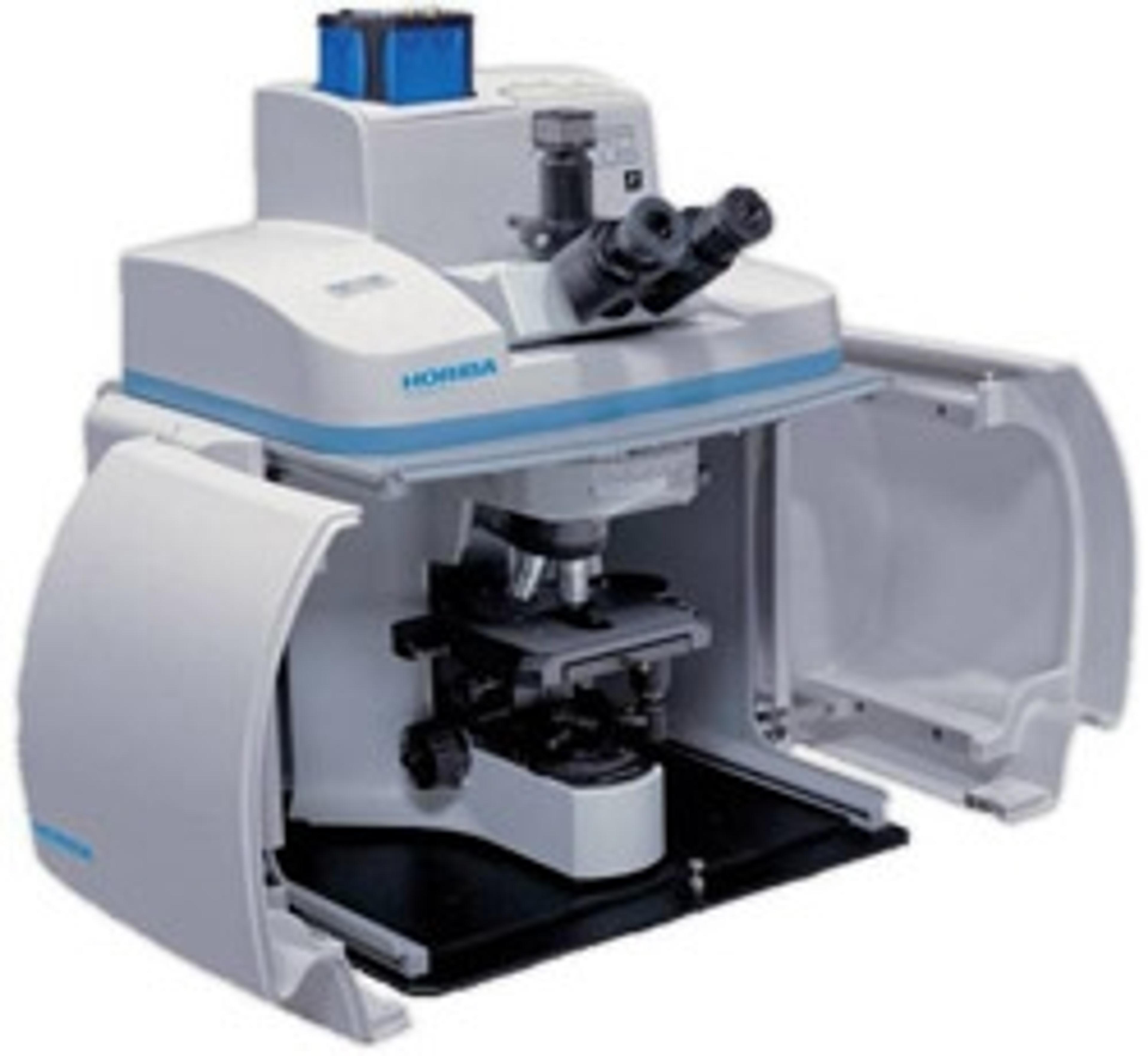
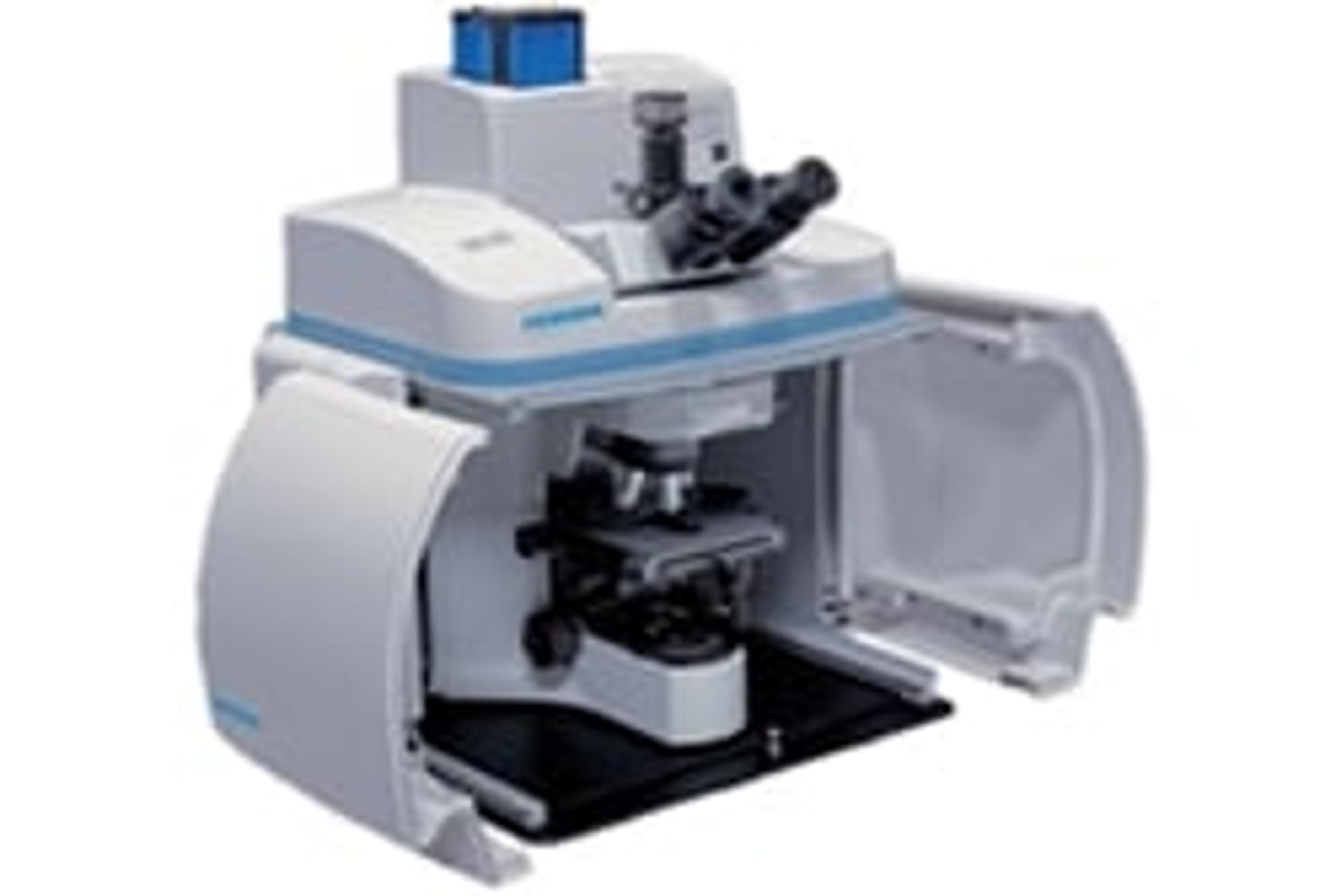
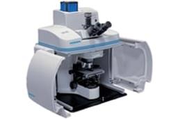
XploRA™ PLUS
Raman imaging has never been so fast! Incorporating unique and powerful functions in a reliable, high performance system, ideally suited to the research and analytical lab, the XploRA PLUS is our best multi-sample, multi-user Raman microscope ever.It is fully confocal, not compromising image quality, spatial or depth resolution. The SWIFT Fast Raman images are the fastest fully confocal Raman images available, typically 10x f…


The supplier does not provide quotations for this product through SelectScience. You can search for similar products in our Product Directory.
Great product that can be relied on!
Polymorphic form detection
Great combination of microscope and Raman spectrometer.
Review Date: 21 May 2021 | HORIBA Scientific
Amazing results! In every run I recieved perfect results.
Diamond like materials
Currently I am a user of this model and I obtained the best results from my samples. All data is reliable and reproducible. I have had a very great experience with this model.
Review Date: 16 Mar 2021 | HORIBA Scientific
Good equipment.
Materials
Easy to use and good results.
Review Date: 30 Oct 2020 | HORIBA Scientific
Solid instrument, excellent price.
Mineralogy
The XploRA PLUS has everything you need to do Raman mineralogical analyses. The LabSpec 6 software is fairly easy to use and intuitive, although there are certainly improvements that could be made. All in all, this is an excellent instrument for the price when compared with other instruments in this class.
Review Date: 8 Jun 2020 | HORIBA Scientific
Great intelligent instrument and software, reliable results with high resolution.
2D materials
The instrument is easily operated and quite efficient. It provides us with reproducible and high-quality Raman results for 2D materials. The software is well-designed for the instrument and has many practical functions for processing and analyzing the spectra. I like the automotive sample platform, which can be controlled with the software. This allows us to precisely test even a point on the 2D material.
Review Date: 4 Jun 2020 | HORIBA Scientific
Raman is providing us a lot of good results
Neurobiology and diagnosis
The instrument is efficient and easy to use but the service and the after-sales care is almost zero, not only for the Raman spectrometer but also for other instruments from Horiba.
Review Date: 20 Jan 2020 | HORIBA Scientific
This is a very versatile system and great product service.
Nanomaterial analysis
The system comes with user-friendly analytical software for the rapid analysis of materials. The data can be analyzed in real time and stored easily for future detailed results.
Review Date: 20 Jan 2020 | HORIBA Scientific
Raman imaging has never been so fast!
Incorporating unique and powerful functions in a reliable, high performance system, ideally suited to the research and analytical lab, the XploRA PLUS is our best multi-sample, multi-user Raman microscope ever.
It is fully confocal, not compromising image quality, spatial or depth resolution. The SWIFT Fast Raman images are the fastest fully confocal Raman images available, typically 10x faster than conventional Raman imaging.
The simplicity and power of the XploRA PLUS is unmatched with an enhanced range of options such as multiple laser wavelengths, EMCCD detection, Raman polarisation and even Raman-AFM combination.
XploRA™PLUS Features:
- SWIFTTM 10x faster Raman imaging
- Improved detection and sensitivity
- Full Confocality for complete image detail
- Full optical microscope so you can see your samples
- Maximum detail, resolution and range for enhanced spectroscopy
- HORIBA’s OneClick easy Raman analysis
- NIST traceable and patented Autocalibration options for validated results
- Ultimate optical stability- robust, reliable, long term operation
- Automated operation offering simple, powerful reliability
- 2 year base unit warranty as standard
Brochures

Find the Raman system best suited to your applications
In this brochure, HORIBA highlights the company’s expertise and innovative solutions in Raman spectrometry, designed for industries like pharmaceuticals, life sciences, environmental science, and materials research.
HORIBA's advanced instruments deliver high sensitivity, resolution, and user-friendly operation for precise molecular analysis. This brochure explains Raman spectroscopy’s ability to measure molecular vibrations, providing insights into chemical composition and structural properties. It also features cutting-edge tools like NanoRaman and automated systems for enhanced imaging and data analysis. With the Raman Academy offering training, resources, and robust customer support, you can maximize your spectroscopy systems.
Advancing polymer research with Raman microscopy
Polymer science requires precise analytical techniques to understand chemical and structural properties at the molecular level. HORIBA’s Raman microscopy solutions provide powerful capabilities for polymer characterization, supporting research, quality control, and process monitoring with unmatched accuracy.
With Raman microscopy, researchers can:
- Characterize raw polymer materials with molecular precision
- Monitor polymerization processes, both inline and outline
- Investigate polymer orientation, crystallization, and structural changes
- Detect defects and analyze compound distribution for quality assurance and traceability
HORIBA explores how its Raman microscopy solutions can enhance your polymer research. Gain expert insights, real-world applications, and practical guidance for optimizing your analysis.
Pharmaceutical and cosmetic product analysis with confocal Raman spectroscopy
In the pharmaceutical and cosmetic industries, maintaining product consistency and quality is essential. The distribution of active compounds within tablets, creams, and emulsions directly impacts their efficacy. Confocal Raman spectroscopy provides a powerful, non-destructive solution for analyzing these formulations at a microscopic level, ensuring optimal compound distribution.
HORIBA describes how confocal Raman microscopy enhances product analysis. Learn about its applications in pharmaceutical tablets, cosmetic creams, and emulsions, and explore the advanced tools and techniques that ensure precise compound distribution.
Graphene characterization with Raman spectroscopy
Graphene’s remarkable electron transport properties, extreme mechanical strength, and high thermal conductivity make it revolutionary for next-generation nanoelectronic devices. With electronic mobilities exceeding 15,000 cm²V⁻¹s⁻¹ and a strength over 200 times that of steel, graphene is poised to play a key role in the future of ultrafast transistors, microcircuits, and computer chips.
However, accurately characterizing graphene — distinguishing layer numbers, assessing disorder, and understanding structural integrity — is crucial for its practical applications. Raman micro-spectroscopy has emerged as an essential tool for graphene research, offering:
- Non-destructive, high-resolution analysis of graphene’s structural properties
- Rapid and reliable identification of layer number and disorder impact
- Precise spectral and spatial resolution for material characterization
- A standard method for graphene-based device development
Explore how Raman spectroscopy enables fast, accurate, and reproducible graphene analysis, accelerating innovation in nanoelectronics.
Enhancing lithium-ion battery research with Raman spectroscopy
The growing demand for more powerful and efficient energy storage has made Lithium-ion batteries (LIBs) a critical focus of research and development. To optimize battery performance, it is essential to understand the structural and chemical changes occurring in cathodes and anodes during charge and discharge cycles.
Raman spectroscopy offers a fast, contactless, and highly informative method for analyzing these changes in real time — without altering the sample. It enables researchers to:
- Investigate structural and electronic modifications in battery materials
- Monitor reversible and irreversible changes during charge/discharge cycles
- Support failure analysis and quality control with automated, high-throughput measurements
- Characterize new materials for next-generation energy storage solutions
Explore how Raman spectroscopy can enhance your LIB research, from fundamental studies to industrial quality control.
The Raman spectroscopy handbook
Explore the power of Raman spectroscopy with this comprehensive handbook from HORIBA, designed for researchers, scientists, and industry professionals. This essential guide explores the fundamentals, applications, and cutting-edge advancements in Raman analysis.
What’s inside?
- Introduction to Raman spectroscopy: Learn how this non-destructive technique reveals molecular structures and material properties
- Principles of Raman scattering: Understand how laser light interacts with molecular bonds to produce unique spectral fingerprints
- Key applications: Explore its use in pharmaceuticals, life sciences, geology, semiconductors, and materials science
- Comparison with other techniques: See how Raman spectroscopy stacks up against FTIR, XRD, and mass spectrometry in speed, sample preparation, and molecular detail
- Advanced Raman systems: Discover innovations like confocal Raman microscopy, hybrid solutions, ultra-fast imaging, and TERS/TRS techniques for enhanced precision
- Practical insights: Get guidance on sample requirements, system components, and the latest technological advancements
Microplastics: HORIBA Scientific's guide to sampling, preparation, analysis, and technology
In this eBook, HORIBA Scientific presents its complete guide to the investigation of microplastics within sediment, biota, food, and water samples. Enclosed is a step-by-step analysis workflow covering microplastics sampling, sample preparation, filtration, data acquisition, and data analysis.
Raman Imaging of Monkey Brain Tissue
Fast and non-invasive methods for clinical and non clinical investigations for biological tissue are more and more required. This application note presents a method combining Raman spectroscopy and microscopy in a fully confocal instrument for the analysis of monkey brain tissue without chemical labeling.

The science of beauty: Exploring characterization techniques in cosmetics
The creation of innovative cosmetics products with demonstrated effects relies on deep scientific knowledge of biological matrices (skin, hair, tooth), the development of efficient and safe active ingredients, optimized formulations, and an instrumental evaluation of the performance.
In this webinar, Florian Formanek, Global Life Science Market Manager at HORIBA will discuss how HORIBA instruments cover a large panel of cosmetic testing applications, from particle size characterization of emulsions, pigments or fillers, through to the molecular analysis of hair chemistry, surface functionalization with smart coatings or tribology analysis, to the assessment of formulation composition and stability. They also cover spreading on substrates or interaction with packaging, to contamination or nanotoxicity studies, to the ex vivo or in vivo objectivation of endogenous skin compounds, topical actives penetration, and the impact of environmental factors (UV exposure, blue light, pollution).
Key learning objectives
- Discover how rapid Raman chemical mapping can provide information on the surface physicochemical behavior of formulations
- Learn how to use fluorescence spectroscopy for the analysis and quality control of raw materials (such as essential oils, natural extracts, hair dyes, or vitamins), as well as for the investigation of endogenous skin markers and actives deposits
- Explore the applications of atomic force microscopy in the nanoscale characterization of materials, coatings, and biological matrices
Who should attend?
- R&D scientists in cosmetics, personal care, chemistry, and pharma companies with activities in beauty and healthcare
- Academics working in the field of skin and hair research (including dermatology and pharmacy)
- Clinical research organizations (CROs)
Certificate of attendance
All webinar participants can request a certificate of attendance, including a learning outcomes summary, for continuing education purposes.
A guide to performance and applications of Raman microscopy
Raman microscopy plays a vital role in material science, chemistry, and life sciences, enabling precise molecular analysis at the microscale. Understanding what makes a Raman microscope truly effective is essential for achieving accurate and reproducible results.
In this in-depth video on Raman microscopy, HORIBA explores the essential characteristics that define a high-performance Raman microscope. Led by expert application scientists, this session delves into crucial evaluation methods, measurement techniques, and real-world demonstrations. Gain valuable insights into optimizing your Raman microscopy setup for precise and reliable analysis.
Over 50 years of Raman spectroscopy
Raman spectroscopy, first discovered by Sir C.V. Raman in 1928, is a powerful analytical technique used to study molecular structures by analyzing light scattering. Initially, its adoption was limited due to weak signal detection and interference from fluorescence and stray light. Early Raman instruments relied on mercury arc lamps and photographic plates, requiring long exposure times. The introduction of lasers in the 1960s transformed the field, paving the way for the first commercial Raman spectrometers in 1966.
In this video, HORIBA celebrates being a leader in Raman spectroscopy innovation for over 50 years, driving advancements in precision and efficiency.
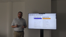
How to tailor Raman microscopy to your application
Raman microscopy is rapidly evolving from a high-end analytical technique to a widely adopted standard across various industries. To fully harness its potential, it’s essential to tailor the approach to your specific application. This involves not only expert insights and best practices but also dedicated tools, accessories, and software solutions designed to enhance performance and efficiency.
In this video, learn how to tailor your Raman microscopy for:
- Polymers
- Microplastics
- Batteries
- Pharmaceuticals
- Materials Science
Understanding the Raman effect
In this video, Thibault Brulé, Raman Product Manager at HORIBA, breaks down the Raman effect with real-world examples. Discover the origins of this powerful phenomenon and learn how Raman spectroscopy provides critical insights into molecular structures and material properties.
Topics covered:
- What is the Raman effect?
- How does it work?
- What kind of information can Raman spectroscopy reveal?

Raman spectroscopy as a practical solution to today's characterization challenges: Your questions answered
Learn about the advances in hardware and software that have made today’s instrumentation accessible to non-Raman experts
HORIBA presents 2020 Young Fluorescence Investigator Award
Dr. Jelle Hendrix, Assistant Professor at the Biomedical Research Institute of Hasselt University in Belgium, was presented with this honor