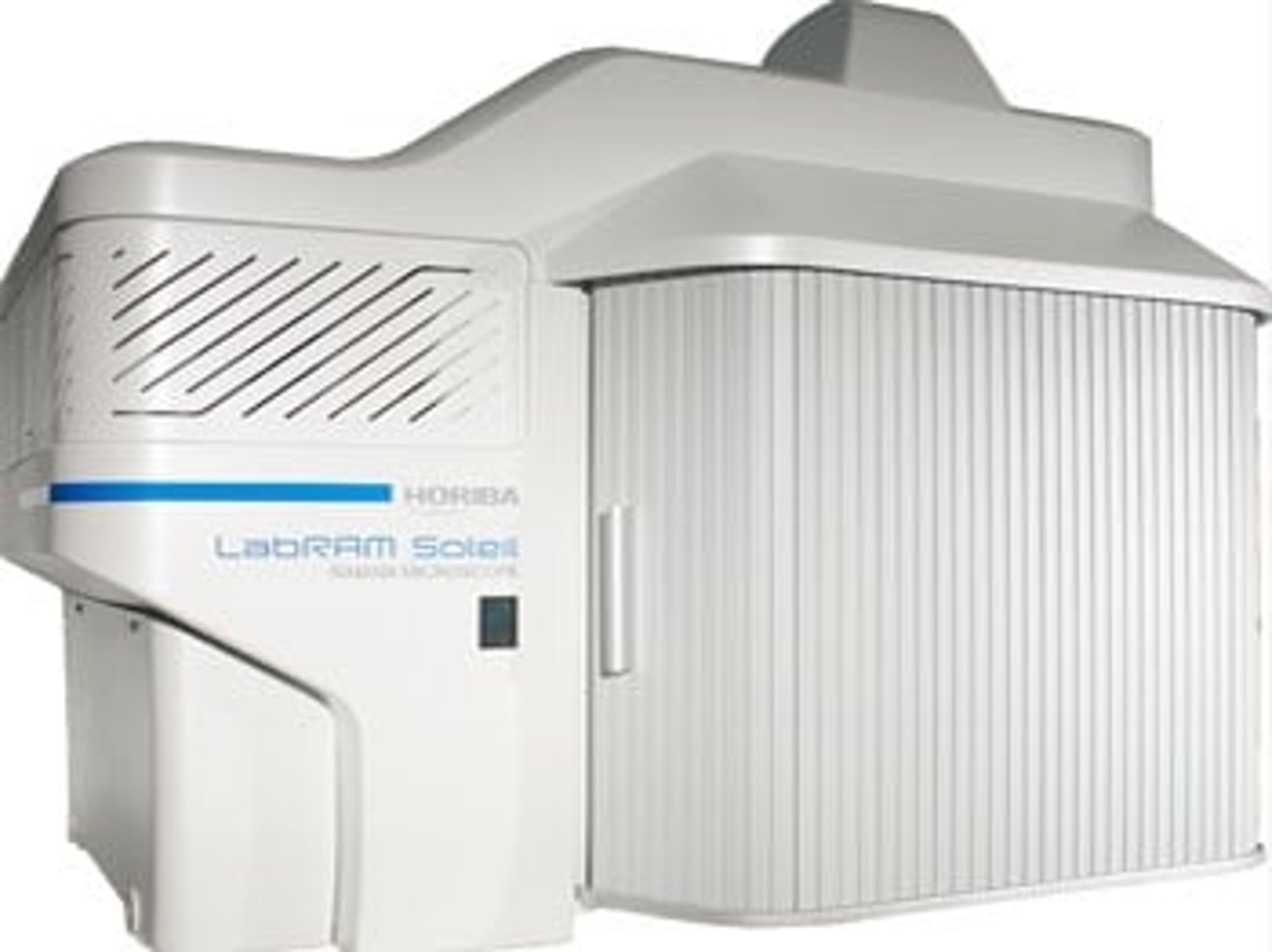
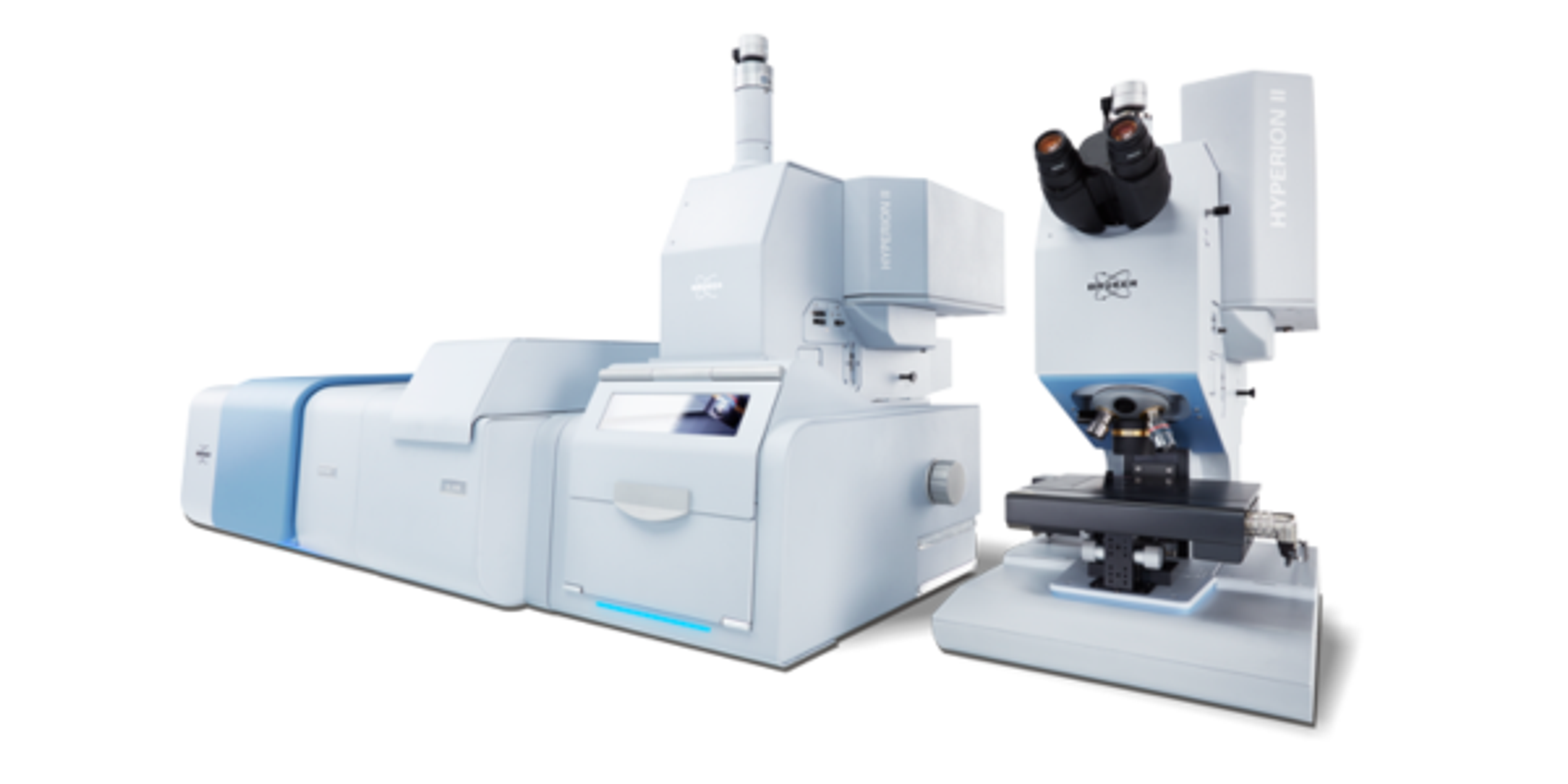
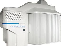
LabRAM Soleil™
Raman multimodal microscope


The supplier does not provide quotations for this product through SelectScience. You can search for similar products in our Product Directory.
Excellent product overall.
Material Laboratory
Easy to use. The provided software is user-friendly. Especially useful for the identification of plastic particles.
Review Date: 11 Dec 2025 | HORIBA Scientific
Great results and accuracy
Analyze metabolites
It was a good experience using this.
Review Date: 30 Jul 2022 | HORIBA Scientific
Great results
Analyze Airobilogical sample
This is nice product for biological research field
Review Date: 5 May 2022 | HORIBA Scientific
Simply essential.
Raman Image
Apart from an excellent after-sales service, the simplicity of use for new training sessions and the reliability of the results make it an indispensable piece of equipment in our laboratory.
Review Date: 16 Dec 2021 | HORIBA Scientific
Nice mapping images.
Materials
Easy to use. We can use anti-alignment if optical is missaligned. It's labor free for optical maintenance.
Review Date: 9 Feb 2021 | HORIBA Scientific
Most reliable and productive analytical instrument of the year!
Material Science
It's an amazing product! The built-in intuitive software is easy to use. It allows characterization of all types of samples, whether they are flat, rough or particulate. Easy to access and ultra fast. Most amazing product for this year.
Review Date: 9 Feb 2021 | HORIBA Scientific
It's a must have instrument in a modern lab
Raman spectroscopy, imaging
A new versatile imager which paves the way for fast and inexpensive chemical imaging.
Review Date: 9 Feb 2021 | HORIBA Scientific
This is a high end research tool, most labs would love to have it.
Raman spectroscopy
This is one of the best in the market product with many advanced features and lot of flexibility for users. Smart system.
Review Date: 9 Feb 2021 | HORIBA Scientific
I can't imagine our lab without ray of sunshine!
Raman Imaging
Fast Raman mapping as a result of flexible imaging options. Cuts my speed of measurements significantly. Very easy intuitive and easy-to-use software. Great after-sales care, in particular from the applications team manager Bridget O'Donnell. Looking forward to more collaborations and working with Horiba and this product. The product is very competitively priced given all its features.
Review Date: 9 Feb 2021 | HORIBA Scientific
Great value of money.
Research
Very powerful equipment with several useful features, and they have the best after sales support for their instruments.
Review Date: 9 Feb 2021 | HORIBA Scientific
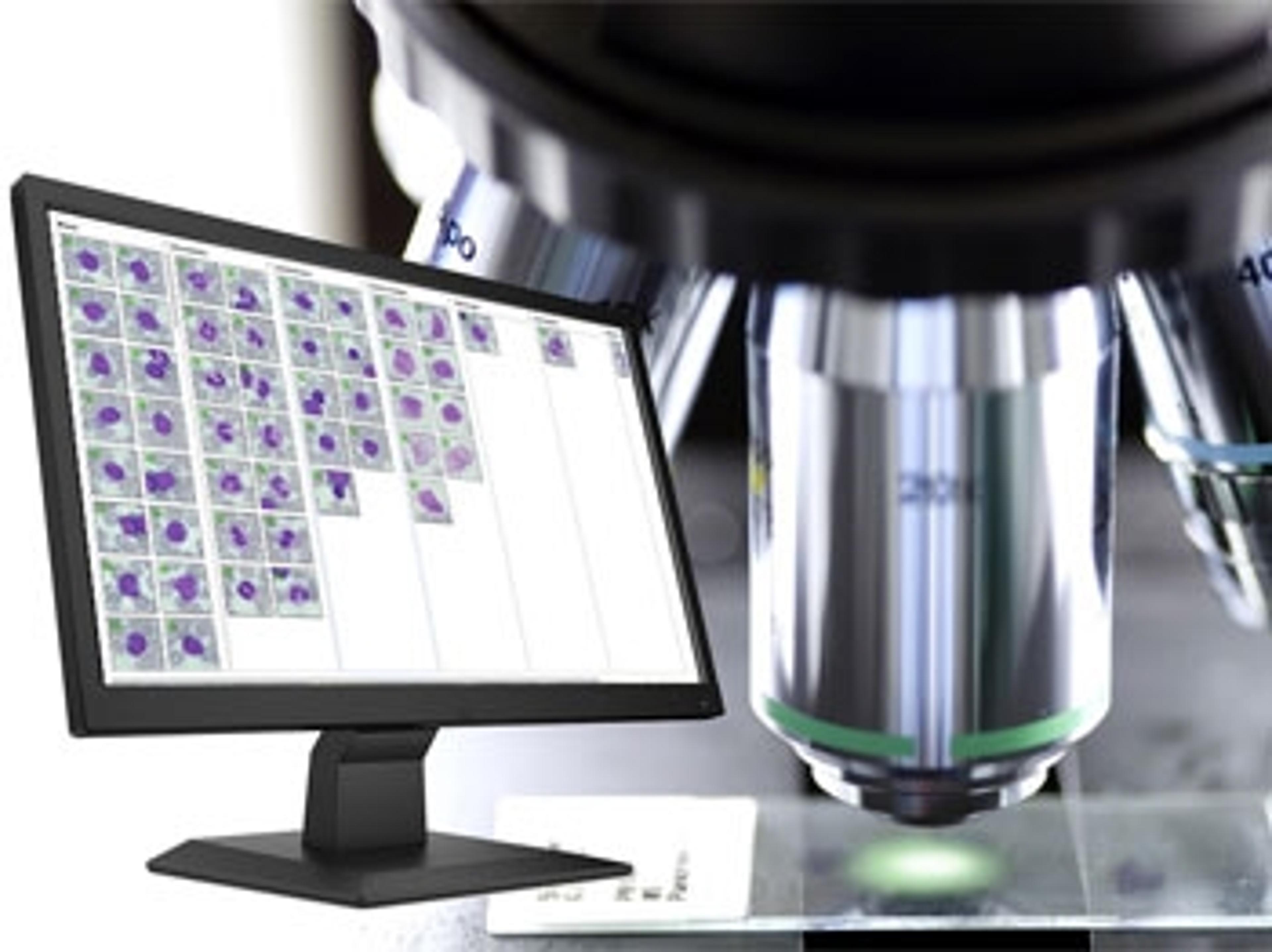
The result of 50 years of innovation by leading instrument manufacturer HORIBA Scientific, the LabRAM Soleil™ multimodal microscope has been designed with application experts to supercharge your analysis and set the new standard for Raman imaging and spectroscopy. It features advanced automation allowing a true self-operation. Ultrafast imaging, up to 100 times faster than a conventional Raman spectrometer, is performed thanks to the innovative SmartSampling™ technology. The patented QScan™ feature offers lightsheet confocal imaging. As shown in our brochure, it presents a robust design and is driven by an intuitive software. Therefore, this Raman microscope is your companion of choice to get your results fast and give you access to new domains of application.
Brochures

Find the Raman system best suited to your applications
In this brochure, HORIBA highlights the company’s expertise and innovative solutions in Raman spectrometry, designed for industries like pharmaceuticals, life sciences, environmental science, and materials research.
HORIBA's advanced instruments deliver high sensitivity, resolution, and user-friendly operation for precise molecular analysis. This brochure explains Raman spectroscopy’s ability to measure molecular vibrations, providing insights into chemical composition and structural properties. It also features cutting-edge tools like NanoRaman and automated systems for enhanced imaging and data analysis. With the Raman Academy offering training, resources, and robust customer support, you can maximize your spectroscopy systems.
Advancing polymer research with Raman microscopy
Polymer science requires precise analytical techniques to understand chemical and structural properties at the molecular level. HORIBA’s Raman microscopy solutions provide powerful capabilities for polymer characterization, supporting research, quality control, and process monitoring with unmatched accuracy.
With Raman microscopy, researchers can:
- Characterize raw polymer materials with molecular precision
- Monitor polymerization processes, both inline and outline
- Investigate polymer orientation, crystallization, and structural changes
- Detect defects and analyze compound distribution for quality assurance and traceability
HORIBA explores how its Raman microscopy solutions can enhance your polymer research. Gain expert insights, real-world applications, and practical guidance for optimizing your analysis.
Pharmaceutical and cosmetic product analysis with confocal Raman spectroscopy
In the pharmaceutical and cosmetic industries, maintaining product consistency and quality is essential. The distribution of active compounds within tablets, creams, and emulsions directly impacts their efficacy. Confocal Raman spectroscopy provides a powerful, non-destructive solution for analyzing these formulations at a microscopic level, ensuring optimal compound distribution.
HORIBA describes how confocal Raman microscopy enhances product analysis. Learn about its applications in pharmaceutical tablets, cosmetic creams, and emulsions, and explore the advanced tools and techniques that ensure precise compound distribution.
Graphene characterization with Raman spectroscopy
Graphene’s remarkable electron transport properties, extreme mechanical strength, and high thermal conductivity make it revolutionary for next-generation nanoelectronic devices. With electronic mobilities exceeding 15,000 cm²V⁻¹s⁻¹ and a strength over 200 times that of steel, graphene is poised to play a key role in the future of ultrafast transistors, microcircuits, and computer chips.
However, accurately characterizing graphene — distinguishing layer numbers, assessing disorder, and understanding structural integrity — is crucial for its practical applications. Raman micro-spectroscopy has emerged as an essential tool for graphene research, offering:
- Non-destructive, high-resolution analysis of graphene’s structural properties
- Rapid and reliable identification of layer number and disorder impact
- Precise spectral and spatial resolution for material characterization
- A standard method for graphene-based device development
Explore how Raman spectroscopy enables fast, accurate, and reproducible graphene analysis, accelerating innovation in nanoelectronics.
Enhancing lithium-ion battery research with Raman spectroscopy
The growing demand for more powerful and efficient energy storage has made Lithium-ion batteries (LIBs) a critical focus of research and development. To optimize battery performance, it is essential to understand the structural and chemical changes occurring in cathodes and anodes during charge and discharge cycles.
Raman spectroscopy offers a fast, contactless, and highly informative method for analyzing these changes in real time — without altering the sample. It enables researchers to:
- Investigate structural and electronic modifications in battery materials
- Monitor reversible and irreversible changes during charge/discharge cycles
- Support failure analysis and quality control with automated, high-throughput measurements
- Characterize new materials for next-generation energy storage solutions
Explore how Raman spectroscopy can enhance your LIB research, from fundamental studies to industrial quality control.
The Raman spectroscopy handbook
Explore the power of Raman spectroscopy with this comprehensive handbook from HORIBA, designed for researchers, scientists, and industry professionals. This essential guide explores the fundamentals, applications, and cutting-edge advancements in Raman analysis.
What’s inside?
- Introduction to Raman spectroscopy: Learn how this non-destructive technique reveals molecular structures and material properties
- Principles of Raman scattering: Understand how laser light interacts with molecular bonds to produce unique spectral fingerprints
- Key applications: Explore its use in pharmaceuticals, life sciences, geology, semiconductors, and materials science
- Comparison with other techniques: See how Raman spectroscopy stacks up against FTIR, XRD, and mass spectrometry in speed, sample preparation, and molecular detail
- Advanced Raman systems: Discover innovations like confocal Raman microscopy, hybrid solutions, ultra-fast imaging, and TERS/TRS techniques for enhanced precision
- Practical insights: Get guidance on sample requirements, system components, and the latest technological advancements
Packaging for the future
In this application note, HORIBA presents its solution to support R&D and QC labs in the challenges faced when designing greener plastic food packaging.
Microplastics: HORIBA Scientific's guide to sampling, preparation, analysis, and technology
In this eBook, HORIBA Scientific presents its complete guide to the investigation of microplastics within sediment, biota, food, and water samples. Enclosed is a step-by-step analysis workflow covering microplastics sampling, sample preparation, filtration, data acquisition, and data analysis.
Raman microscopy applied to polymer characterization: An overview
Raman microscopy is an excellent tool to address the polymer research. Raman microscopy can be used to characterize raw materials, to inline or outline monitor polymerization process, to investigate orientation and crystallization changes, and also to control the quality and traceability of genuine products, by understanding defects and compounds distribution. This application note present how HORIBA Raman microscopy solutions can support the polymer chemical and structural understanding.
Raman spectroscopy: Rapid QC of healthcare products
The development of Raman spectroscopy has come a long way since its discovery in 1928. Its combination with optical microscopy in the latest, next-generation confocal Raman microscopes has enabled fast, non-destructive, non-invasive hyperspectral imaging of samples without the need for sample preparation in most cases.
In this application compendium, we look at some of the wide-ranging applications of Raman microscopy to address various formulation challenges encountered by the pharmaceutical and cosmetics industries. In particular, we consider HORIBA Scientific’s latest confocal Raman imaging microscope, the LabRAM Soleil™, designed to make Raman imaging faster and easier than ever before.
Download the free eBook to discover how Raman microscopy can yield information on molecular structures, crystal phases, polymorphisms and much more, as we cover:
- Characterization of pharmaceuticals, including compound distribution, chemical homogeneity and more
- Characterization of cosmetics for perfect formulations
- Raman spectroscopy as an essential tool in pharma

The science of beauty: Exploring characterization techniques in cosmetics
The creation of innovative cosmetics products with demonstrated effects relies on deep scientific knowledge of biological matrices (skin, hair, tooth), the development of efficient and safe active ingredients, optimized formulations, and an instrumental evaluation of the performance.
In this webinar, Florian Formanek, Global Life Science Market Manager at HORIBA will discuss how HORIBA instruments cover a large panel of cosmetic testing applications, from particle size characterization of emulsions, pigments or fillers, through to the molecular analysis of hair chemistry, surface functionalization with smart coatings or tribology analysis, to the assessment of formulation composition and stability. They also cover spreading on substrates or interaction with packaging, to contamination or nanotoxicity studies, to the ex vivo or in vivo objectivation of endogenous skin compounds, topical actives penetration, and the impact of environmental factors (UV exposure, blue light, pollution).
Key learning objectives
- Discover how rapid Raman chemical mapping can provide information on the surface physicochemical behavior of formulations
- Learn how to use fluorescence spectroscopy for the analysis and quality control of raw materials (such as essential oils, natural extracts, hair dyes, or vitamins), as well as for the investigation of endogenous skin markers and actives deposits
- Explore the applications of atomic force microscopy in the nanoscale characterization of materials, coatings, and biological matrices
Who should attend?
- R&D scientists in cosmetics, personal care, chemistry, and pharma companies with activities in beauty and healthcare
- Academics working in the field of skin and hair research (including dermatology and pharmacy)
- Clinical research organizations (CROs)
Certificate of attendance
All webinar participants can request a certificate of attendance, including a learning outcomes summary, for continuing education purposes.
A guide to performance and applications of Raman microscopy
Raman microscopy plays a vital role in material science, chemistry, and life sciences, enabling precise molecular analysis at the microscale. Understanding what makes a Raman microscope truly effective is essential for achieving accurate and reproducible results.
In this in-depth video on Raman microscopy, HORIBA explores the essential characteristics that define a high-performance Raman microscope. Led by expert application scientists, this session delves into crucial evaluation methods, measurement techniques, and real-world demonstrations. Gain valuable insights into optimizing your Raman microscopy setup for precise and reliable analysis.
Over 50 years of Raman spectroscopy
Raman spectroscopy, first discovered by Sir C.V. Raman in 1928, is a powerful analytical technique used to study molecular structures by analyzing light scattering. Initially, its adoption was limited due to weak signal detection and interference from fluorescence and stray light. Early Raman instruments relied on mercury arc lamps and photographic plates, requiring long exposure times. The introduction of lasers in the 1960s transformed the field, paving the way for the first commercial Raman spectrometers in 1966.
In this video, HORIBA celebrates being a leader in Raman spectroscopy innovation for over 50 years, driving advancements in precision and efficiency.
How to tailor Raman microscopy to your application
Raman microscopy is rapidly evolving from a high-end analytical technique to a widely adopted standard across various industries. To fully harness its potential, it’s essential to tailor the approach to your specific application. This involves not only expert insights and best practices but also dedicated tools, accessories, and software solutions designed to enhance performance and efficiency.
In this video, learn how to tailor your Raman microscopy for:
- Polymers
- Microplastics
- Batteries
- Pharmaceuticals
- Materials Science
Understanding the Raman effect
In this video, Thibault Brulé, Raman Product Manager at HORIBA, breaks down the Raman effect with real-world examples. Discover the origins of this powerful phenomenon and learn how Raman spectroscopy provides critical insights into molecular structures and material properties.
Topics covered:
- What is the Raman effect?
- How does it work?
- What kind of information can Raman spectroscopy reveal?
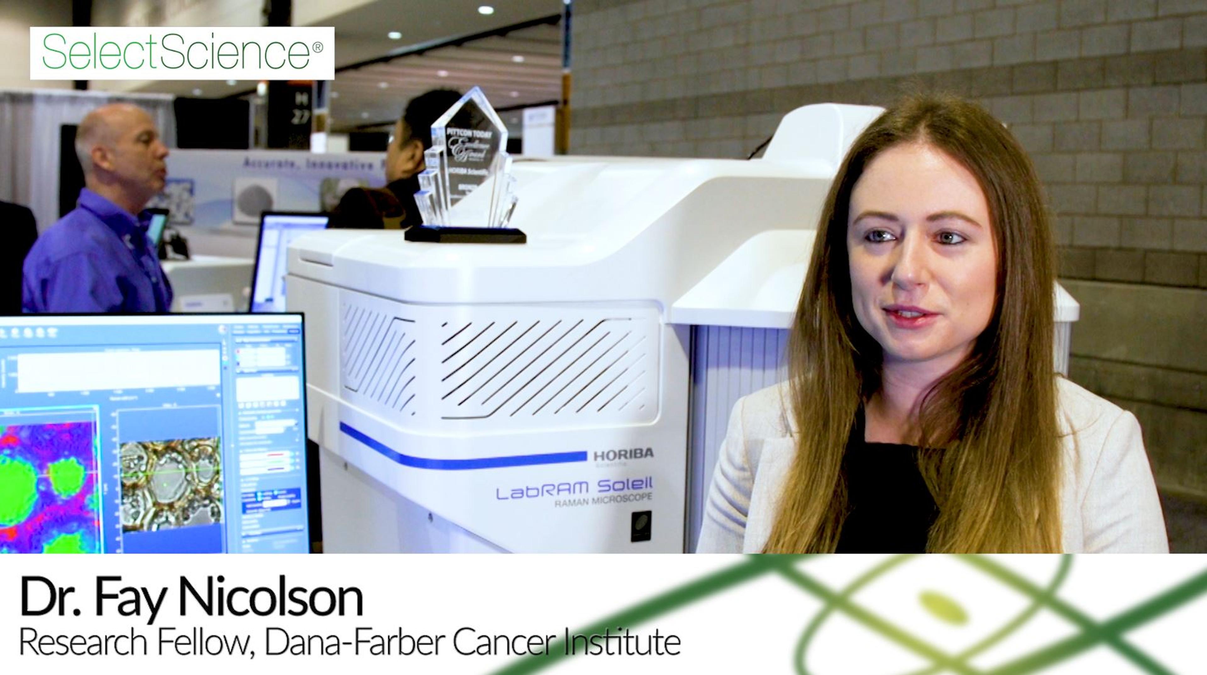
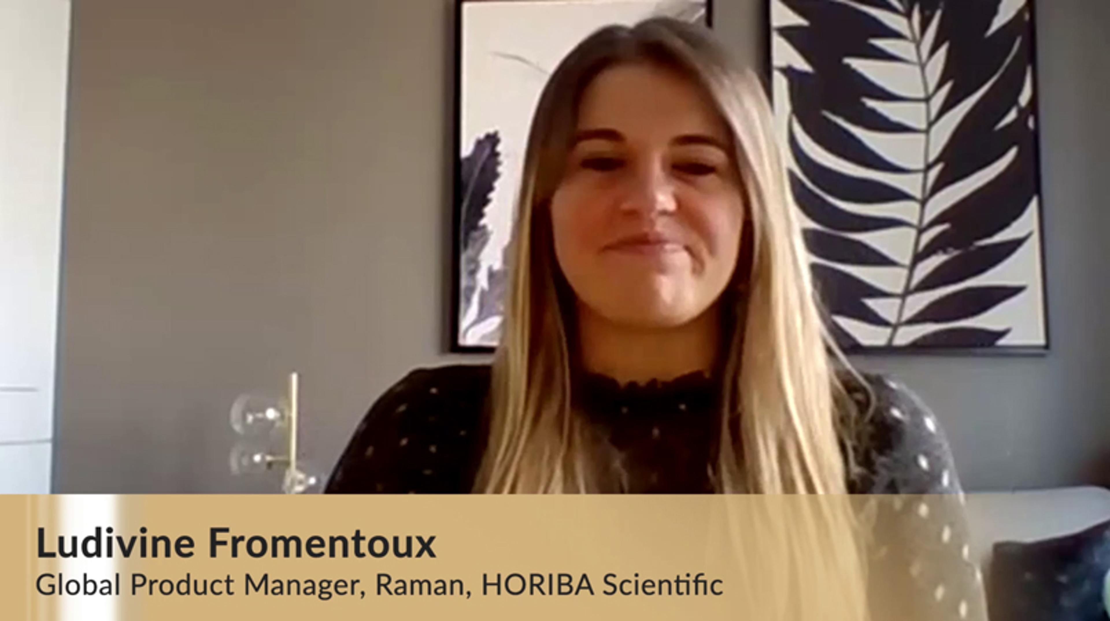
Life Sciences Video of the Year highlights Raman spectroscopy imaging in cancer research
The 2021 Scientists' Choice Award for Life Sciences Video of the Year has gone to an interview with Dr. Fay Nicolson, Dana-Farber Cancer Institute and Harvard Medical School, featuring technology by HORIBA Scientific.
Here, Ludivine Fromentoux, Global Product Manager, Raman, HORIBA Scientific, shares her thoughts on the award-winning video and explains how communication like this enable the company to better understand its customers’ needs.
Watch the winning video here: How to detect cancer in vivo using nanoparticles
How to detect cancer *in vivo* using nanoparticles
In this video, Dr. Fay Nicolson, from the Dana-Farber Cancer Institute and Harvard Medical School, shares how she uses nanoparticles to precisely image cancer in pre-clinical animal models. Nicolson explains why the lack of harmful, ionizing radiation makes Raman spectroscopy the technique of choice over other existing methods such as PET and CT, and discusses how the new LabRAM Soleil ensures fast, reliable spectral information, with the promise of translating this technology into the clinic.
This interview was filmed at Pittcon 2020 - watch other highlights from the show here >>

Raman spectroscopy as a practical solution to today's characterization challenges: Your questions answered
Learn about the advances in hardware and software that have made today’s instrumentation accessible to non-Raman experts

Free masterclass on Raman microscopy for pharma applications — watch on demand
In this SelectScience webinar, explore how Raman spectroscopy can be used to accelerate pharmaceutical studies

Micro-XRF & Raman microscopy: A winning combo for your elemental and chemical analyses
Watch this on-demand webinar to gain an understanding of how elemental and molecular characterization can be combined to help solve issues across many applications

Raman microscopy: Comprehensive characterization of polymers
Learn how to obtain accurate, non-destructive, information on polymers in this on-demand webinar

Raman microscopy: Comprehensive characterization of polymers
Join us on Monday, September 28, for this expert-led webinar on the intricacies of Raman technology

Video interviews: Cutting-edge research and technology in cancer, neuroscience, forensics and more
Meet the researchers making waves in science and learn about the technologies innovating their research


Pittcon 2020: 14 tech highlights to provide advanced analytical research solutions
Top innovations and technology news from the laboratory science conference and expo in Chicago, U.S.A.