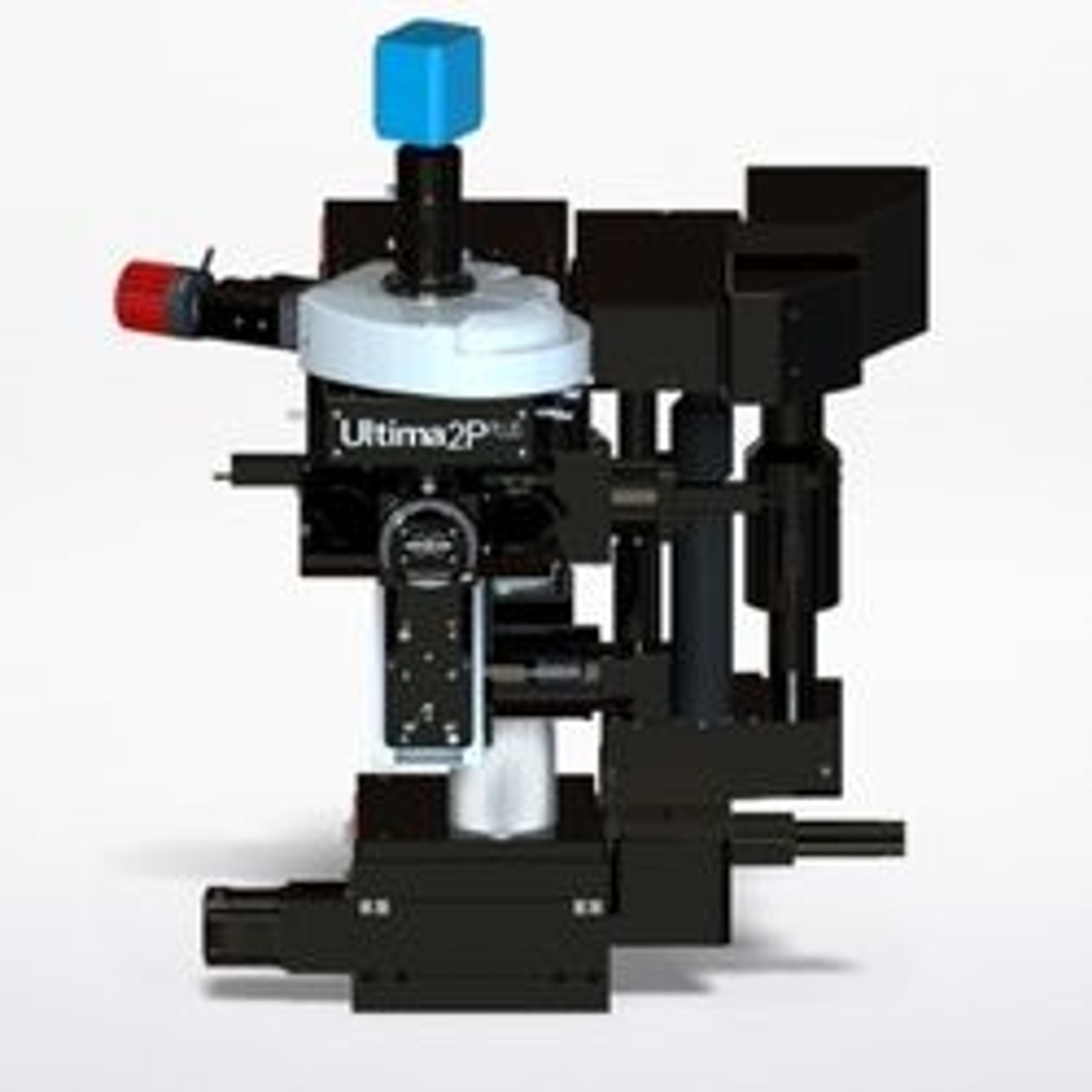
Ultima 2Pplus
With new advances in field of view, sensitivity, wavelength, and sample accommodation, the Ultima 2Pplus delivers an ideal combination of flexibility, resolution, imaging depth and speed, allowing simultaneous imaging, stimulation and electrophysiology protocols with greater efficiency and effectivity. The system is designed specifically for intravital imaging, with fully motorized control of the objective X-Y-Z position, as…

Ultima 2Pplus Two-Photon Microscope


The supplier does not provide quotations for this product through SelectScience. You can search for similar products in our Product Directory.
Ultima 2Pplus is a complete all-optical multiphoton workstation for imaging and optical manipulations. --- Bruker's Ultima 2Pplus has been at the forefront of many important scientific discoveries for nearly two decades and is a direct result of close collaboration and laboratory experience with leading neuroscientists around the world. This multiphoton workstation is giving researchers the necessary tools for deep, fast, and large field-of-view (50% larger to facilitate modern optogenetics experiments) imaging. In order to record cells with exact timing and spatial precision, the Ultima 2Pplus is equipped for simultaneous imaging and stimulation experiments in different planes. Capable of two-photon (2P) and three-photon (3P) microscopy, where 2 and 3 photons respectively are absorbed by the same fluorophore simultaneously, our technology is useful for imaging living biological tissues or animals at high resolution and in real-time. Furthermore, recent advancements include modulated focus enabling almost instantaneous imaging in different planes and voltage experiments, where membrane potentials from multiple neurons are simultaneously recorded, will have a transformative effect on neuroscience research.
Brochures

Ultima 2Pplus all-optical multiphoton workstation
In this product brochure, Bruker Fluorescence Microscopy presents the Ultima 2Pplus. With new advances in field of view, sensitivity, wavelength, and sample accommodation, the Ultima 2Pplus delivers an ideal combination of flexibility, resolution, imaging depth and speed, allowing simultaneous imaging, stimulation and electrophysiology protocols with greater efficiency and effectivity. The system is designed specifically for intravital imaging, with fully motorized control of the objective X-Y-Z position, as well as two axes of rotation for precise imaging orientation.
Studying human cortical organoids in the mouse retrosplenial cortex with two-photon imaging and electrophysiology
Here, Bruker Fluorescence Microscopy presents research from Madison Wilson and Martin Thunemann. Organoids are becoming an increasingly useful tool to study various neurological phenomena. Recently, a collaboration between neuroscientists and engineers at Boston University, University of California San Diego, and Salk Institute successfully integrated human cortical organoids in the adult mouse retrosplenial cortex. Two-photon (2P) imaging showed vascularization of the transplanted organoids and electrophysiology recordings showed a response to visual stimuli. For this study, Madison Wilson and Martin Thunemann share first authorship and both are excited by the implications of their collaborative study on evaluating the development, maturation, and functional integration of human neuronal networks within the mouse brain.
Monoamine oxidase enzymes contribute to mitochondrial oxidant stress and methamphetamine-induced neurodegeneration
Here, Bruker presents research from Dr. Steven Graves. Dr. Steven Graves is interested in investigating the pathogenesis of neurodegenerative diseases and drug-induced neurotoxicity. More specifically, he explores methamphetamine-induced neurodegeneration and the implications this has for developing neurodegenerative diseases such as Parkinson’s Disease. Methamphetamine increases cytosolic dopamine, which is metabolized by monoamine oxidase (MAO) enzymes. Historically, it was believed that cytosolic dopamine would auto-oxidize into a reactive quinone or be deaminated by MAO enzymes resulting in the generation of free electrons that were thought to contribute to cytosolic hydrogen peroxide production. However, during Dr. Graves’ work with Dr. D. James Surmeier at Northwestern University using multiphoton microscopy, they discovered that the electrons generated by MAO metabolism of dopamine were not released into the cytosol; rather, these electrons were transferred to the mitochondrial intermembrane space, thereby supporting the electron transport chain but also increasing oxidant stress.
Astrocytes play a starring role in sleep regulation
Here, Bruker Fluorescence Microscopy highlights Dr. Ashley Ingiosi's research. Dr. Ingiosi’s research focuses on the role of astrocytes, a type of glial cell in the brain, in sleep and sleep homeostasis. Sleep plays a critical role in overall mammalian health, although many aspects of sleep regulation are not yet understood. When Dr. Ingiosi was choosing her graduate project at the University of Michigan, she wanted to contribute novel advancements to her field. She noticed that most of what was known about sleep was based on the study of neurons, however, there was another brain cell type - glia - that was potentially important to sleep and there were not many in vivo studies looking at these non-neuronal cell types in the brain.
Defining the barriers to effective anti-tumor immunity in poor prognosis cancers and engineering cell therapies to break them down
Here, Bruker presents Dr. Paolo Provenzano research. Dr. Provenzano’s research program focuses on poor prognosis cancers including pancreatic cancer, which has one of the lowest survival rates of all cancers, as well as the poor prognosis subgroup of metastatic breast cancer. His lab takes a two-pronged approach to their research. The first prong is defining the barriers, both chemical and physical, to effective anti-tumor immunity - in other words, defining why certain areas of a tumor do not allow infiltration of T cells. This is a concept that Dr. Provenzano referred to as immune exclusion.
Imaging hippocampal microcircuitry using glass microperiscopes
Multiphoton microscopy (2P) overcomes many of the limitations that conventional microscopy faces by being able to image deeply, quickly, and in 3D — providing compelling and useful data in a variety of disciplines. A combination of ultra-fast pulsed lasers, lenses with high numerical apertures, and highly sensitive detectors provide fast Z imaging and makes Bruker’s 2P technology a go-to technique for revealing dynamic biological processes deep within living tissues. Here, Bruker summarizes the webinar content it presented with Michael Goard, PhD., an Associate Professor for the Department of Molecular, Cellular & Developmental Biology at UC Santa Barbara. His lab investigates the neural circuitry underlying perceptual, spatial, and cognitive abilities in the mouse cortex and hippocampus by using two-photon microscopy, microprisms, and microperiscopes.
<em>In vivo</em> imaging of microglia as the brain’s thermostat for tuning neuronal activity
Understanding the interaction between neurons and glia is critical to answering the most pertinent questions in neuroscience research. To understand brain cell dynamics, researchers must be able to image brain sections deeply and in vivo. Bruker’s Ultima Investigator™ multiphoton microscope is well-suited for neuroscience applications such as this, featuring a base system specifically optimized for in vivo studies while supporting deep, fast, and sensitive imaging. Here, Bruker summarizes the presentation given by Dr. Mario Merlini, assistant professor at the Institute of Blood and Brain at the University of Caen-Normandie, about his use of multiphoton microscopy and other techniques to address crucial research questions regarding the role of microglial cells in tuning neuronal activity in live mice.