Studying human cortical organoids in the mouse retrosplenial cortex with two-photon imaging and electrophysiology
18 Jul 2023Here, Bruker Fluorescence Microscopy presents research from Madison Wilson and Martin Thunemann. Organoids are becoming an increasingly useful tool to study various neurological phenomena. Recently, a collaboration between neuroscientists and engineers at Boston University, University of California San Diego, and Salk Institute successfully integrated human cortical organoids in the adult mouse retrosplenial cortex. Two-photon (2P) imaging showed vascularization of the transplanted organoids and electrophysiology recordings showed a response to visual stimuli. For this study, Madison Wilson and Martin Thunemann share first authorship and both are excited by the implications of their collaborative study on evaluating the development, maturation, and functional integration of human neuronal networks within the mouse brain.
Related products
Request Quote for All Products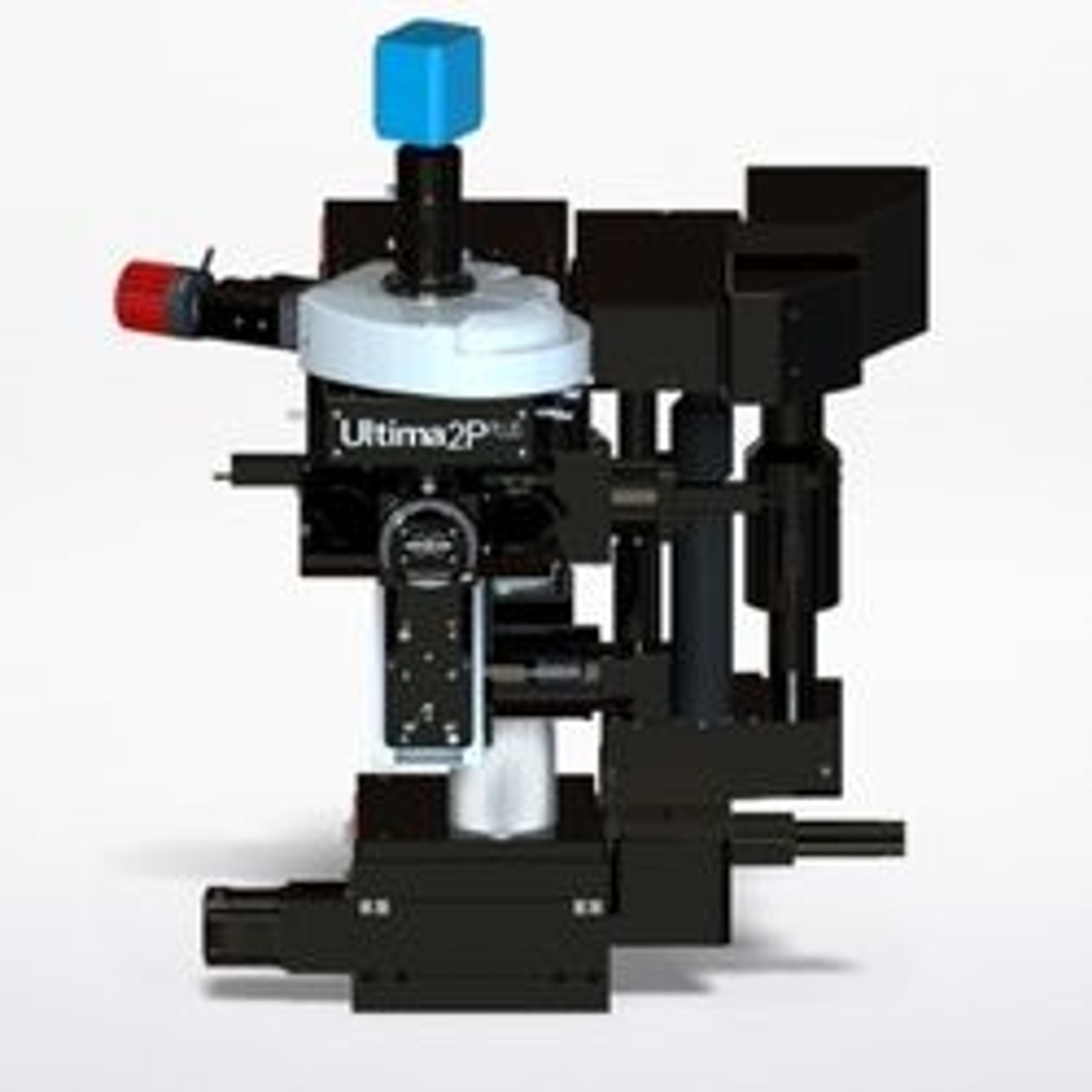
Ultima 2Pplus
Bruker Fluorescence MicroscopyWith new advances in field of view, sensitivity, wavelength, and sample accommodation, the Ultima 2Pplus delivers an ideal combination of flexibility, resolution, imaging depth and speed, allowing simultaneous imaging, stimulation and electrophysiology protocols with greater efficiency and effectivity. The system is designed specifically for intravital imaging, with fully motorized control of the objective X-Y-Z position, as well as two axes of rotation for precise imaging orientation.