Ultima 2Pplus all-optical multiphoton workstation
17 Jul 2023In this product brochure, Bruker Fluorescence Microscopy presents the Ultima 2Pplus. With new advances in field of view, sensitivity, wavelength, and sample accommodation, the Ultima 2Pplus delivers an ideal combination of flexibility, resolution, imaging depth and speed, allowing simultaneous imaging, stimulation and electrophysiology protocols with greater efficiency and effectivity. The system is designed specifically for intravital imaging, with fully motorized control of the objective X-Y-Z position, as well as two axes of rotation for precise imaging orientation.
Related products
Request Quote for All Products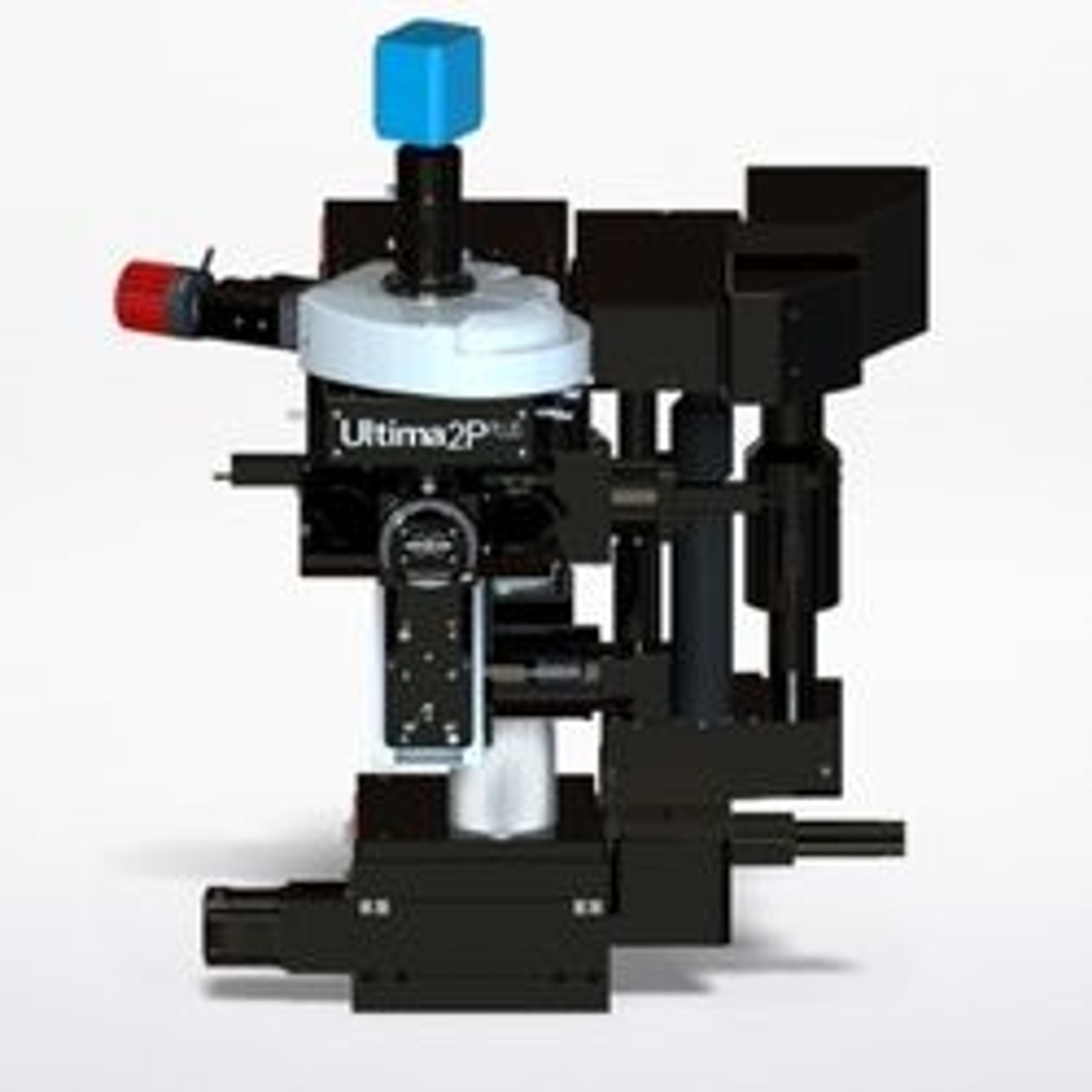
Ultima 2Pplus
Bruker Fluorescence MicroscopyWith new advances in field of view, sensitivity, wavelength, and sample accommodation, the Ultima 2Pplus delivers an ideal combination of flexibility, resolution, imaging depth and speed, allowing simultaneous imaging, stimulation and electrophysiology protocols with greater efficiency and effectivity. The system is designed specifically for intravital imaging, with fully motorized control of the objective X-Y-Z position, as well as two axes of rotation for precise imaging orientation.