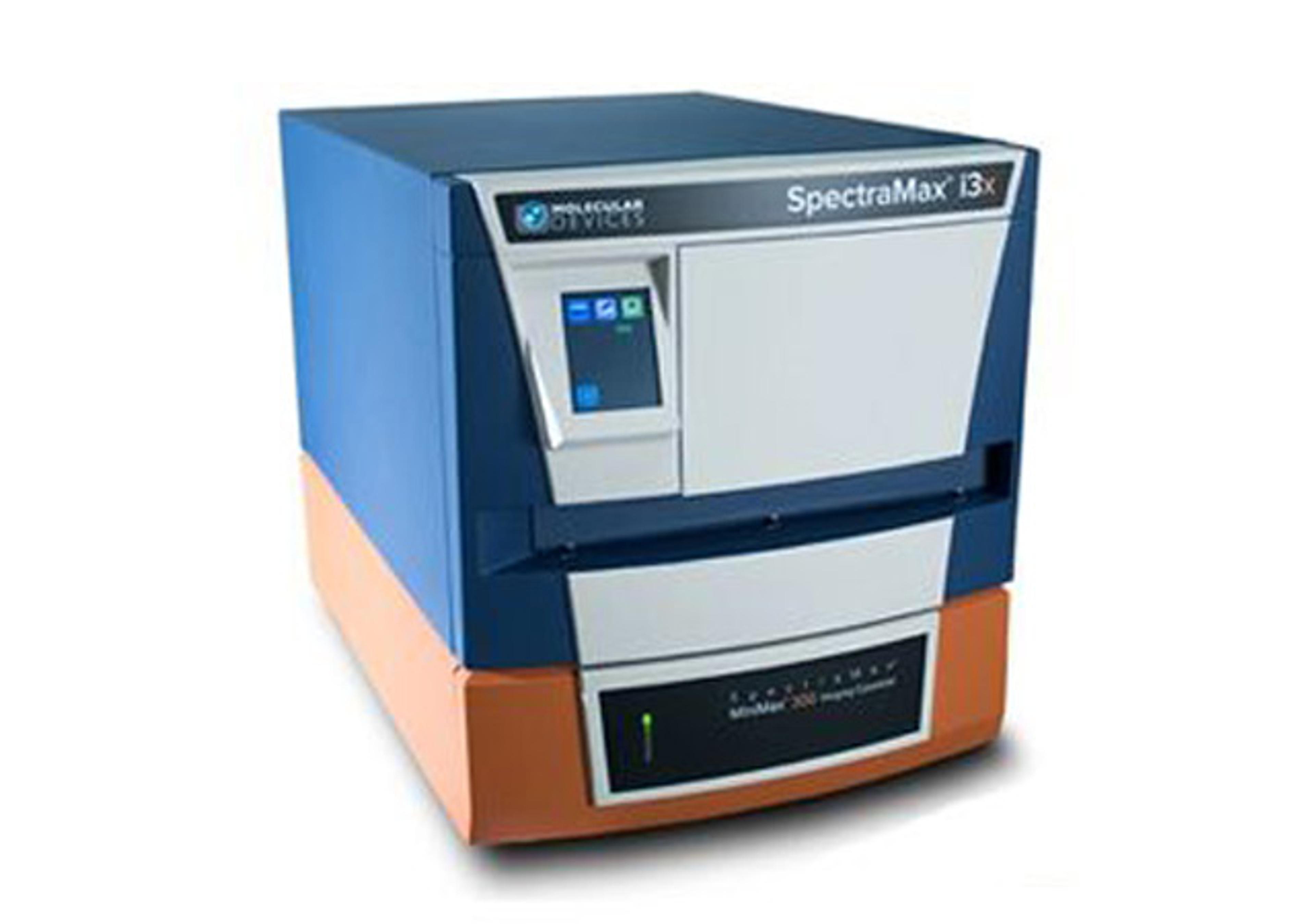
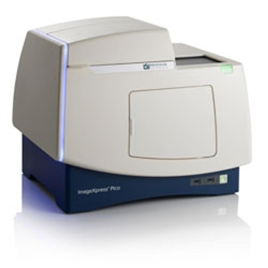
ImageXpress Pico Automated Cell Imaging System
Affordable cellular imaging and analysis system for individual biology labs - go from sample to results in minutes


Receive your quote directly from the manufacturer.
Great, reproducible results, easy to use.
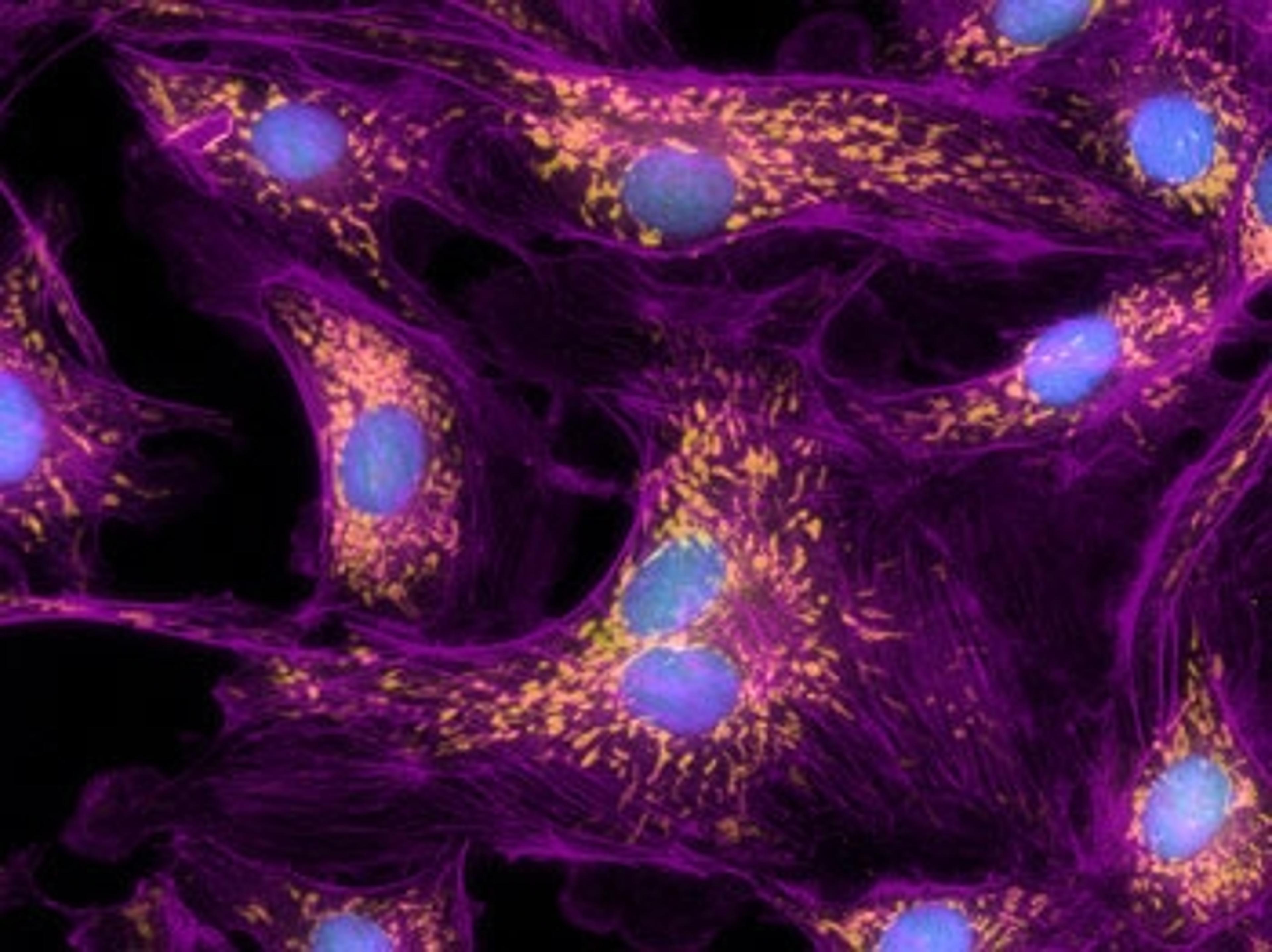
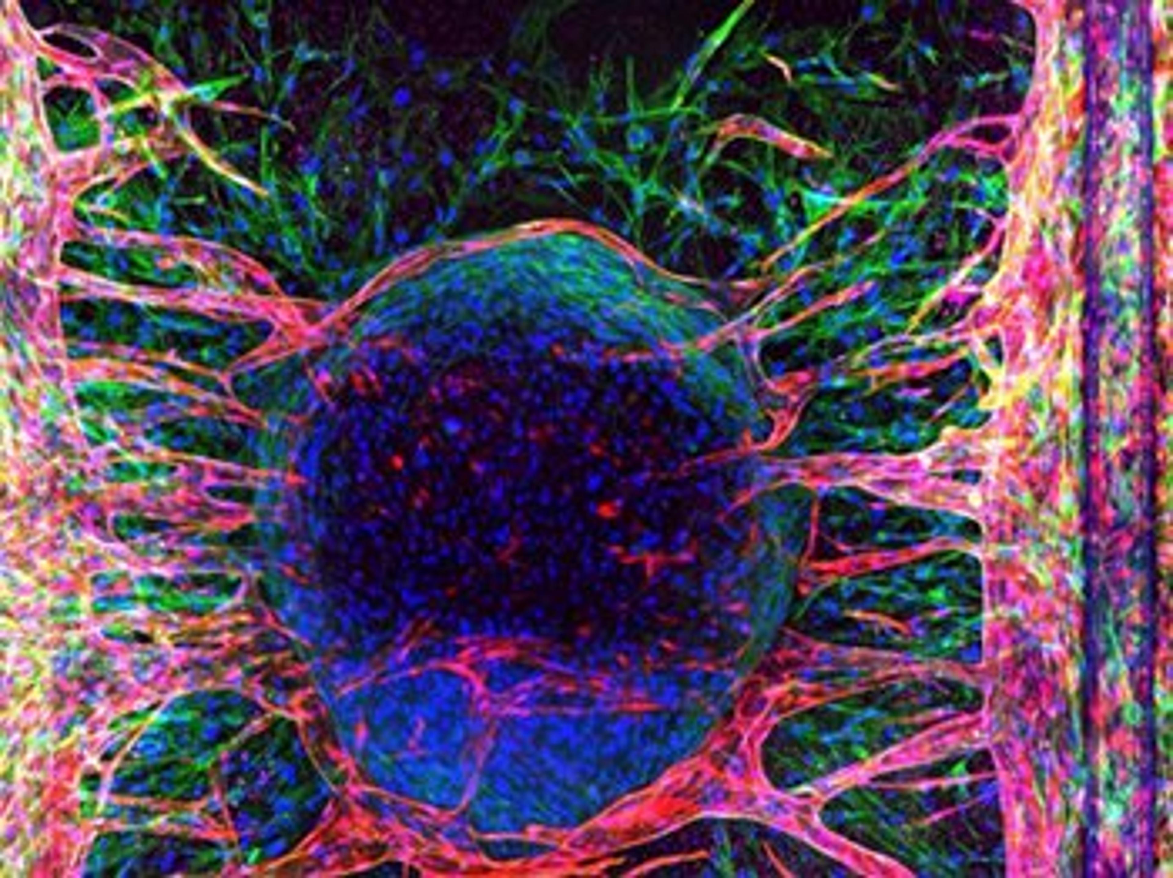
High content image analysis of subcellular localization
We have been using this instrument for two months now and have greatly improved our workflow through its reproducible imaging capabilities. Integrating the automated plate imaging has been simple and straightforward.
Review Date: 23 Nov 2020 | Molecular Devices®
User-friendly software that makes it easy for non-specialists to use.
Cell imaging
The major plus point for our lab of the Pico is its ease of use for non-imaging specialists: the CRX software is pretty intuitive and the ability to analyze data remotely is good. Indeed, the majority of our lab has no imaging experience and can use it competently. Additional plus points for us are its footprint compared to other imaging systems and its affordability. As with all imaging systems, appropriate IT systems are needed to handle the large amounts of data generated. Imaging is comparable to other systems.
Review Date: 15 Sept 2020 | Molecular Devices®
The ImageXpress® Pico Automated Cell Imaging System combines high-resolution imaging and powerful analysis, with a streamlined workflow. It includes CellReporterXpress® software for acquisition and analysis, and features a comprehensive portfolio of preconfigured protocols. The automated setup of analysis parameters shortens the learning curve, so you can start running experiments quickly. If you’re in the market for fluorescence imaging or digital microscopy, the ease and affordability of an automated imager is now within reach.
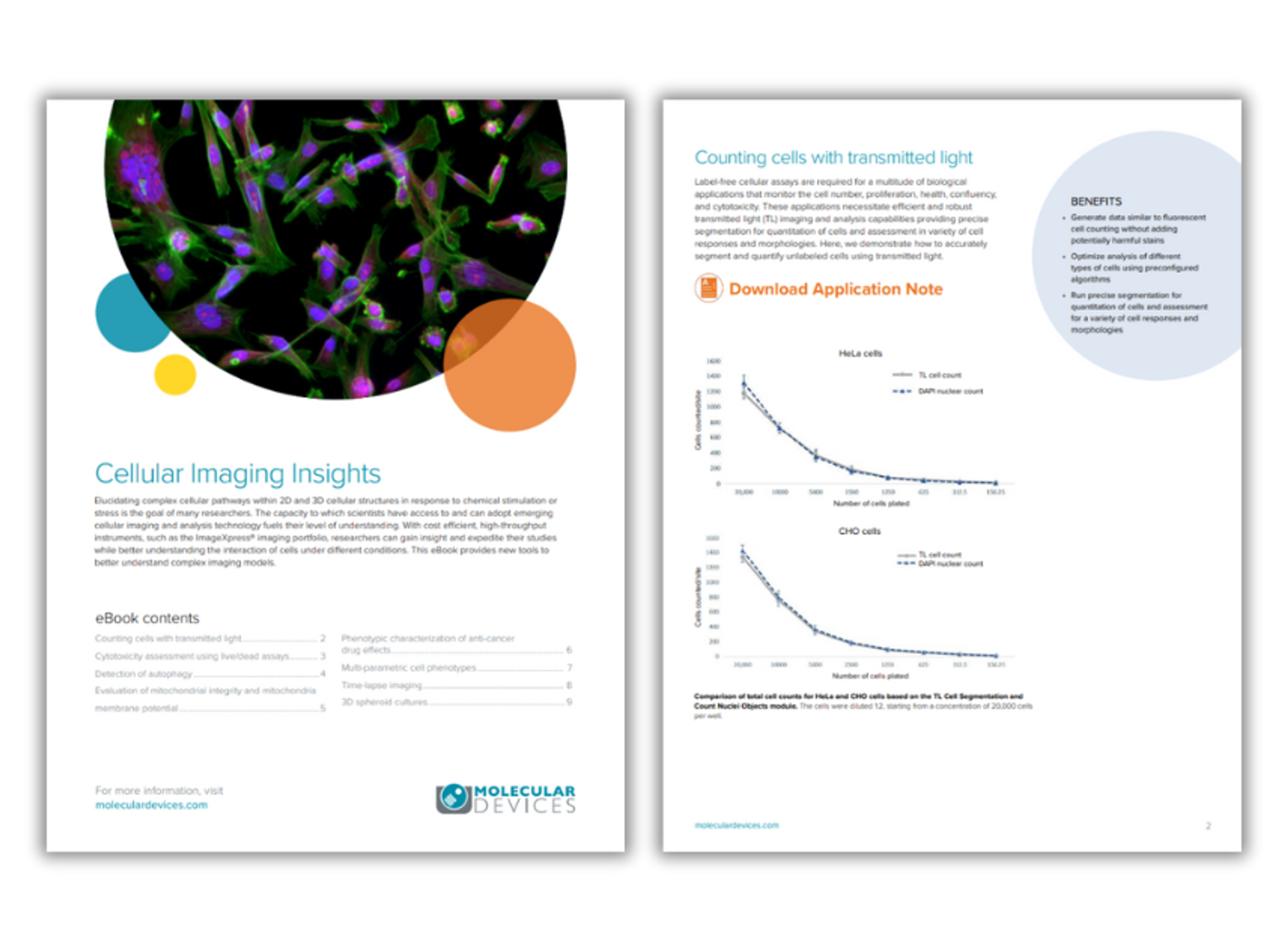
Cellular imaging insights
The goal of many researchers is to elucidate the complex cellular pathways within 2D and 3D cellular structures in response to chemical stimulation or stress. The capacity to which scientists have access to and can adopt emerging cellular imaging and analysis technology fuels their level of understanding. In this eBook, Molecular Devices provides an overview of the new tools that can be used to better understand complex imaging models.
Resource details:
- Document type: Application notes
- Page count: 10
- Read time: 15 mins
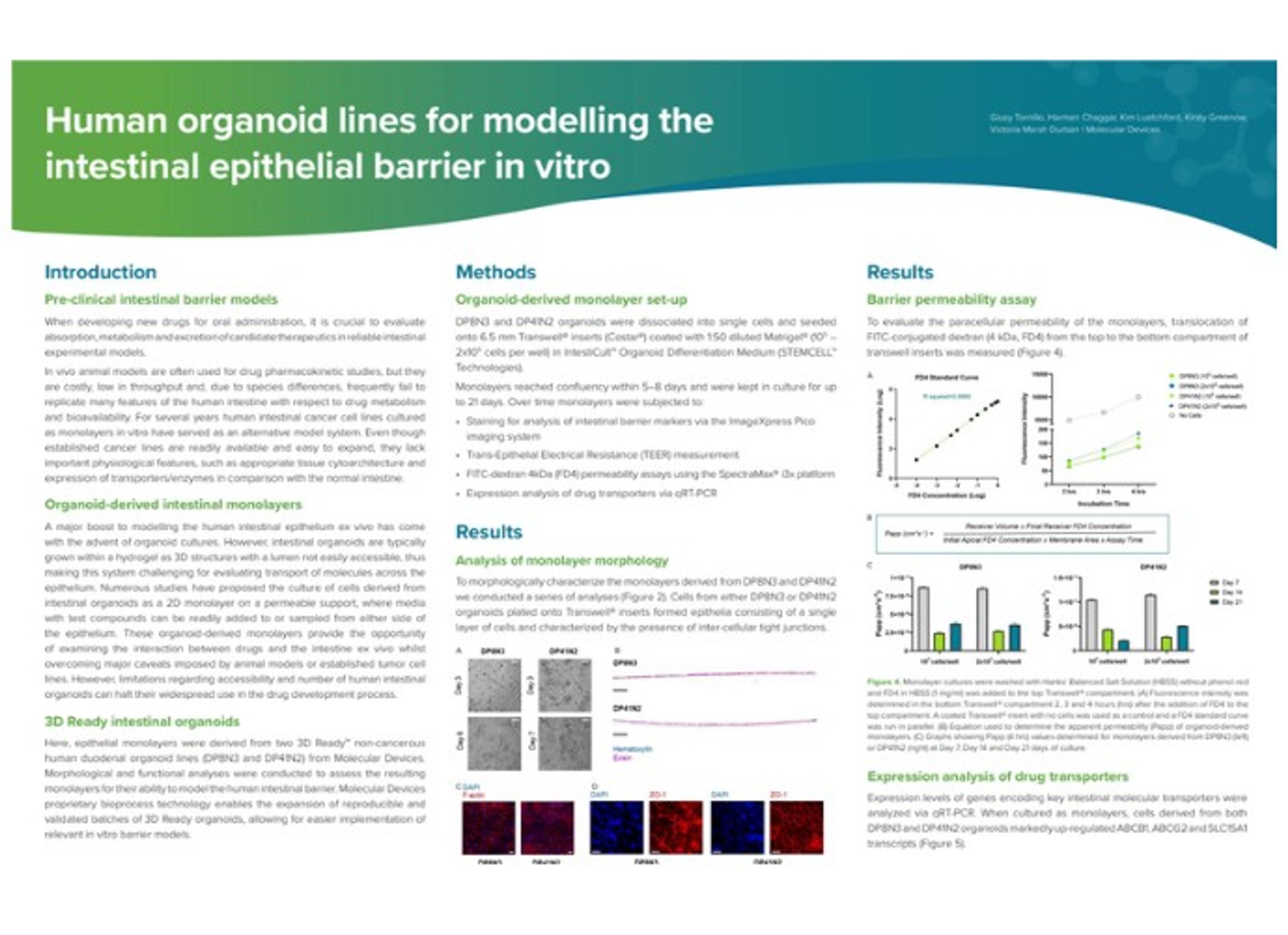
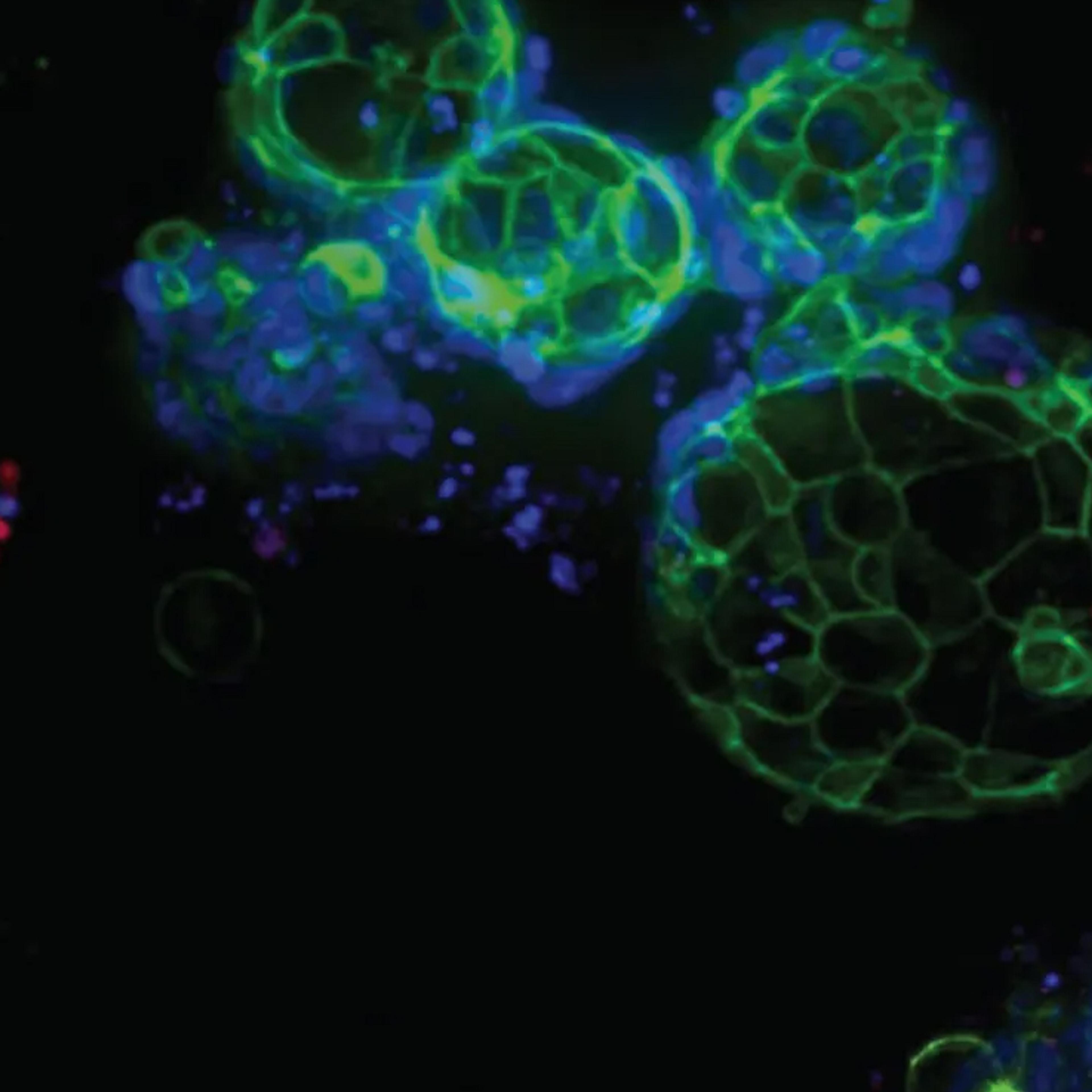
Human organoid lines for modelling the intestinal epithelial barrier in vitro
In this application note, epithelial monolayers were derived from two 3D-Ready non-cancerous human duodenal organoid lines from Molecular Devices and morphological and functional analyses were conducted to assess the resultant monolayers' ability to model the human intestinal barrier.
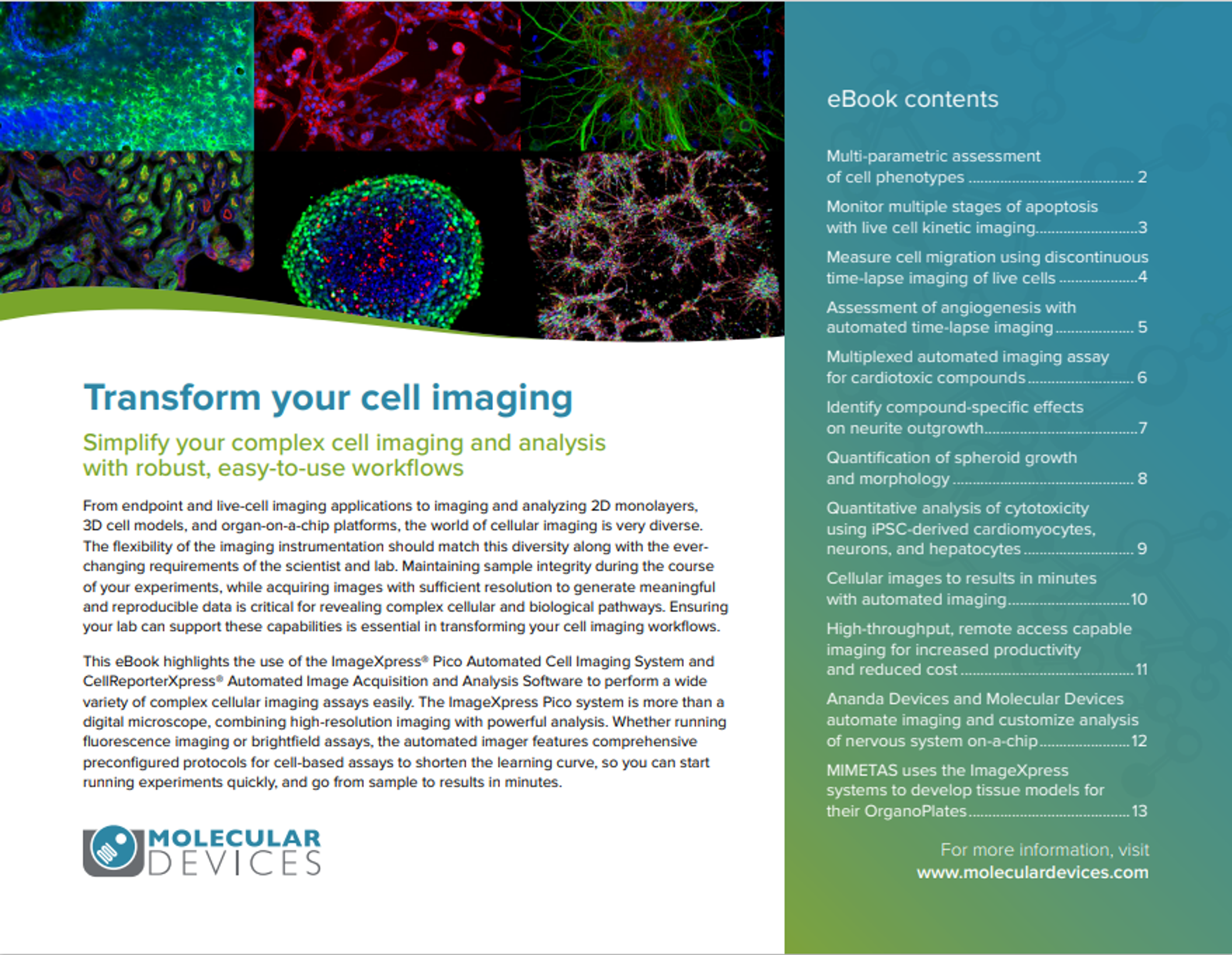
Transform your cell imaging
From endpoint and live-cell imaging applications to imaging and analyzing 2D monolayers, 3D cell models, and organ-on-a-chip platforms, the world of cellular imaging is very diverse. This eBook highlights the use of the ImageXpress® Pico Automated Cell Imaging System and CellReporterXpress® Automated Image Acquisition and Analysis Software to perform a wide variety of complex cellular imaging assays easily.

Assessment of angiogenesis with automated time-lapse imaging
In this application note, Molecular Devices demonstrates the utility of the ImageXpress® Pico Automated Cell Imaging System in the quantitative assessment of angiogenesis over time. Download now to discover the benefits this system can provide.
Multiplexed automated imaging assay for cardiotoxic compounds using the ImageXpress Pico system
In this application note, find a study by Molecular Devices, which uses human induced pluripotent stem cell (iPSC)-derived cardiomyocytes to develop functional and morphological readouts for testing effects of different compounds in a multi-parametric assay format. Download to discover what they found by using the ImageXpress® Pico system.
Monitor cell proliferation and cell cycle in real time
In this application note, Molecular Devices use real-time quantitative cell imaging assays with the ImageXpress® Pico Automated Cell Imaging System to monitor complex effects of anti-cancer compounds in cell models. Download to find out the results of their study, and to see why the ImageXpress® Pico system helps accelerate their cell-based assay development.
Improve nuclear translocation assay results using image deconvolution
In this application note, the effects of TNF-α treatment time were tested on the nuclear translocation of NF-κB in HeLa cells. Cells were imaged with the ImageXpress® Pico Automated Cell Imaging System and CellReporterXpress® Image Acquisition and Analysis Software.
Monitor multiple stages of apoptosis with live cell kinetic imaging
The study of apoptosis is a critical aspect of drug discovery and development. In this application note, two apoptosis assays were run to study the cytotoxic effects of anti-cancer compounds on HeLa cells. Long-term time-lapse imaging was performed on the ImageXpress® Pico Automated Cell Imaging System.
Monitor cell proliferation and cell cycle in real time
There is an increased need to expand the variety and complexity of cell-based assays for biological research and drug discovery. Live-cell assays allow monitoring of cell responses in real time and provide important insights about compound treatment effects and biological complexity. In this application note, Molecular Devices used real-time quantitative cell imaging assays with the ImageXpress® Pico Automated Cell Imaging System to monitor complex effects of anti-cancer compounds in cell models.
Measure cell migration using discontinuous time-lapse imaging of live cells
In this application note, Molecular Devices demonstrates a scaled-up method that illustrates how cell migration imaging and real-time analysis can be performed in a 96- or 384-well microplate to enable high-throughput cell motility experiments.
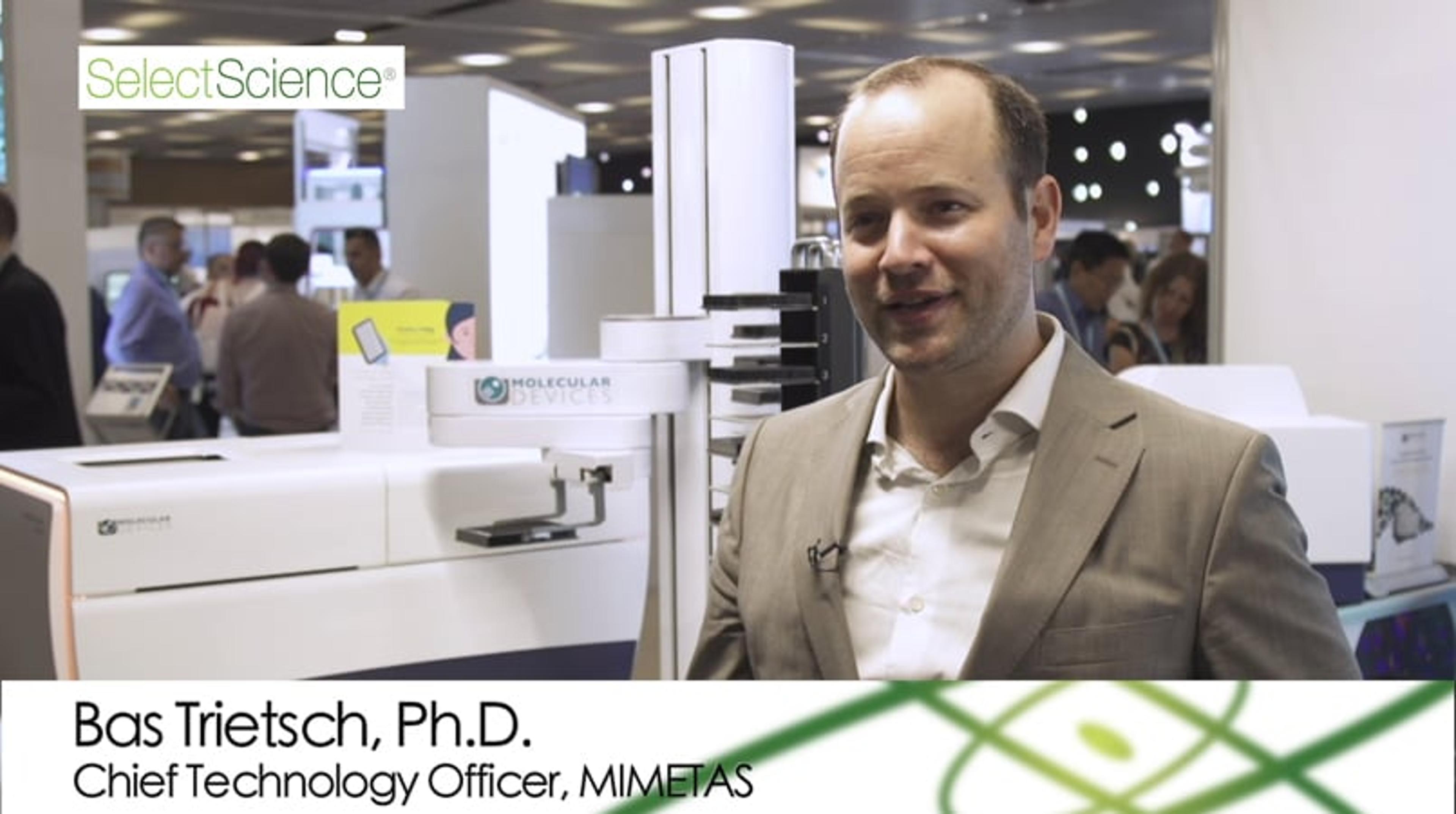
Angiogenesis Research: High-Content Imaging Systems Help Unlock the Full Potential of 3D Tissue Models
Angiogenesis is an important field of research and a focus for cancer therapeutics. In this interview, Dr. Bas Trietsch, CTO, MIMETAS, introduces a new solution for the study of angiogenesis; the OrganoPlate® Graft, an in vitro cell culture microplate platform that allows vascularization of 3D tissues. Hear how Molecular Devices’ ImageXpress Pico Automated Cell Imager and ImageXpress Micro Confocal High-Content Imaging System play a vital role in the development and analysis of 3D tissue models built on the OrganoPlate® Graft.
Video filmed at SLAS Europe 2019 – visit the SelectScience special feature for more videos from the event.
ImageXpress Pico: Automated Cell Imaging System
The ImageXpress Pico from Molecular Devices is a compact, automated cell imager. Its small size makes it ideal for imaging cells in your own lab, including assays from cell counting to neurite outgrowth, with consistent reproducible results. The Pico allows data analysis with a single software package with prefigured protocols that generates heat maps, scatter plots and movies from plate to cell level.
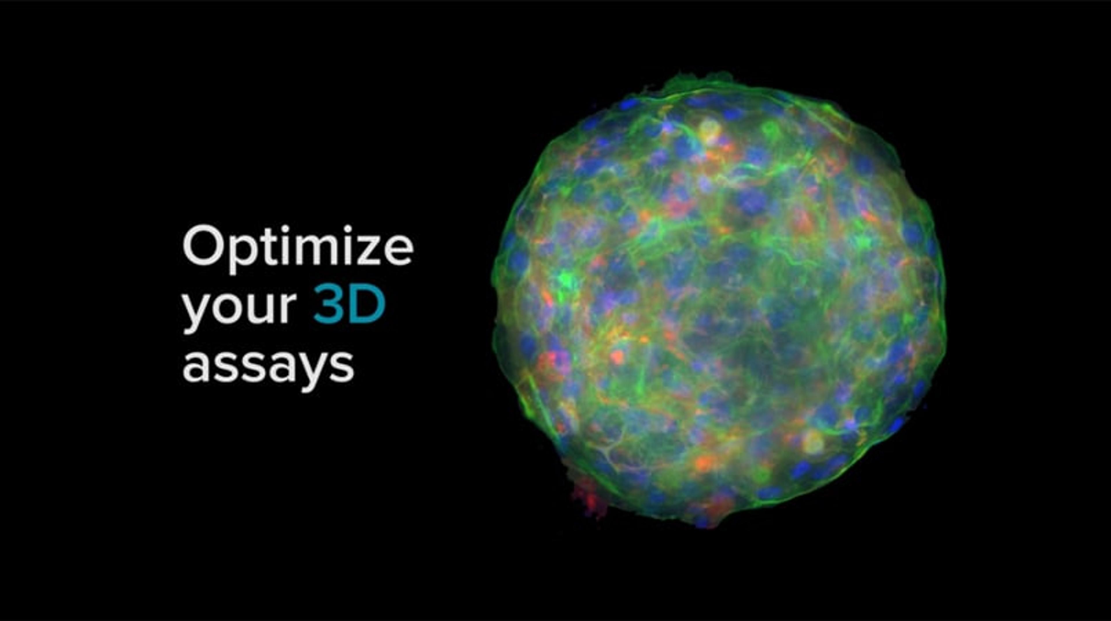
Optimize Your 3D Assays with ImageXpress
Optimize your 3D assay and make your next breakthrough in cellular imaging - with intelligent, innovative imaging solutions.

Molecular Devices opens Organoid Innovation Center
The modular, automated 3D cell culture and image analysis lab enables customers to streamline and scale organoid interrogation, advancing drug discovery research

Ananda Devices, Molecular Devices collaborate to automate imaging and customize analysis of nervous system on-a-chip
Neuroscientists can now screen over 3,000 neurons from a NeuroHTS microplate in under 30 minutes with the ImageXpress Pico Automated Cell Imaging System
Everything you need to know about the ImageXpress Pico Automated Cell Imaging System
We put your most pressing questions to the team behind Molecular Devices latest cellular imaging and analysis system to find out how it could make an impact in your lab
Harnessing the latest imaging technology to advance angiogenesis research
Learn how one biotechnology company is using cutting-edge imaging to improve the visualization of angiogenesis in its 3D tissue models