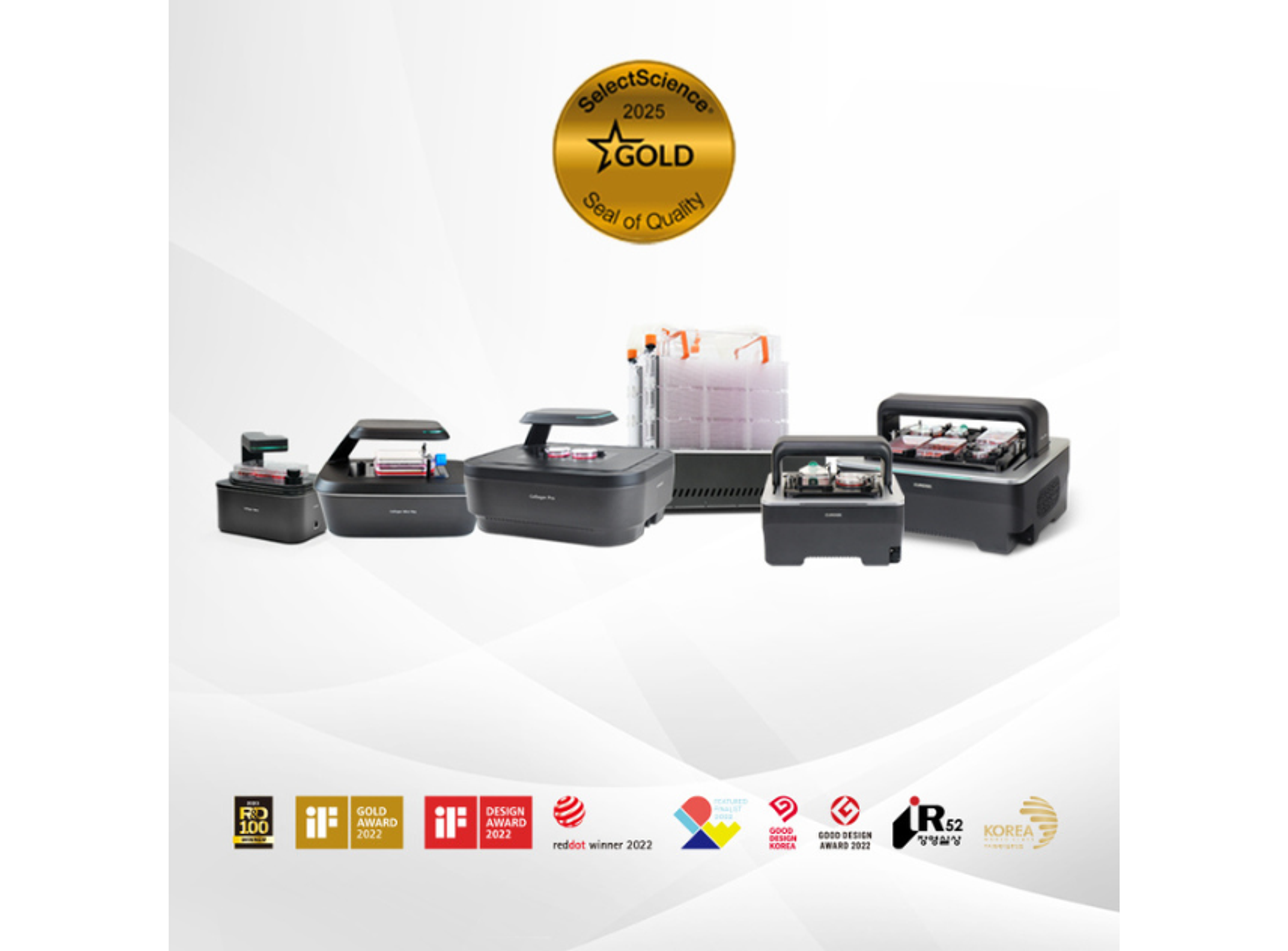
Celloger® M26, High-throughput Live Cell Imaging System from Curiosis
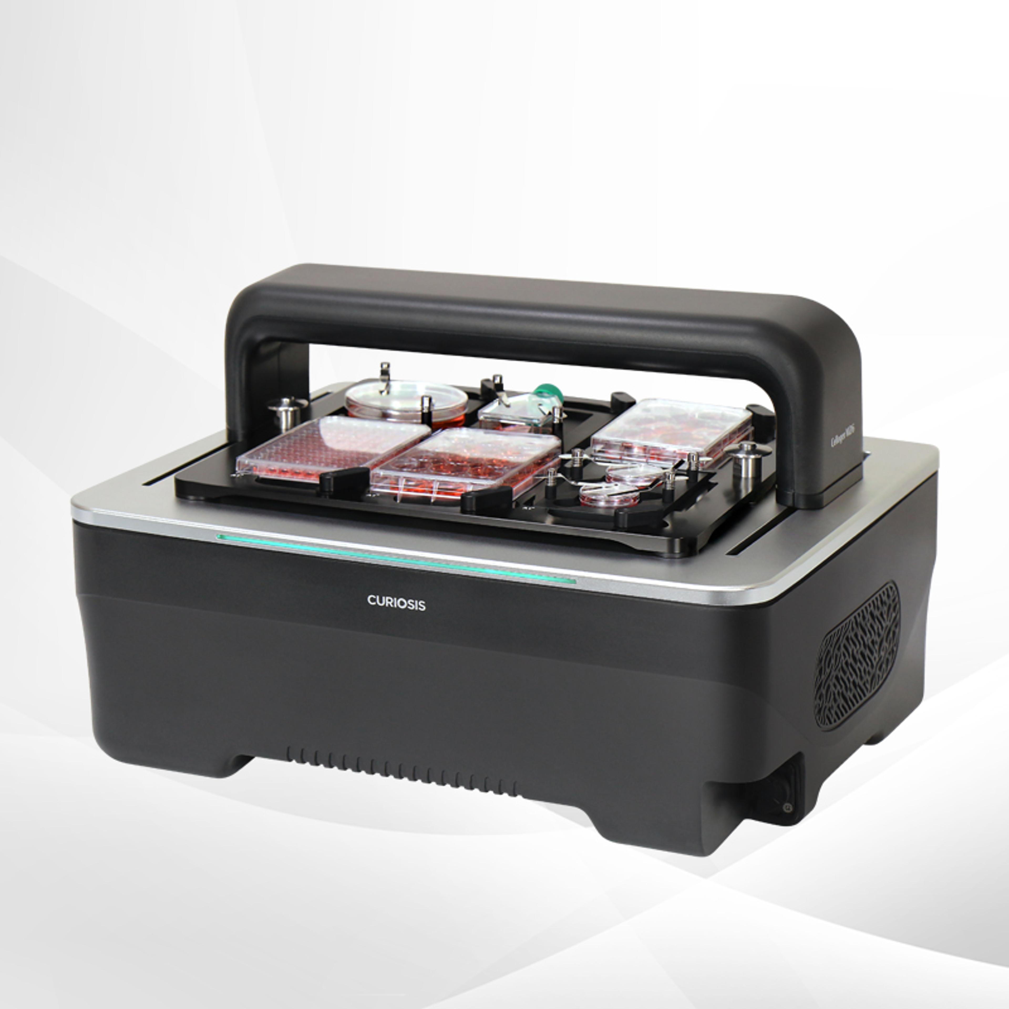
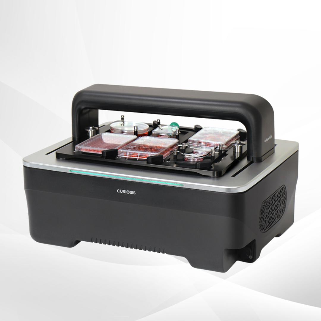
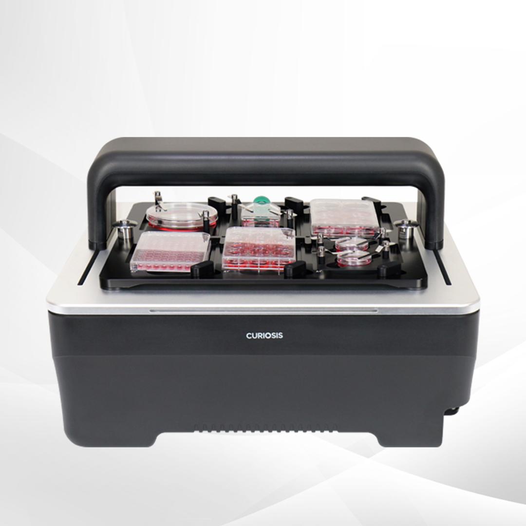
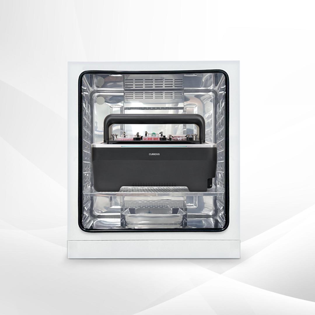
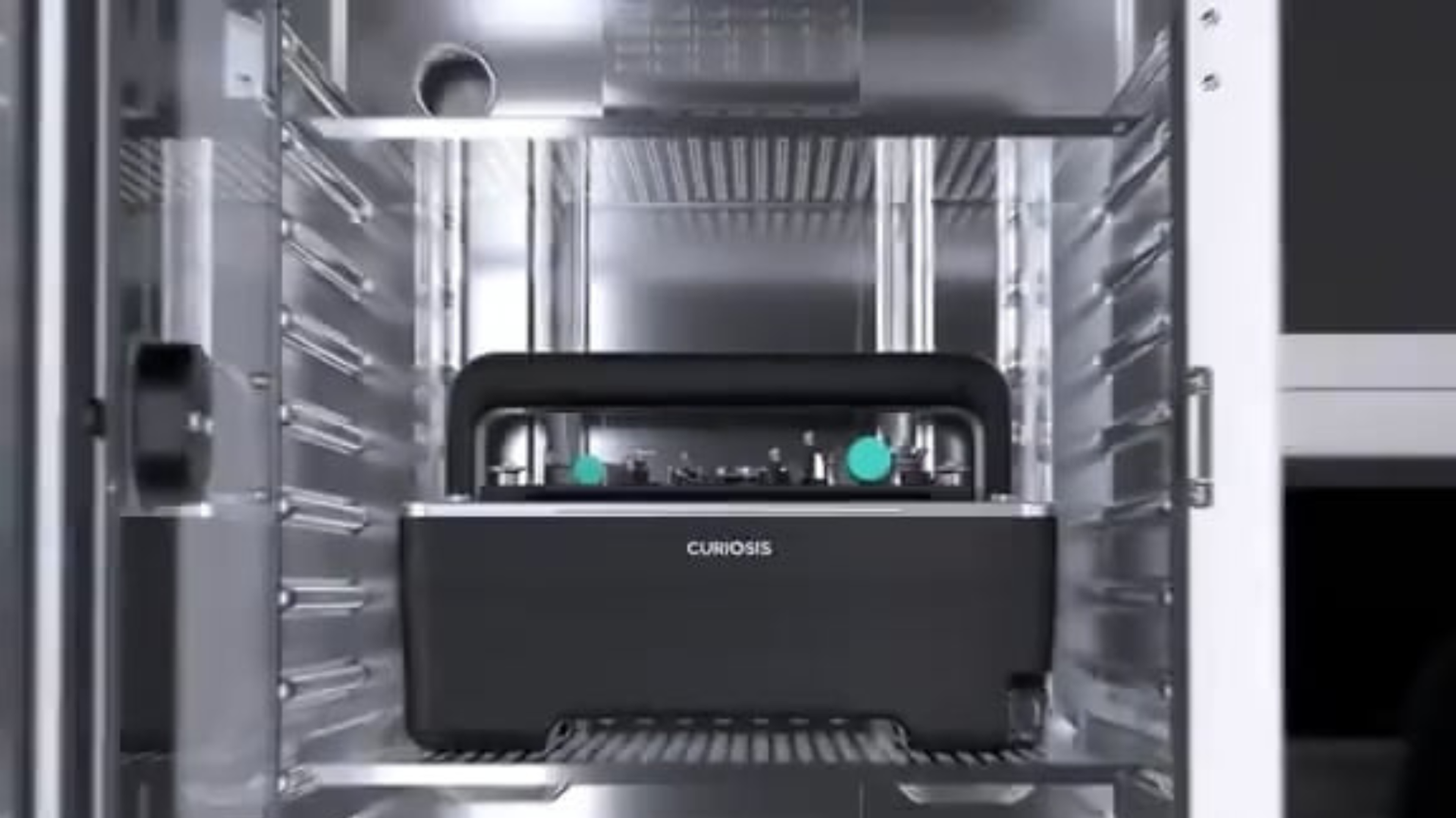
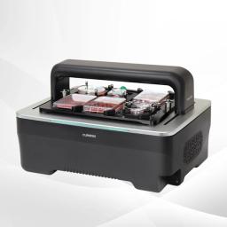
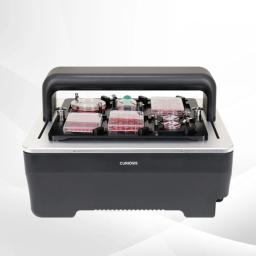
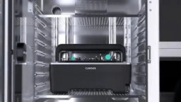
Celloger® M26 is a high-throughput live cell imaging system for real-time monitoring inside the incubator. It offers synchronized illumination, ultra high-resolution imaging, interchangeable lenses, and brightfield/fluorescence modes, enabling simultaneous imaging of up to six plates for large-scale cellular analysis.

Receive your quote directly from the manufacturer.
Celloger® M26 is a high-throughput live cell imaging system based on brightfield and 2 fluorescence channels (Green & Red) microscopy. The fully motorized XYZ stage (camera moving type) with synchronized illumination enables uniform and reliable imaging across multiple wells and plates inside the incubator. Designed for large-scale, uninterrupted workflows, M26 supports simultaneous high-content monitoring while maintaining stable culture conditions.
Key Features
- High-throughput live cell imaging performance
- Fully motorized XYZ stage (camera moving type)
- Synchronized illumination for uniform data acquisition
- User-interchangeable objectives (4× / 10× / 20×)
- Brightfield + 2 fluorescence channels (Green & Red)
- High sensitivity 5.0 MP CMOS image sensor
- Auto focusing for stable long-term imaging
- Advanced analysis methods: stitching, Z-stacking, time-lapse, real-time recording
- Compatible with various vessels: up to 96-well plate, flask, dish, slide
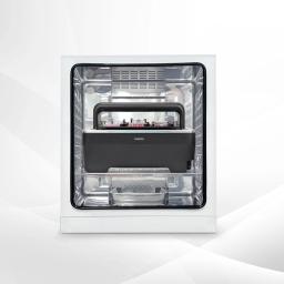
- Incubator-compatible design (recommended incubator volume: ≥230 L)
Applications
- High-content screening
- Drug screening
- Stem cell research
- Dose-response analysis
- Cell proliferation monitoring
- Cell morphology assessment
- Long-term time-lapse imaging
- Multipoint parallel experiments
Specifications
- Imaging modes: Brightfield, 2 fluorescence channels (Green & Red)
- Objectives: 4× / 10× / 20× (user-interchangeable lenses)
- Fluorescence modes (customizable):
Green (EX: 470/40, EM: 540/50)
Red (EX: 562/40, EM: 641/75) - Stage movement: Fully motorized XYZ (camera moving type)
- Image sensor: High sensitivity 5.0 MP CMOS
- Analysis methods: Single/multicolor, stitching, Z-stacking, time-lapse, real-time recording
- Focusing: Auto focusing
- Culture vessels: Well plate up to 96-well, flask, dish, slide
- Software: Scan App (acquisition), Analysis App (analysis)
- File export format: TIFF, AVI, JPEG, PNG
- Operating environment: 10–40℃, 20–95% humidity
- Power supply: 100–240V, 50/60Hz
- PC requirements (O/S): Windows 10 and above
- Incubator specification: Above 230 L (recommended)
- Dimensions (mm): W 485 × D 440 × H 313
- Weight: Approx. 14.44 kg (excluded vessel holder)
Celloger M26, a high-throughput live imaging system
The Celloger® M26 is a high-throughput live cell imaging system designed for continuous, real-time monitoring of cells directly within incubator environments.
Discover how the system combines brightfield and fluorescence imaging modes to support comprehensive analysis of cell morphology and function. Synchronized illumination ensures consistent image capture, while interchangeable lenses provide flexibility across different experimental requirements.
Capable of accommodating multiple plate formats and imaging up to six plates simultaneously, the platform is designed to support high-throughput workflows. Explore how its high-resolution imaging capabilities enable researchers to observe dynamic cellular processes without interrupting culture conditions, improving experimental continuity and data quality.
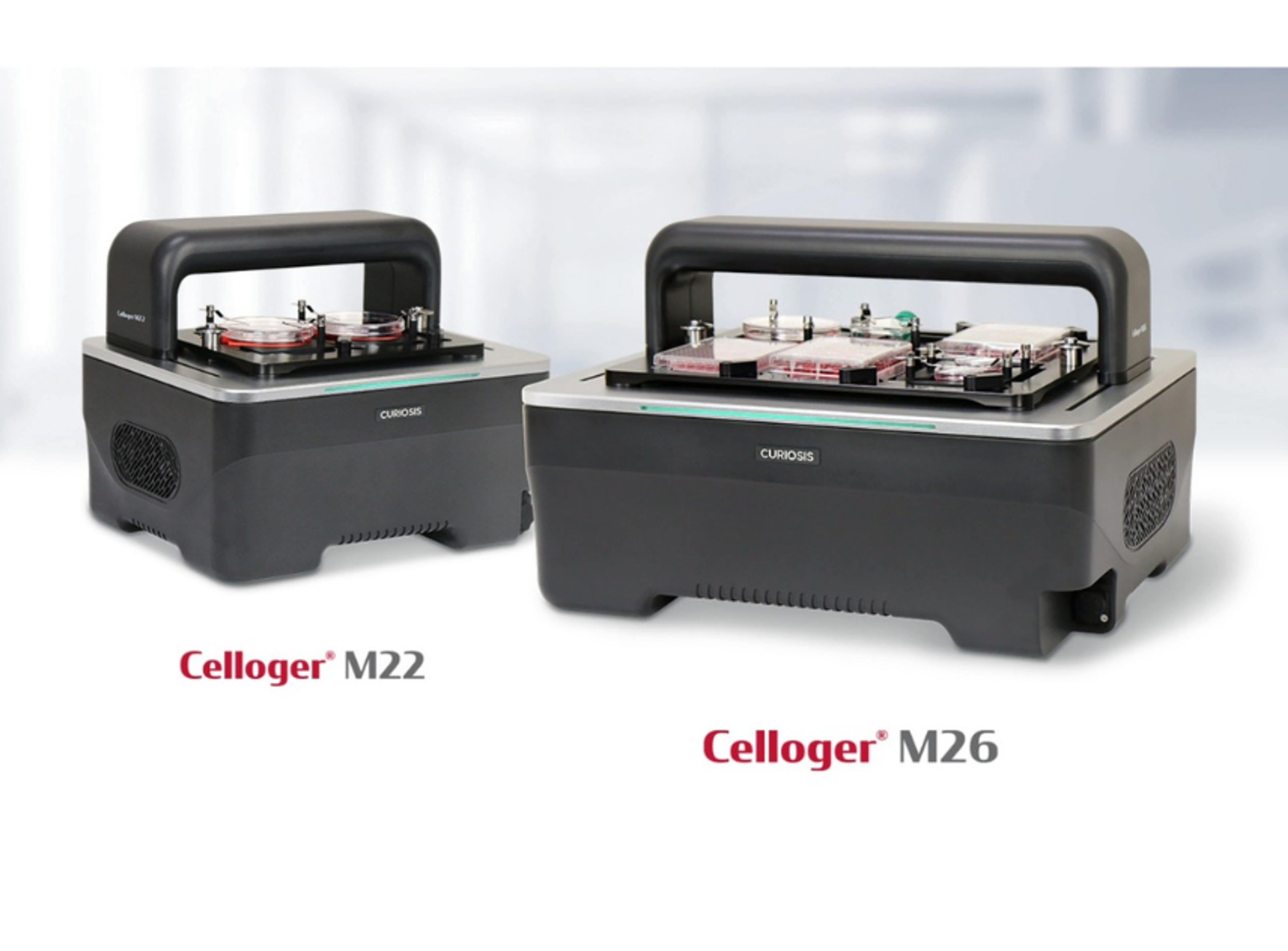
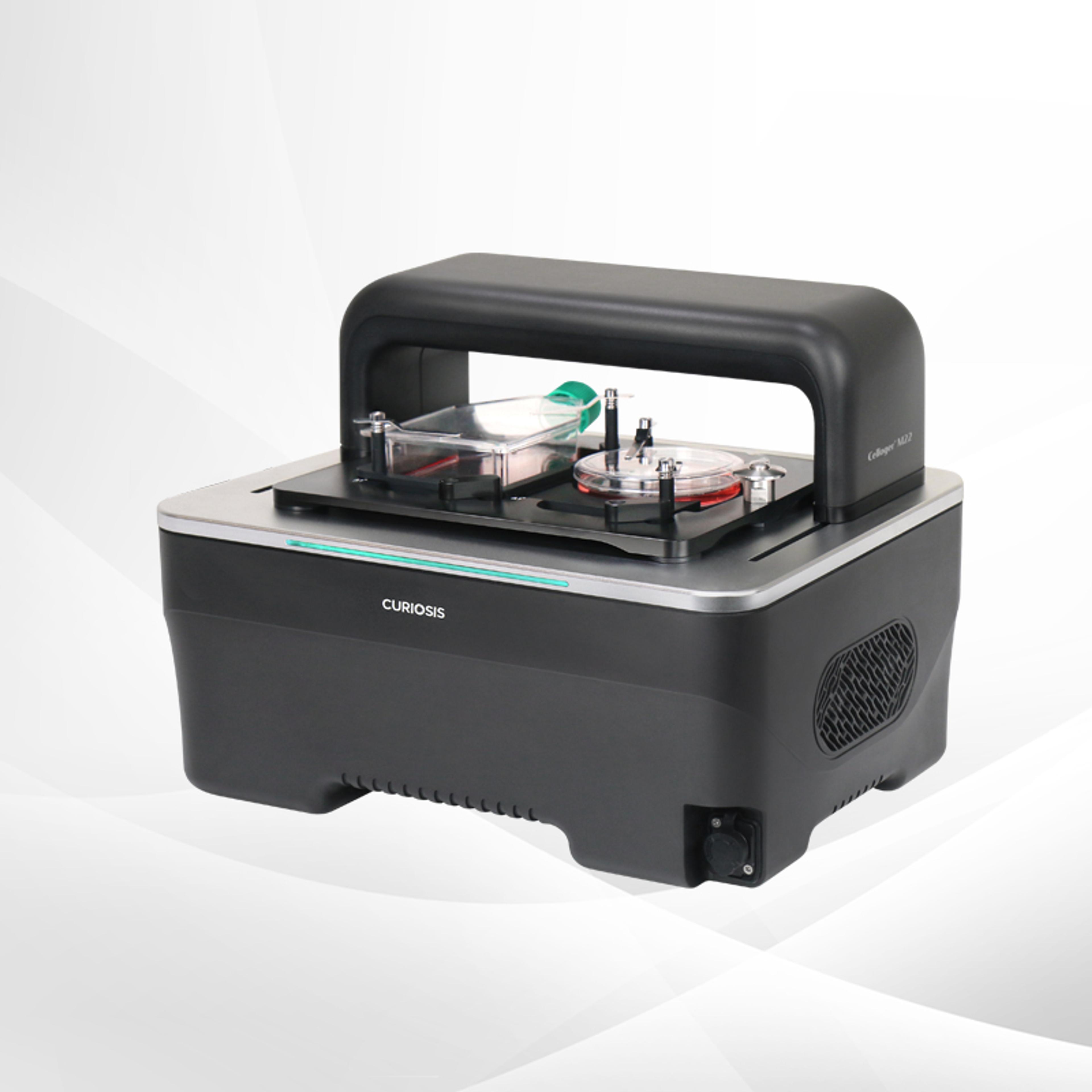
Flexible high-throughput live-cell imaging workflows for complex 3D cell models with the Celloger M22 and M26
Automated imaging systems support real-time analysis of spheroids and organoids in scalable drug discovery workflows