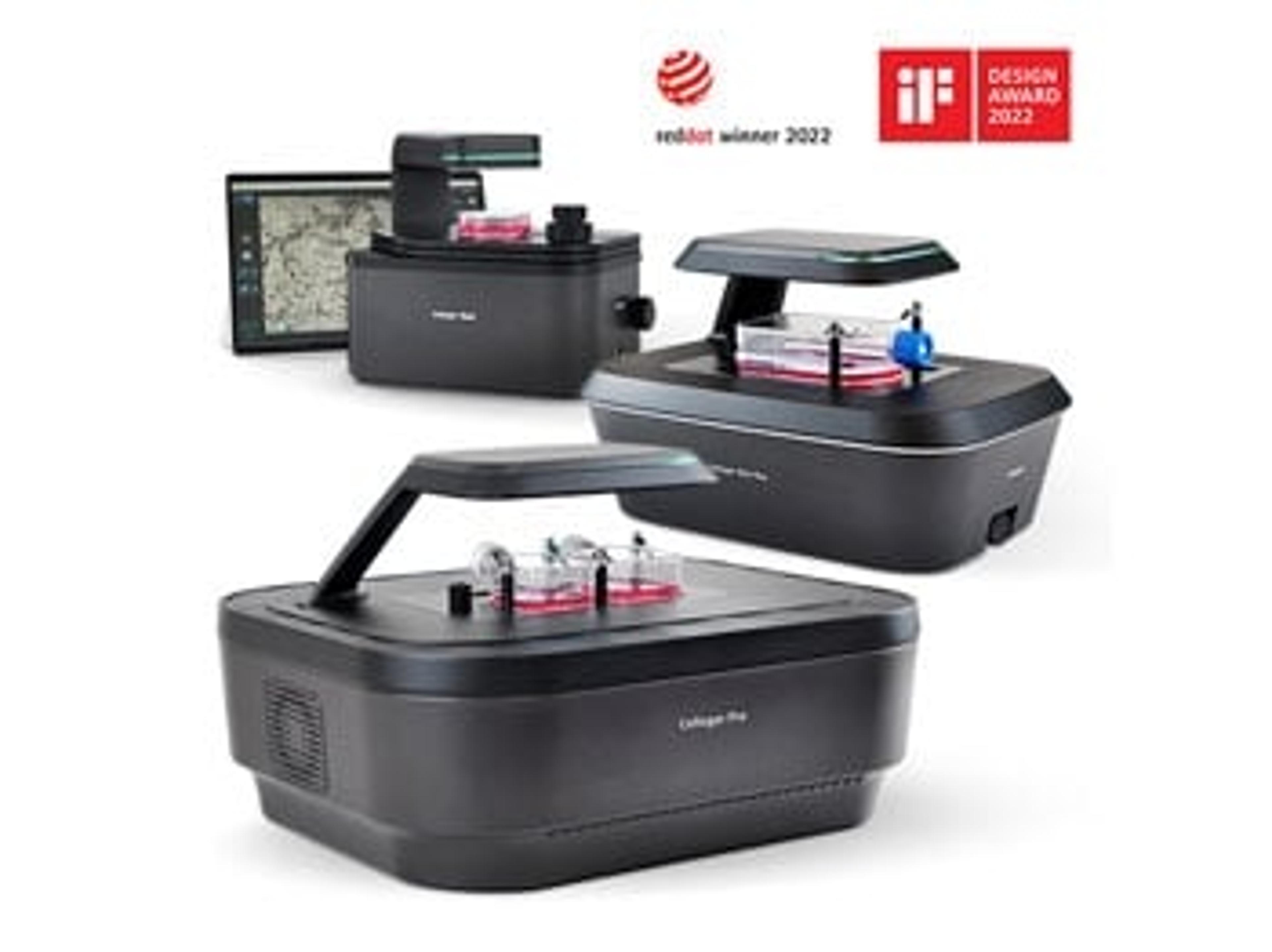
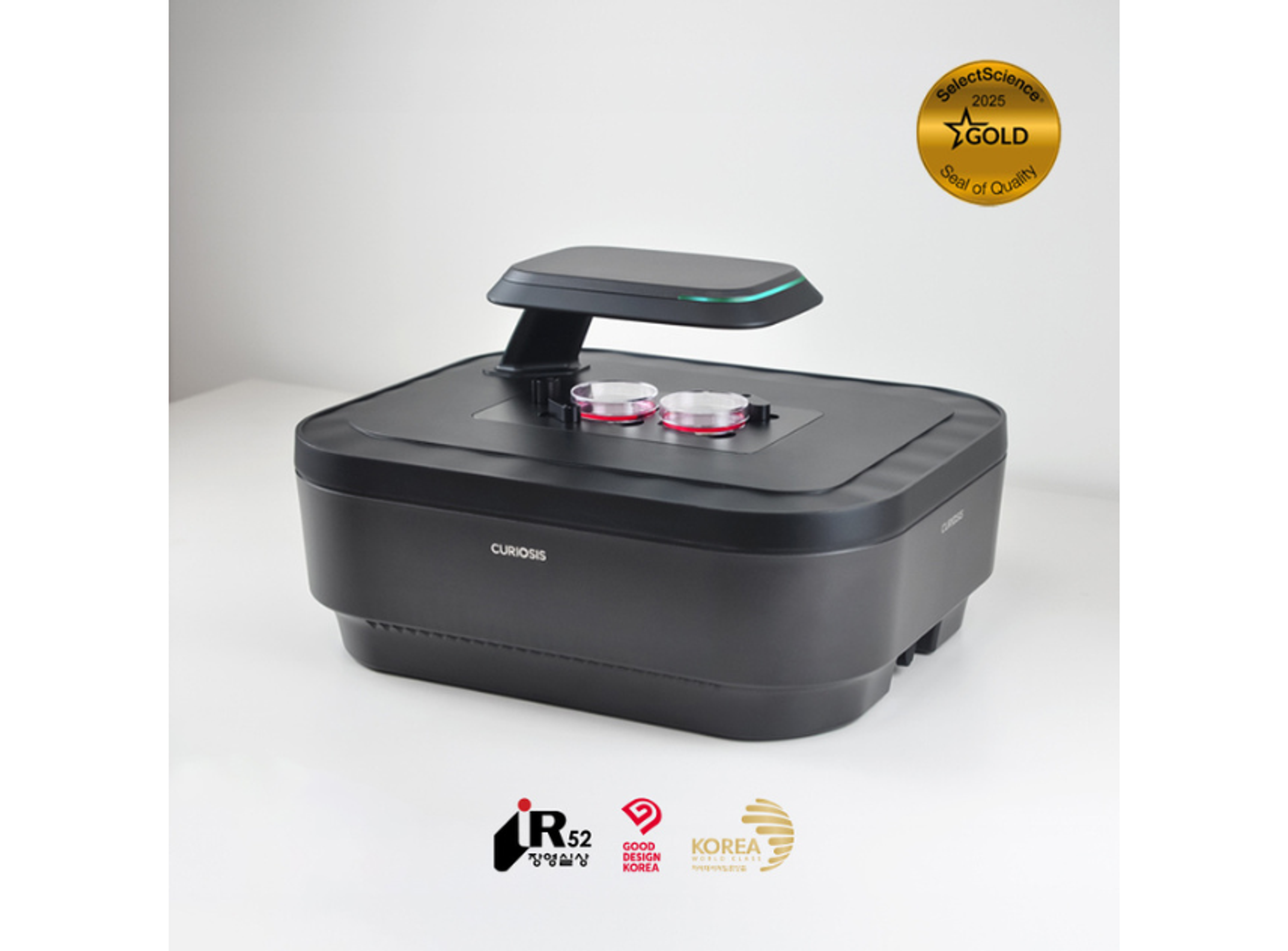
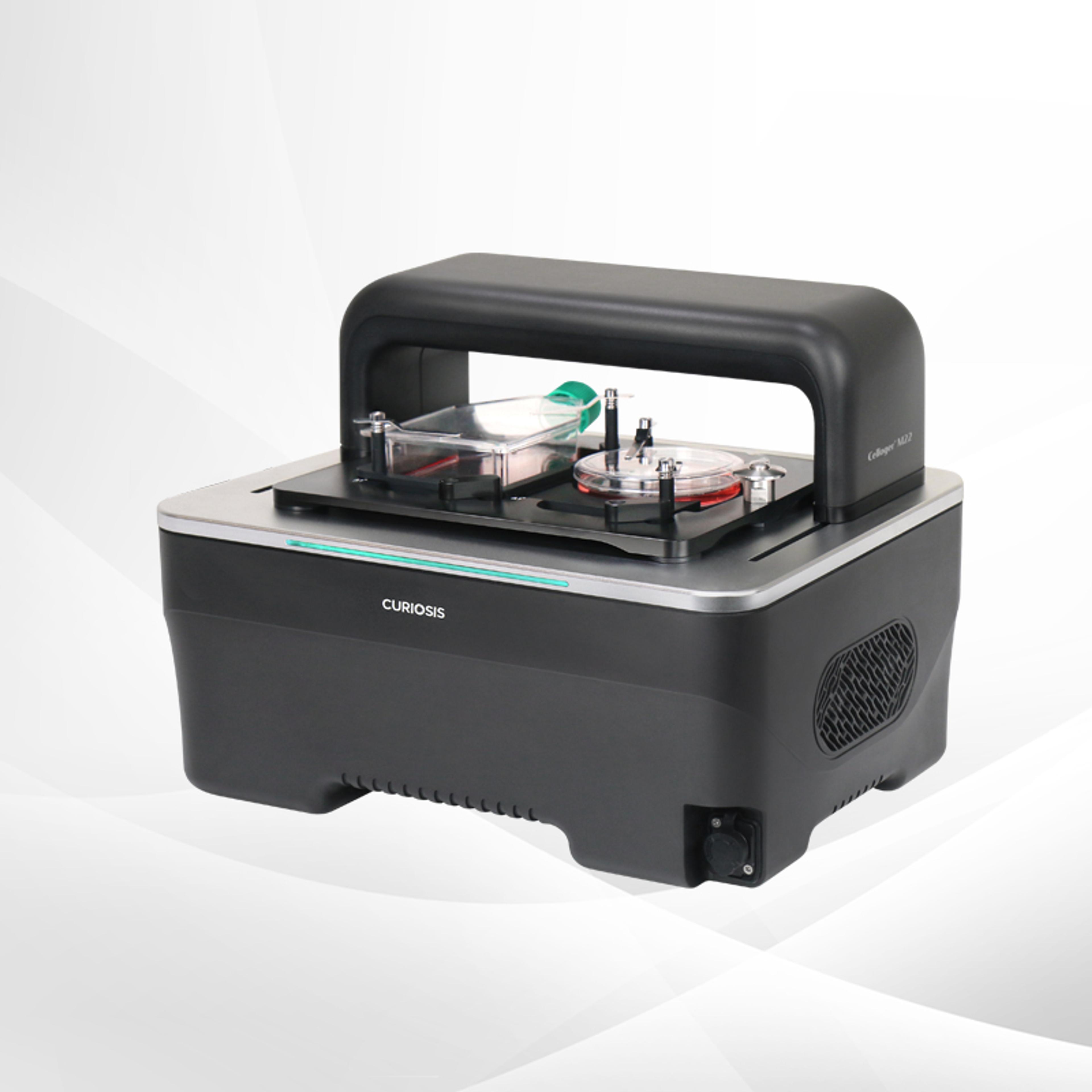
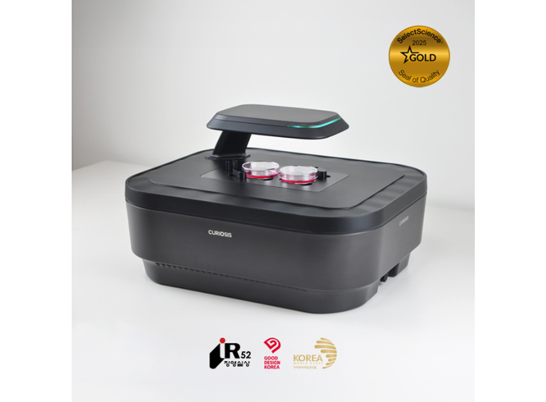
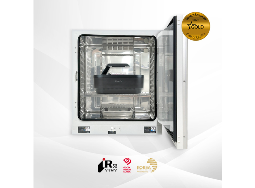
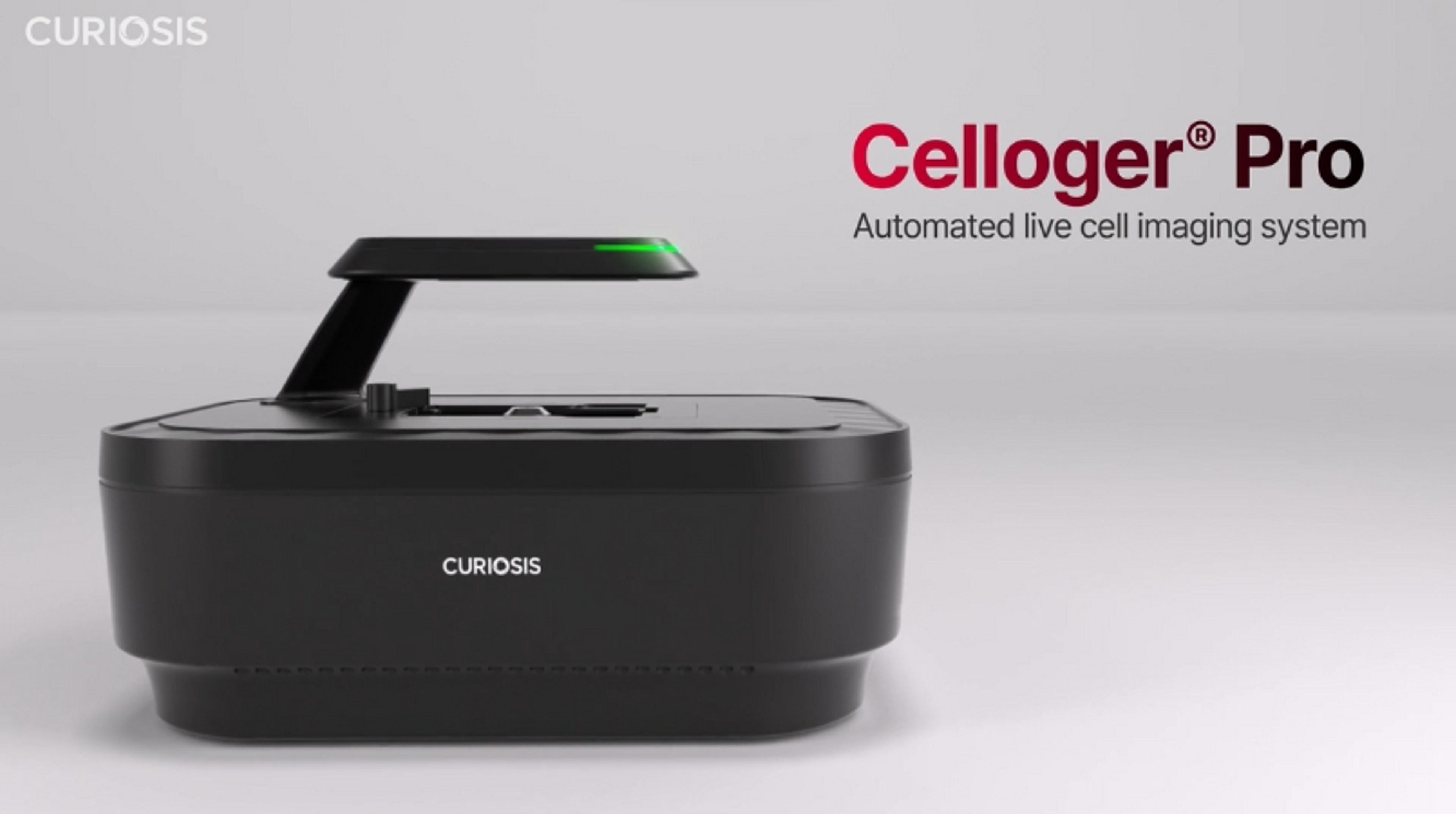
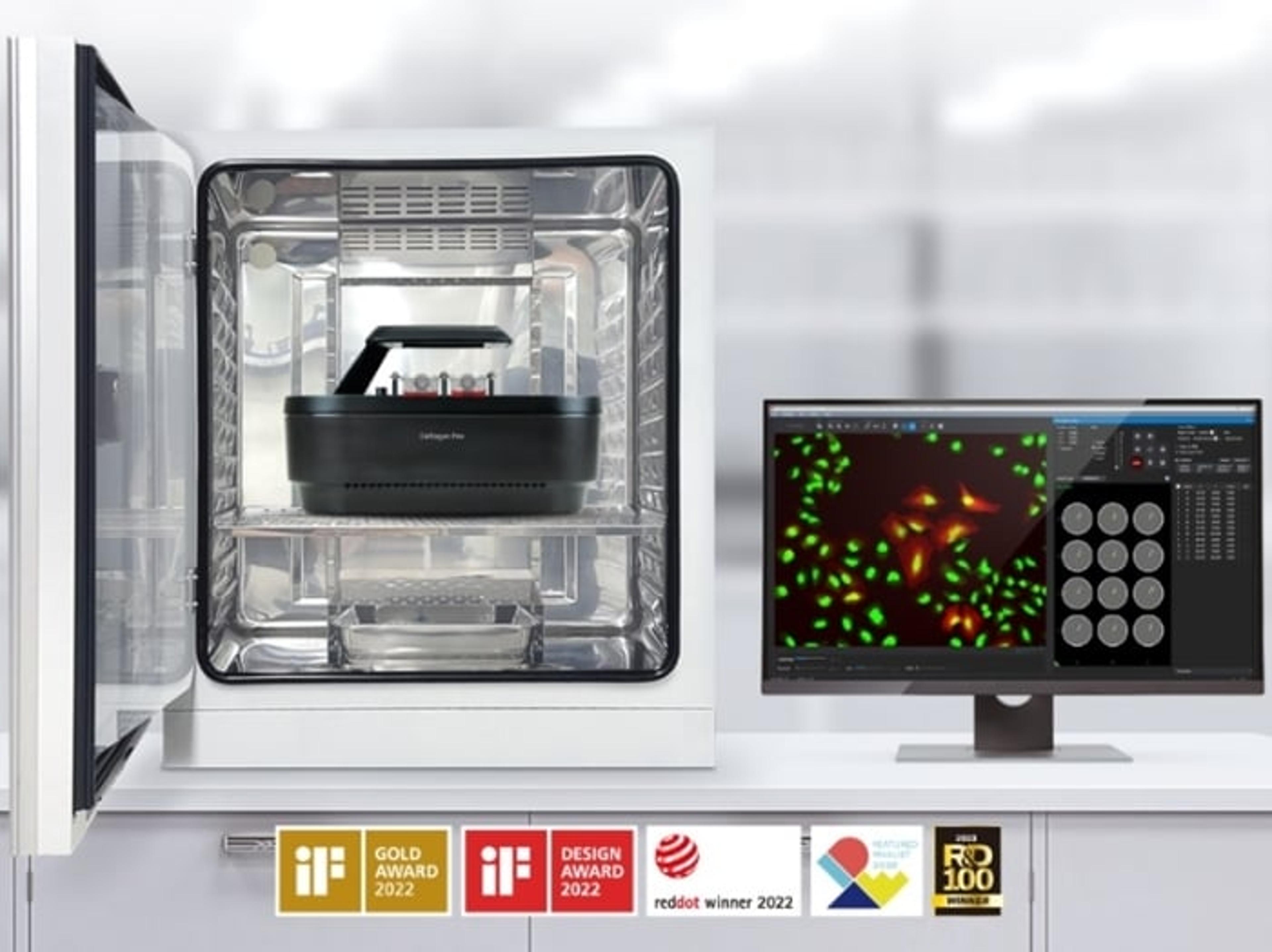
Celloger® Pro, Automated Live Cell Imaging System from Curiosis
With its streamlined workflow and versatile capabilities, Celloger® Pro is a valuable tool for researchers seeking efficient, accurate, and cost-effective cellular analysis.


Receive your quote directly from the manufacturer.
We were able to easily image and quantify 3D models derived from liver cancer cell lines and human tissue–derived cells.
3D cell model research
Using Celloger® Pro, we were able to easily image and quantify 3D models derived from liver cancer cell lines and human tissue–derived cells. Its stable performance, high-quality imaging, and built-in analysis tools consistently provided the data we needed.
Review Date: 25 Nov 2025 | CURIOSIS
Celloger® Pro made it easy to monitor spheroids under different conditions and track changes in real time.
Cancer biology
Celloger® Pro made it easy to monitor spheroids under different conditions and track changes in their size in real time. After adding fluorescent reagents, we could clearly visualize how the fluorescence signal was distributed within the spheroids. Overall, the real-time fluorescence imaging and analysis features provided exactly the data we needed.
Review Date: 25 Nov 2025 | CURIOSIS
I was especially impressed by the high image quality and clarity of the images.
Cancer biology
I performed an exosome uptake experiment with C2C12 cells and obtained the expected results, and I was especially impressed by the high image quality and clarity of the images.
Review Date: 25 Nov 2025 | CURIOSIS
The built-in area measurement and automated wound-healing analysis tools were very convenient.
Cell movement
It was very convenient that the analysis software included area measurement tools and automated functions for wound-healing analysis. I also appreciated that it was compatible not only with standard well plates but also with other types of culture vessels.
Review Date: 24 Nov 2025 | CURIOSIS
The Celloger scan and analysis software was easy to use, and the image quality was very good.
Vascular cell aging and disease research
The Celloger scan and analysis software was easy to use, and the image quality was very good.
Review Date: 24 Nov 2025 | CURIOSIS
The Celloger® Pro system has really boosted my research.
Immunotherapy research
The Celloger® Pro system has really boosted my research. Instead of spending time on tedious imaging work myself, I can rely on the system to handle it automatically. The image quality has been excellent, and the software is very intuitive and easy to work with.
Review Date: 24 Nov 2025 | CURIOSIS
Using the Celloger® Pro for NK cell–mediated cytotoxicity experiments has been very helpful.
Immunotherapy research
Using the Celloger® Pro for NK cell–mediated cytotoxicity experiments has been very helpful. By using two fluorescent markers, I was able to clearly visualize the interactions between NK cells and target cells, and also assess the extent of NK cell–mediated killing. This made it much easier to understand the dynamics of the assay in real time.
Review Date: 24 Nov 2025 | CURIOSIS
I was able to perform 24-hour time-lapse imaging with a 384-well plate without any misalignment.
Organoid cytotoxicity
I was able to perform 24-hour time-lapse imaging with a 384-well plate without any misalignment in the X, Y, or Z coordinates, which made long-term experiments very reliable. The edges and corners of the plate were captured with the same brightness as the center, so I could use all of the wells, which was both practical and economical. The process of drug-induced cell death was clearly visualized over time. Because images could be taken without opening and closing the incubator, I didn’t have to worry about fogging or condensation on the plate lid, which often interferes with imaging on conventional microscopes. Being able to control the device externally was also very convenient, and the intuitive software made it easy to manage and organize the data.
Review Date: 24 Nov 2025 | CURIOSIS
It enables real-time imaging within incubation chambers.
In-vitro vascularization
It enables real-time imaging within incubation chambers, featuring an integrated image processing system for user-friendly operation.
Review Date: 24 Nov 2025 | CURIOSIS
Easy to use, quick running time
Organoid research
Easy to use, quick running time. Providing free analysis software would be beneficial.
Review Date: 16 Apr 2025 | CURIOSIS
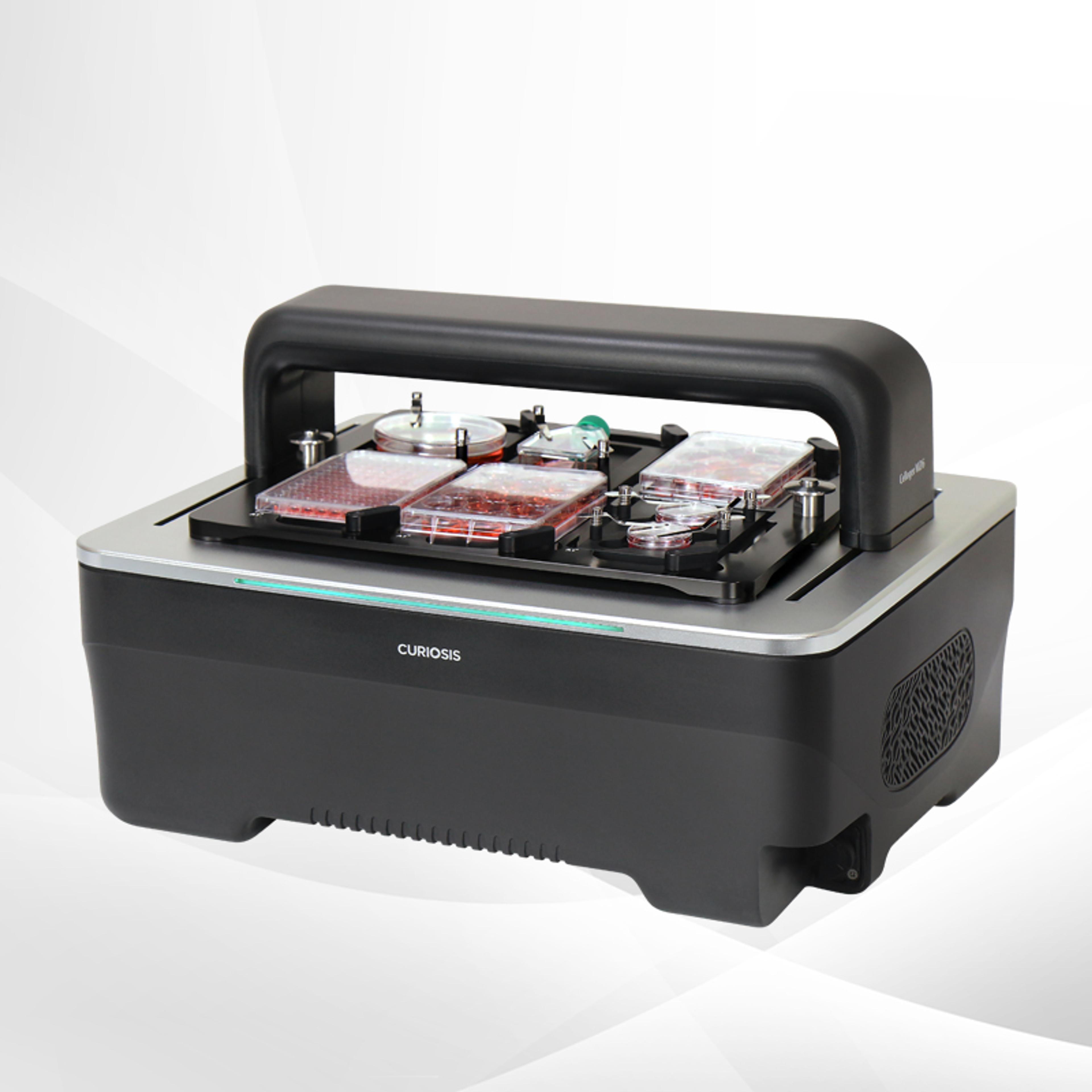
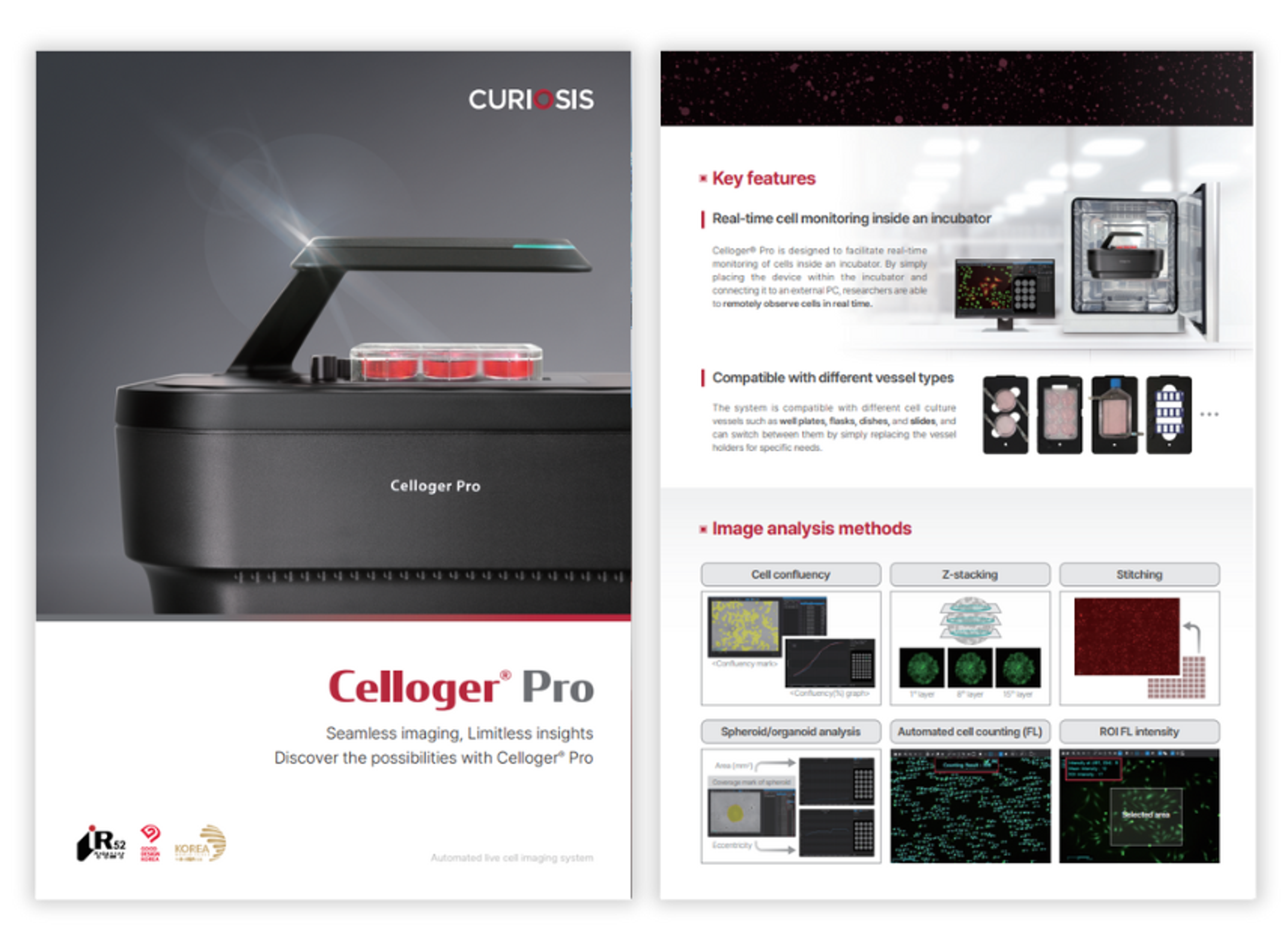
Celloger® Pro is an innovative live cell imaging system that redefines the research capabilities. With its exceptional image quality and unmatched convenience, it empowers researchers with the advanced features. By enabling real-time cell monitoring inside the incubator, it allows for seamless observation and tracking of cellular dynamics without disrupting the natural growth environment. The system's dual fluorescence and bright-field microscopy enable simultaneous visualization of multiple markers, while the multi-point time-lapse imaging captures dynamic cellular events across different locations. Celloger Pro’s user-friendly interface and intuitive tools make image acquisition and analysis a seamless experience. Additionally, its ability to capture high-resolution images and to effortlessly create videos, eliminates the labor-intensive tasks associated with live cell imaging processes.
Key features
- Real-time cell monitoring inside incubator
- Dual fluorescence microscopy
- Multi-point time-lapse imaging
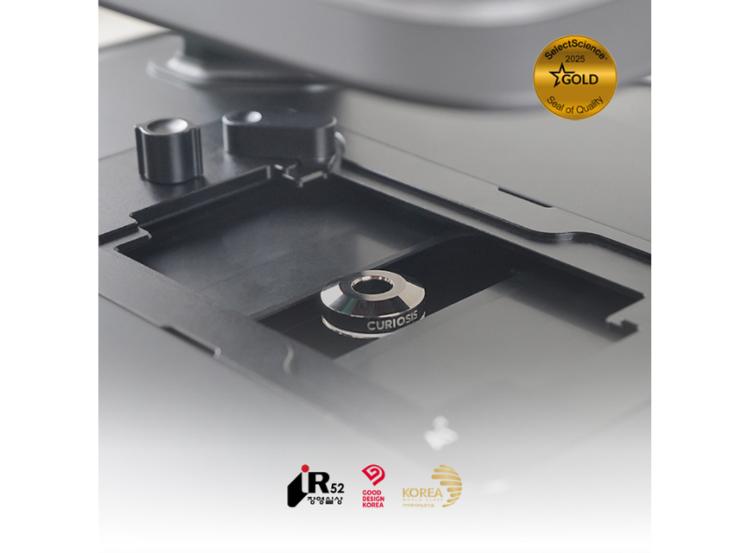
- User interchangeable lens
- Easy-to-use user interface and tools
Applications
- Spheroid assay
- Neurite outgrowth
- Cytotoxicity
- Scratch wound assay
Specification
- Imaging modes: Brightfield, Dual fluorescence (Green & Red)
- Objective lens: 2X, 4X, 10X (User-interchangeable)
- Fluorescence: Green - Excitation (470/40x), Emission (540/50m) / Red - Excitation (562/40x), Emission (641/75m)
- Stage: Fully motorized XYZ (Fixed stage, camera moving type)
- Camera: High sensitivity 5.0 MP CMOS
- Imaging positions: Multiple
- Focus: Autofocus, Manual focus
- Dimensions: 250 x 338 x 412 mm
- Weight: 9kg
Brochures
Celloger Pro product brochure
In this product brochure, CURIOSIS introduces Celloger® Pro, which boasts dual-color fluorescence and bright-field imaging capabilities for capturing high-resolution images. The system employs innovative merging techniques and improved scanning methods to reduce scanning time, enhancing the clarity and efficiency of cellular dynamics analysis. Designed for real-time monitoring of cells within an incubator, Celloger Pro can be placed in the incubator and connected to an external PC, enabling remote observation of cells. The time-lapse function allows for scheduled image capture and easy conversion to time-lapse videos. The system offers user-interchangeable objective lenses, including 2X, 4X, and 10X options, providing flexibility for researchers' specific study needs.
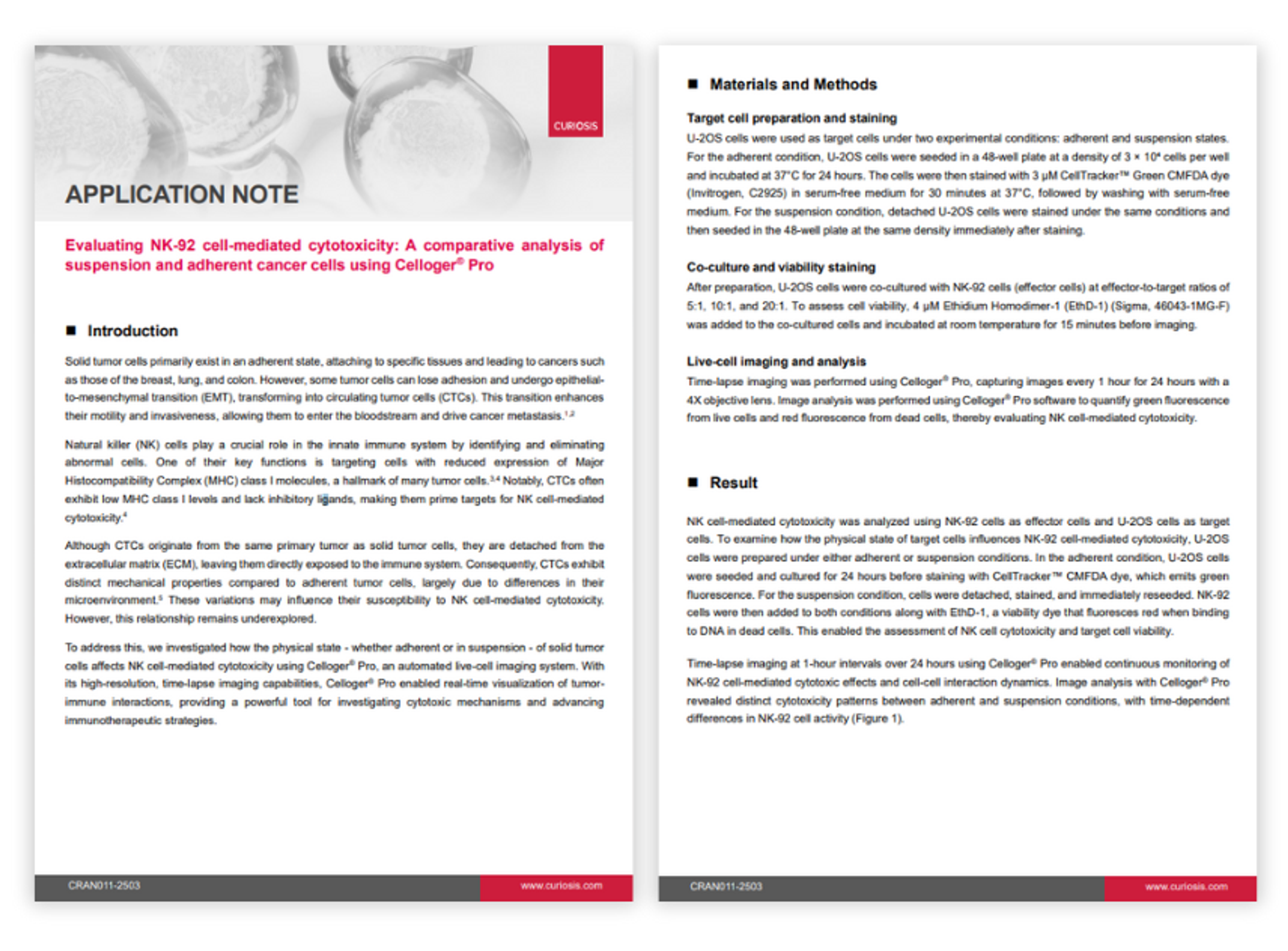
Evaluating NK-92 cell-mediated cytotoxicity: A comparative analysis of suspension and adherent cancer cells using Celloger Pro
In this application note, CURIOSIS explores how the physical state of solid tumor cells affects NK cell-mediated cytotoxicity using Celloger® Pro, an automated live-cell imaging system. With its high-resolution, time-lapse imaging capabilities, Celloger® Pro enabled real-time visualization of tumor-immune interactions, providing a powerful tool for investigating cytotoxic mechanisms and advancing immunotherapeutic strategies.
Resource details:
Resource type: Application notes
Page count: 6
Read time: 9 mins
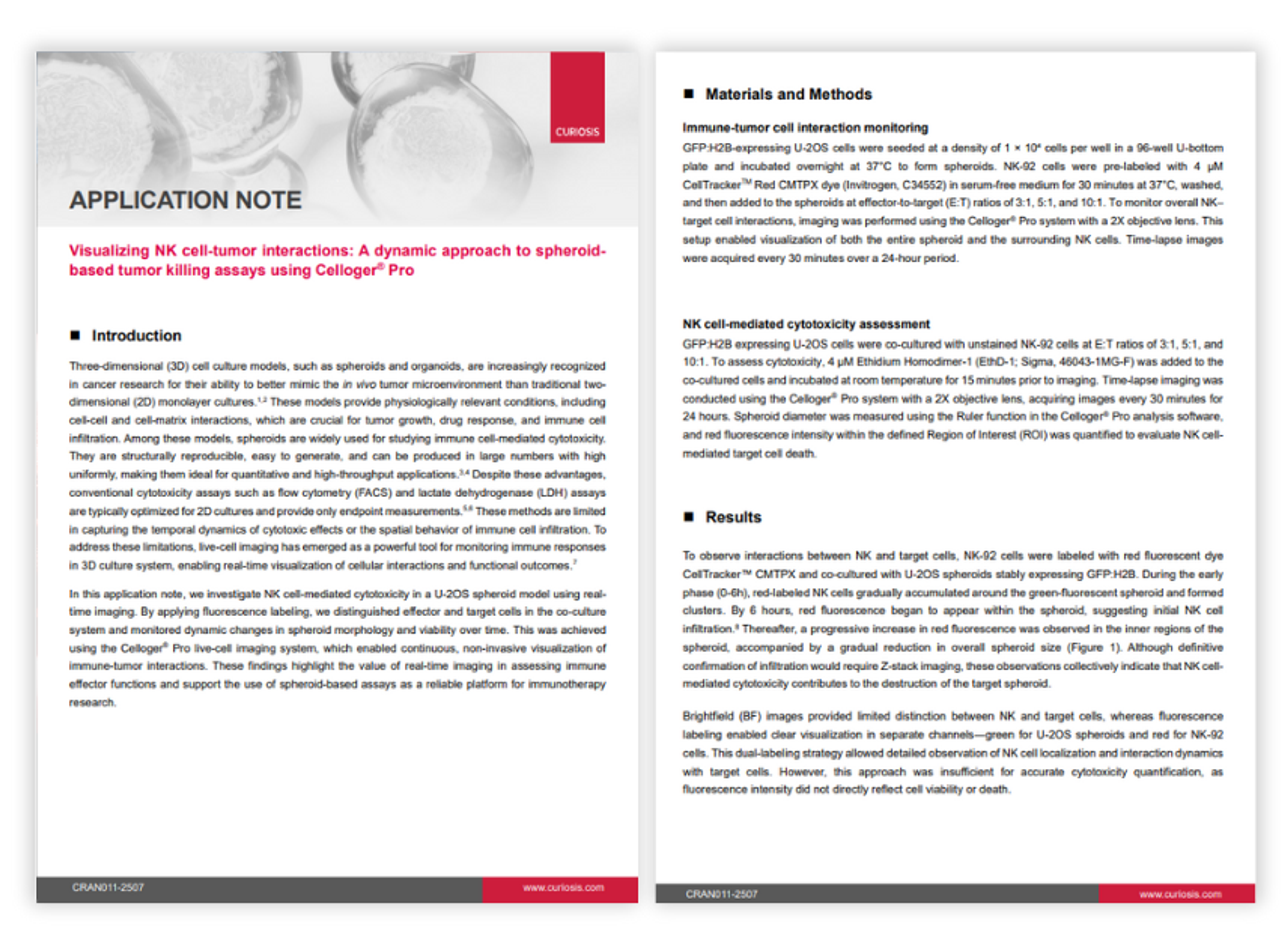
Visualizing NK cell-tumor interactions: A dynamic approach to spheroid-based tumor killing assays using Celloger Pro
In this application note, CURIOSIS investigates NK cell-mediated cytotoxicity in a U-2OS spheroid model using real-time imaging. This was achieved using the Celloger® Pro live-cell imaging system, which enabled continuous, non-invasive visualization of immune-tumor interactions. These findings highlight the value of real-time imaging in assessing immune effector functions and support the use of spheroid-based assays as a reliable platform for immunotherapy research.
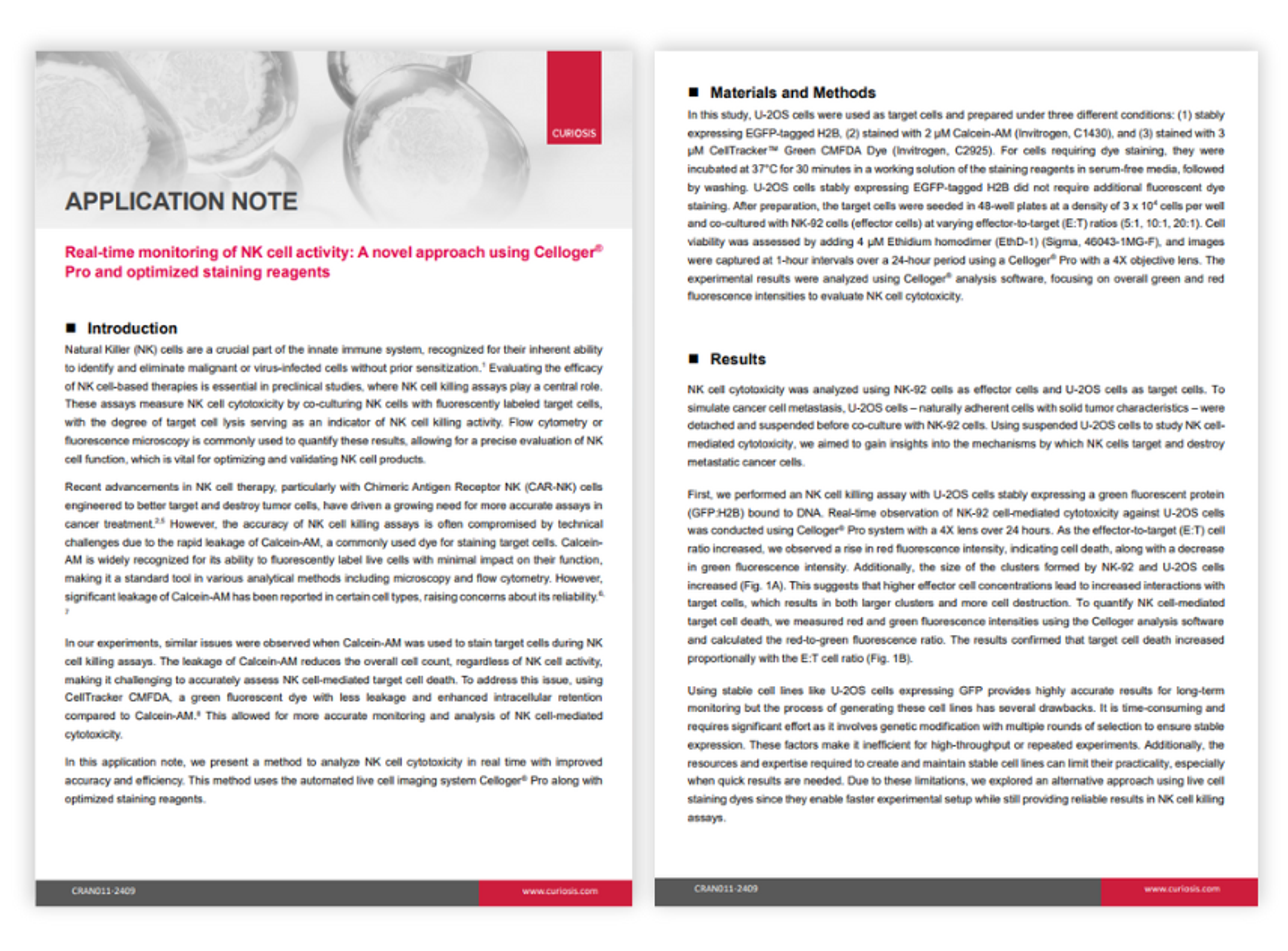
Real-time monitoring of NK cell activity: A novel approach using Celloger Pro and optimized staining reagents
In this application note, CURIOSIS demonstrates a method to analyze natural killer (NK) cell cytotoxicity in real time with improved accuracy and efficiency. This method uses the automated live cell imaging system Celloger® Pro along with optimized staining reagents.
Resource details:
Resource type: Application Notes
Page count: 6
Read time: 9 mins
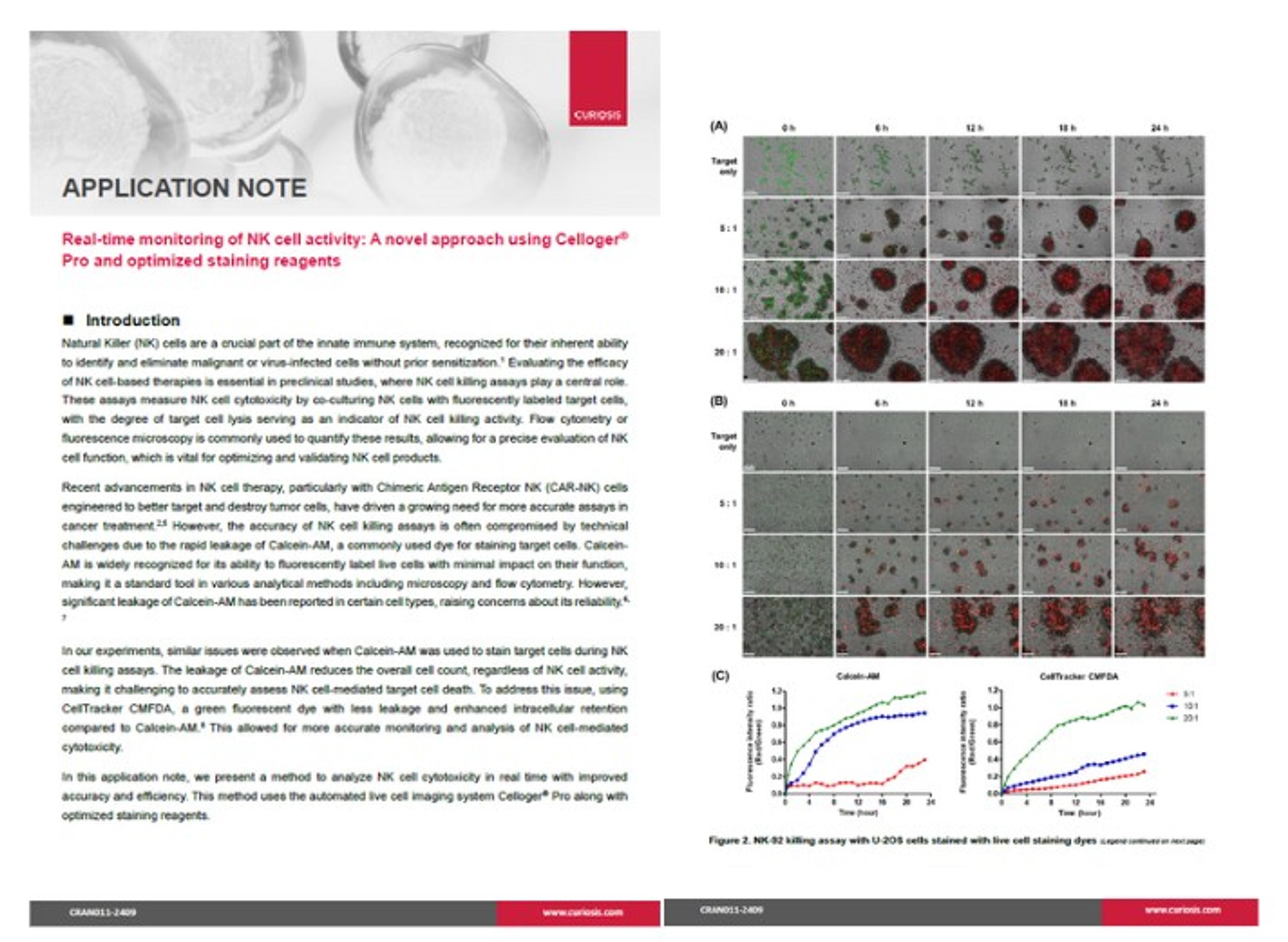
Real-time monitoring of NK cell activity using Celloger® Pro and optimized staining reagents
This application note presents a method to analyze NK cell cytotoxicity in real time with improved accuracy and efficiency.
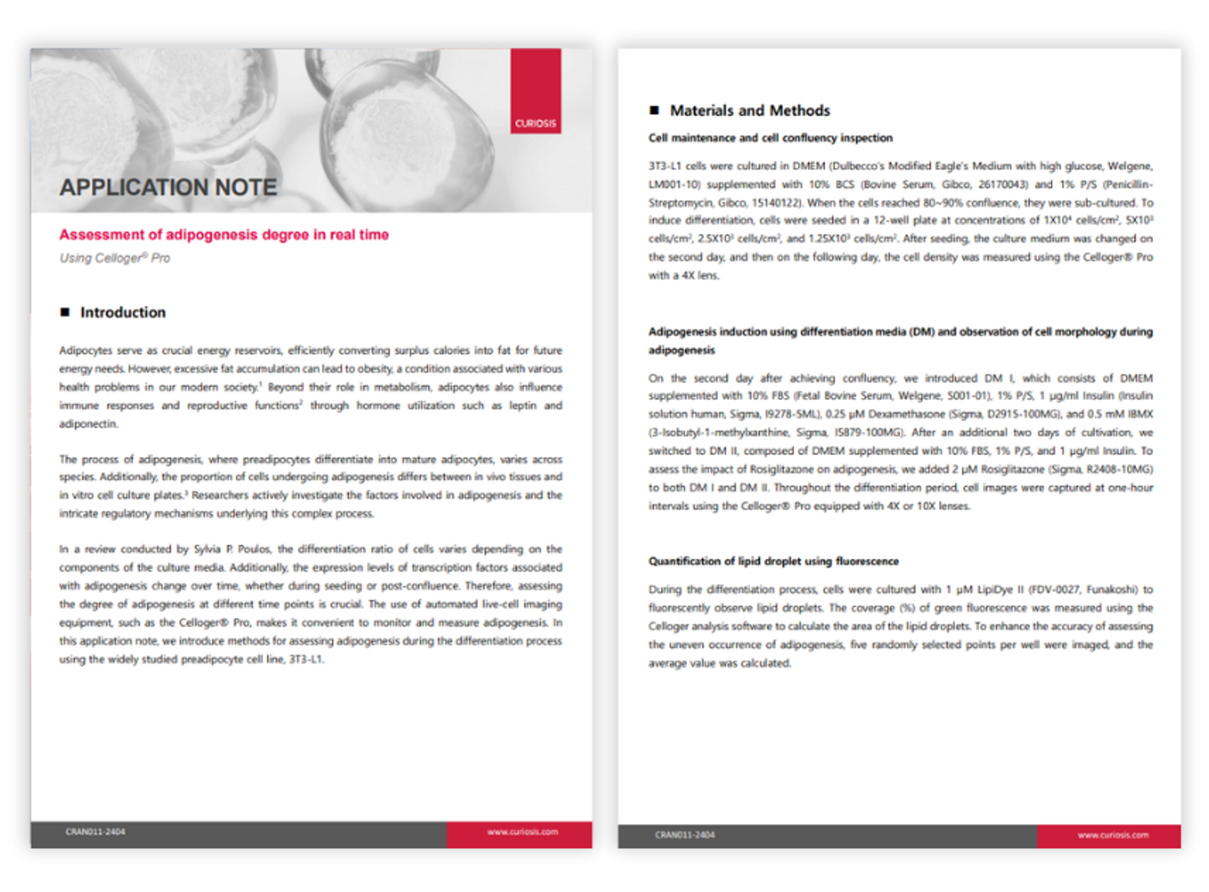
Assessment of adipogenesis degree in real time
Adipocytes serve as crucial energy reservoirs, efficiently converting surplus calories into fat for future energy needs. However, excessive fat accumulation can lead to obesity, a condition associated with various health problems in our modern society. Beyond their role in metabolism, adipocytes also influence immune responses and reproductive functions through hormone utilization such as leptin and adiponectin. Therefore, assessing the degree of adipogenesis at different time points is crucial. The use of automated live-cell imaging equipment, such as the Celloger® Pro, makes it convenient to monitor and measure adipogenesis. Curiosis introduces methods for assessing adipogenesis during the differentiation process using the widely studied preadipocyte cell line, 3T3-L1.
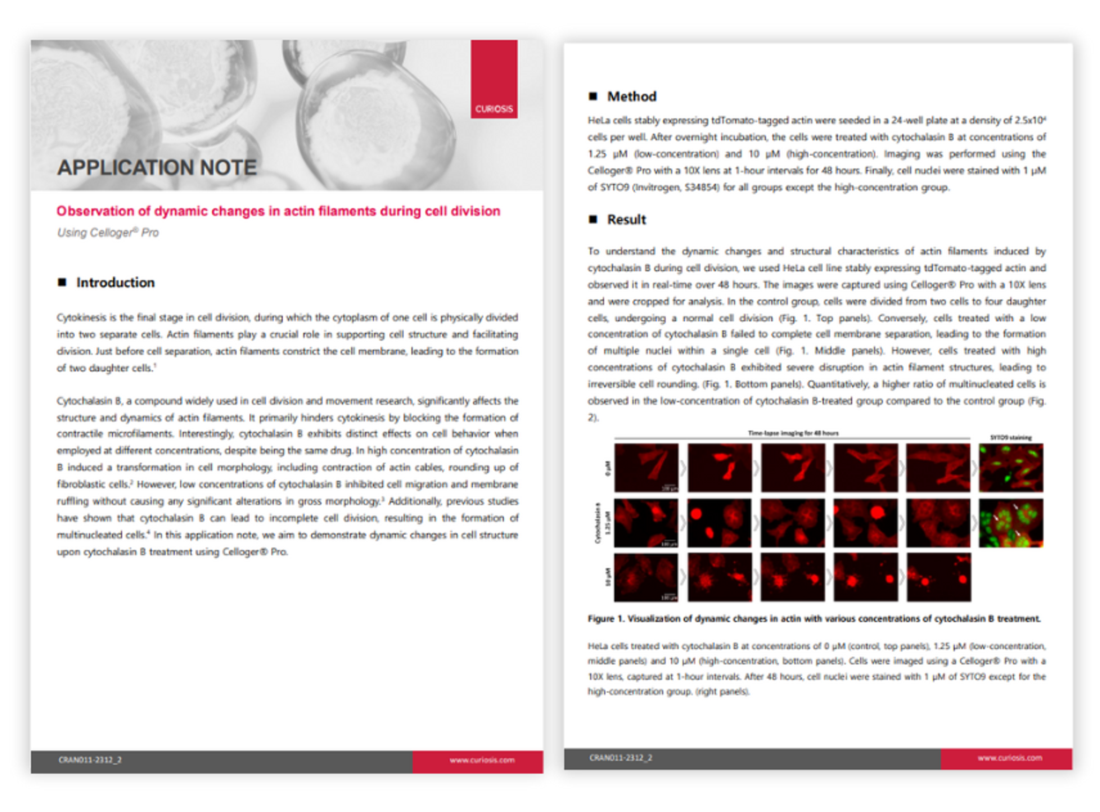
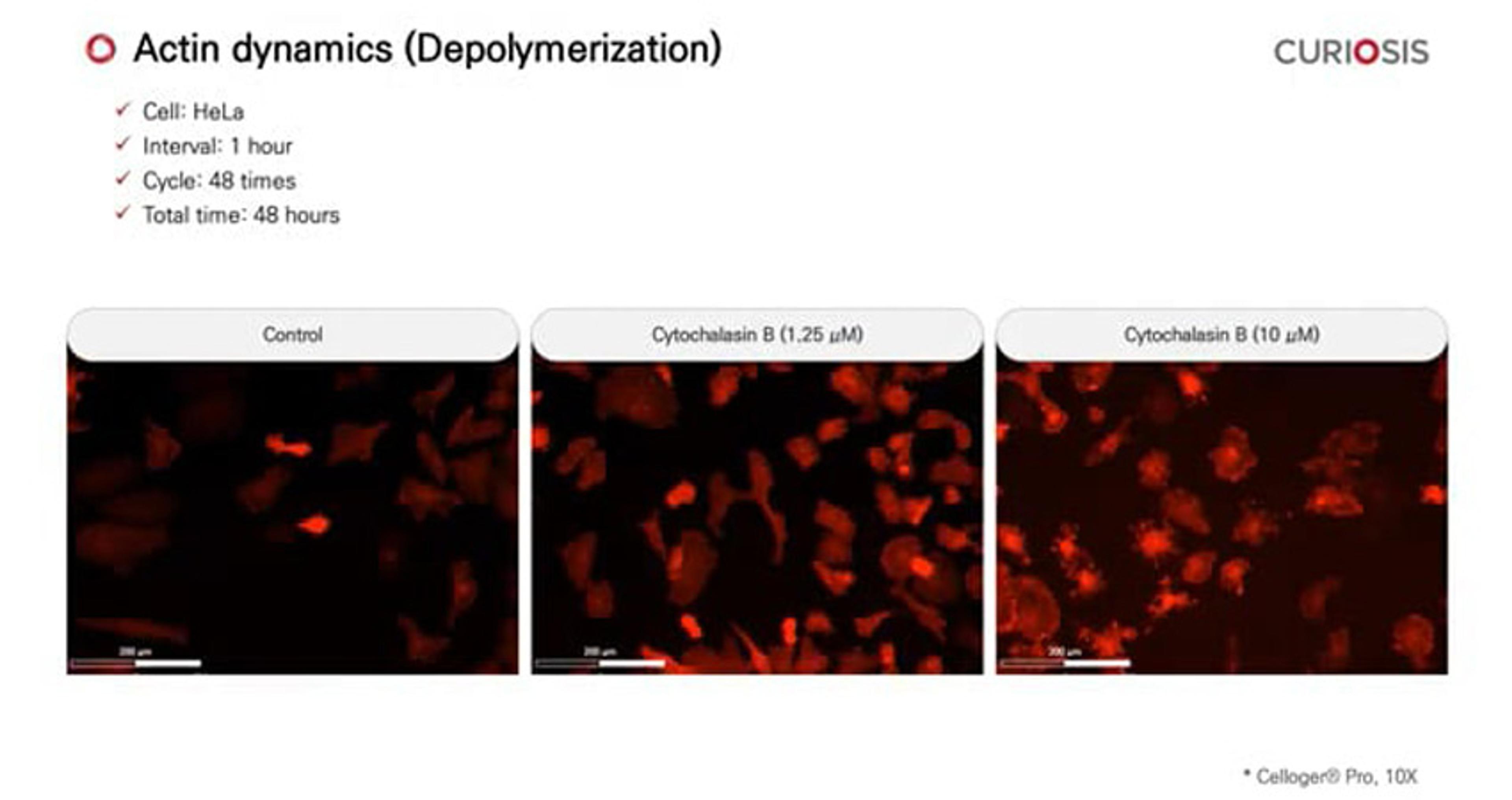
Observation of dynamic changes in actin filaments during cell division
Cytokinesis is the final stage in cell division, during which the cytoplasm of one cell is physically divided into two separate cells. Actin filaments play a crucial role in supporting cell structure and facilitating division. Just before cell separation, actin filaments constrict the cell membrane, leading to the formation of two daughter cells. Cytochalasin B, a compound widely used in cell division and movement research, significantly affects the structure and dynamics of actin filaments. It primarily hinders cytokinesis by blocking the formation of contractile microfilaments. Additionally, previous studies have shown that cytochalasin B can lead to incomplete cell division, resulting in the formation of multinucleated cells. Curiosis demonstrates dynamic changes in cell structure upon cytochalasin B treatment using the Celloger® Pro live cell imaging system. By enabling real-time cell monitoring inside the incubator, it allows for seamless observation and tracking of cellular dynamics without disrupting the natural growth environment.
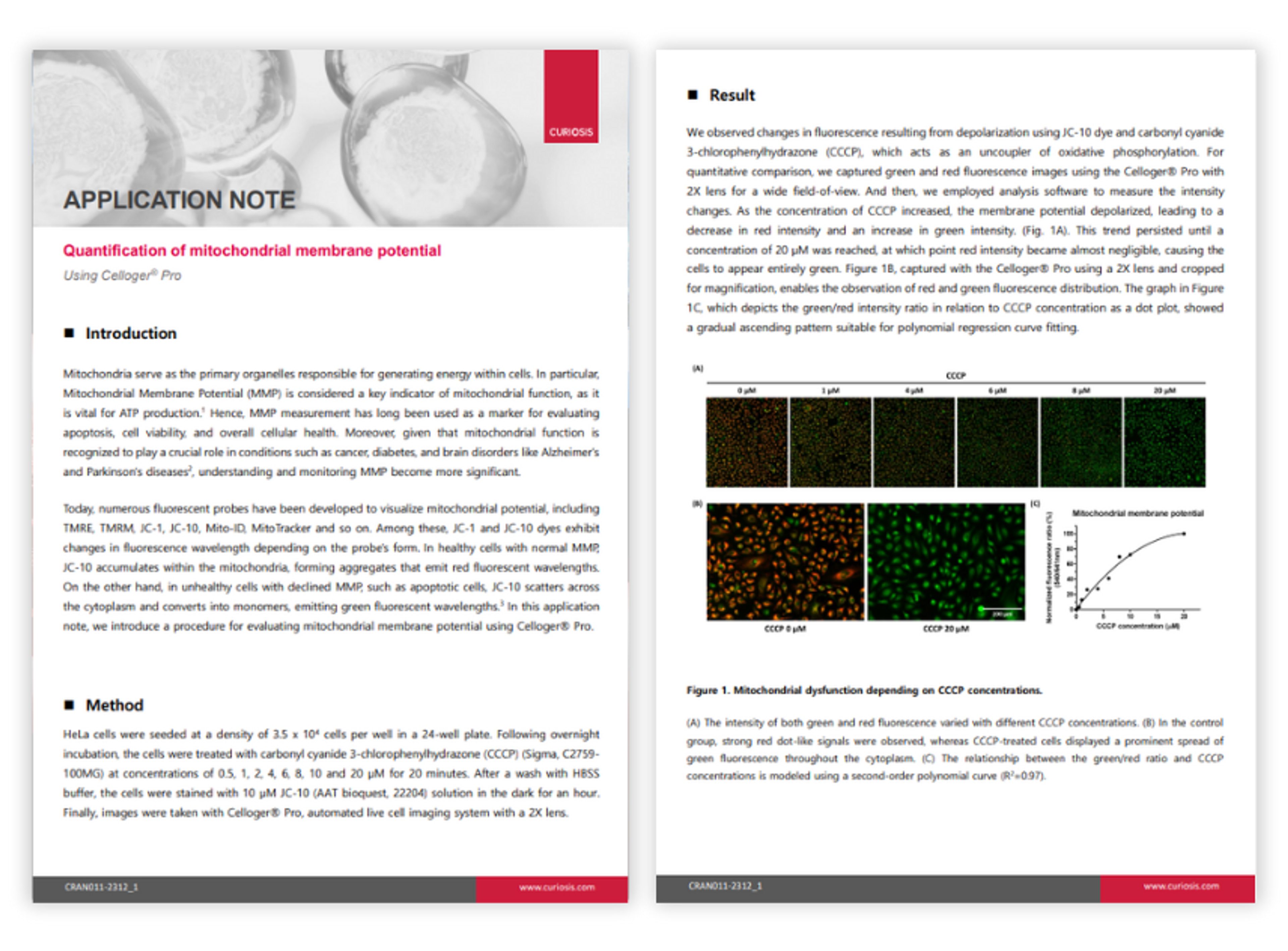
Quantification of mitochondrial membrane potential
Mitochondria serve as the primary organelles responsible for generating energy within cells. In particular, mitochondrial membrane potential (MMP) is considered a key indicator of mitochondrial function, as it is vital for adenosine triphosphate (ATP) production. Hence, MMP measurement has long been used as a marker for evaluating apoptosis, cell viability, and overall cellular health. In healthy cells with normal MMP, JC-10 accumulates within the mitochondria, forming aggregates that emit red fluorescent wavelengths. In unhealthy cells with declined MMP, such as apoptotic cells, JC-10 scatters across the cytoplasm and converts into monomers, emitting green fluorescent wavelengths. Curiosis introduces a procedure for evaluating mitochondrial membrane potential using Celloger® Pro live cell imaging system. The system's dual fluorescence and bright-field microscopy enable simultaneous visualization of multiple markers, while the multi-point time-lapse imaging captures dynamic cellular events across different locations.
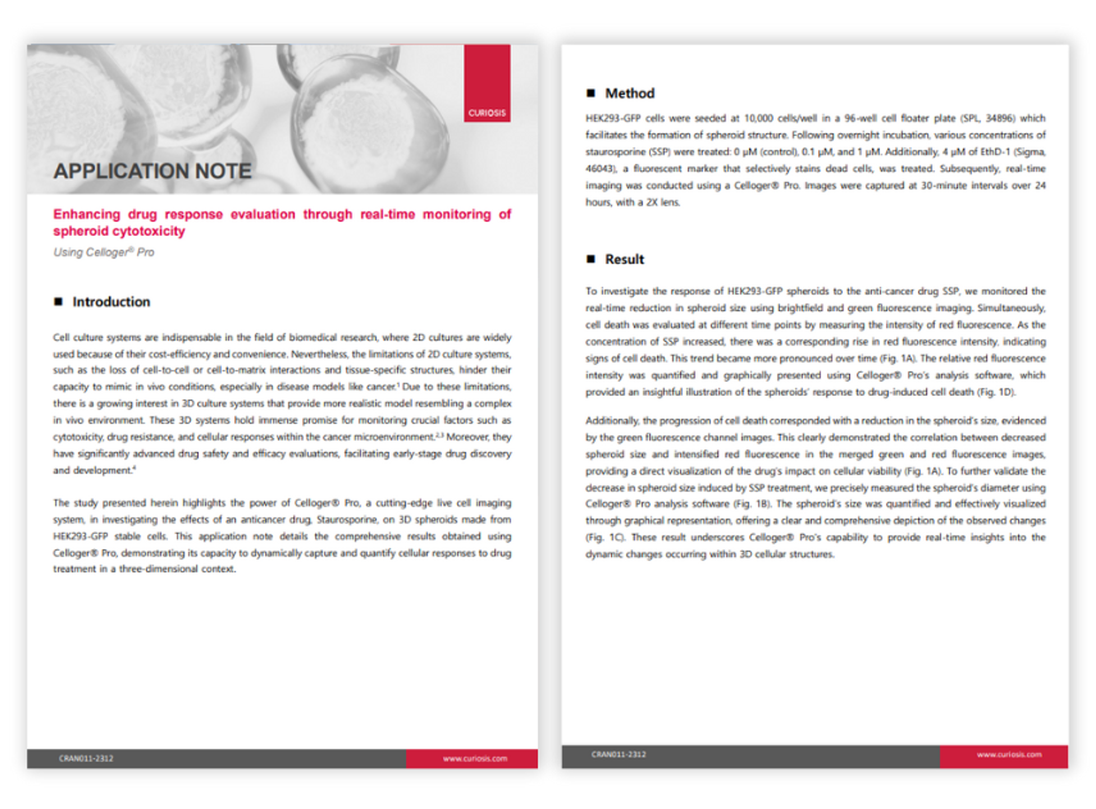
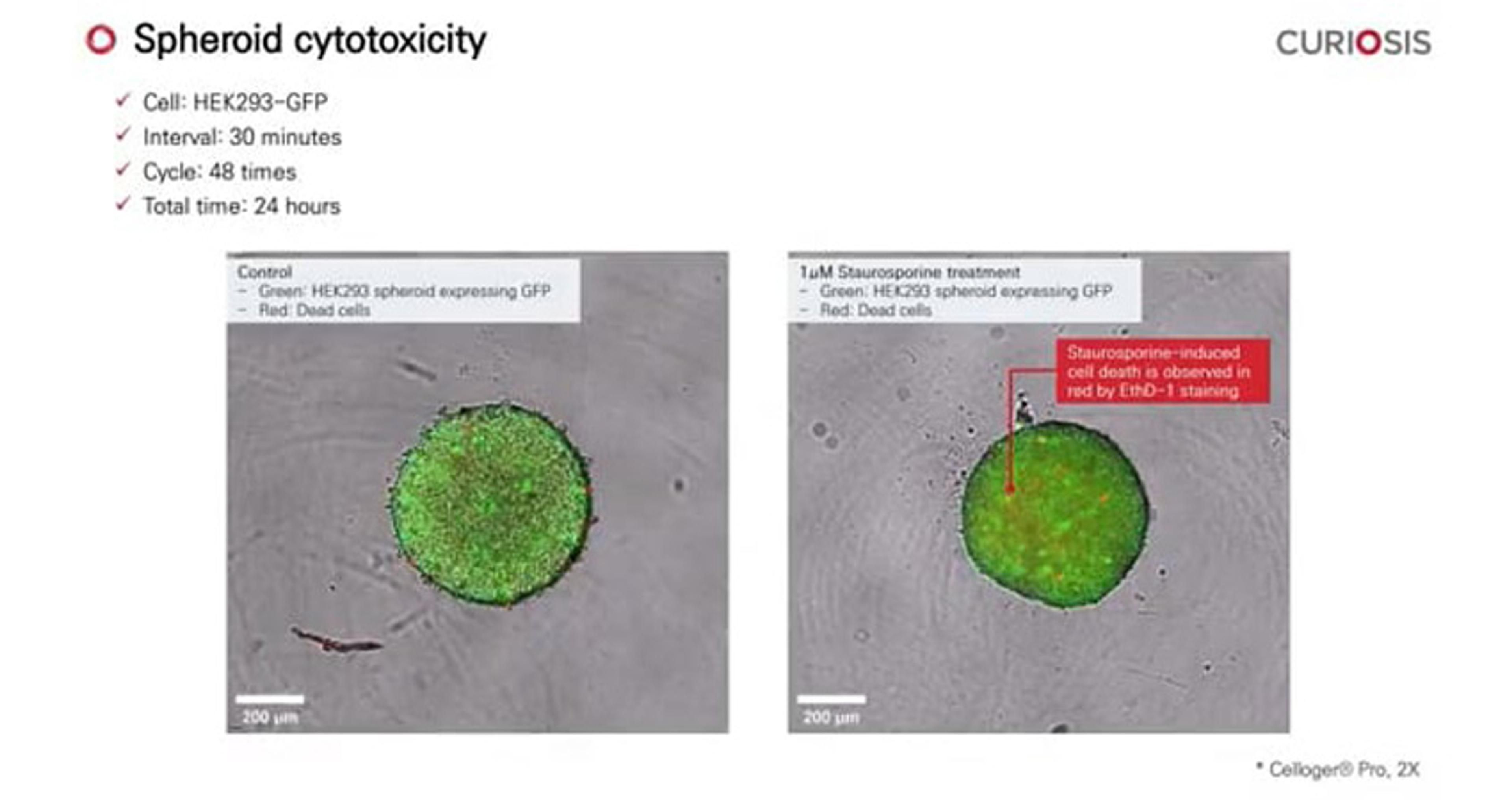
Enhancing drug response evaluation through real-time monitoring of spheroid cytotoxicity
In this application note, CURIOSIS presents the Celloger® Pro, a cutting-edge live cell imaging system, used in a study to investigate the effects of the anticancer drug Staurosporine on 3D spheroids made from HEK293-GFP stable cells. The results showcase Celloger Pro's capability to dynamically capture and quantify cellular responses to drug treatment in a three-dimensional context, contributing to advancements in drug safety and efficacy evaluations for early-stage drug discovery and development.
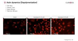
Celloger Pro application: Actin dynamics
In this video, CURIOSIS demonstrates how the Celloger® Pro can be used to observe the actin depolymerization process. HeLa cells stably expressing tdTomato-tagged actin were treated with cytochalasin B at low (1.25 μM) and high (10 μM) concentrations. Time-lapse images were taken at 1-hour intervals over 48 hours using a Celloger® Pro with a 10X objective.
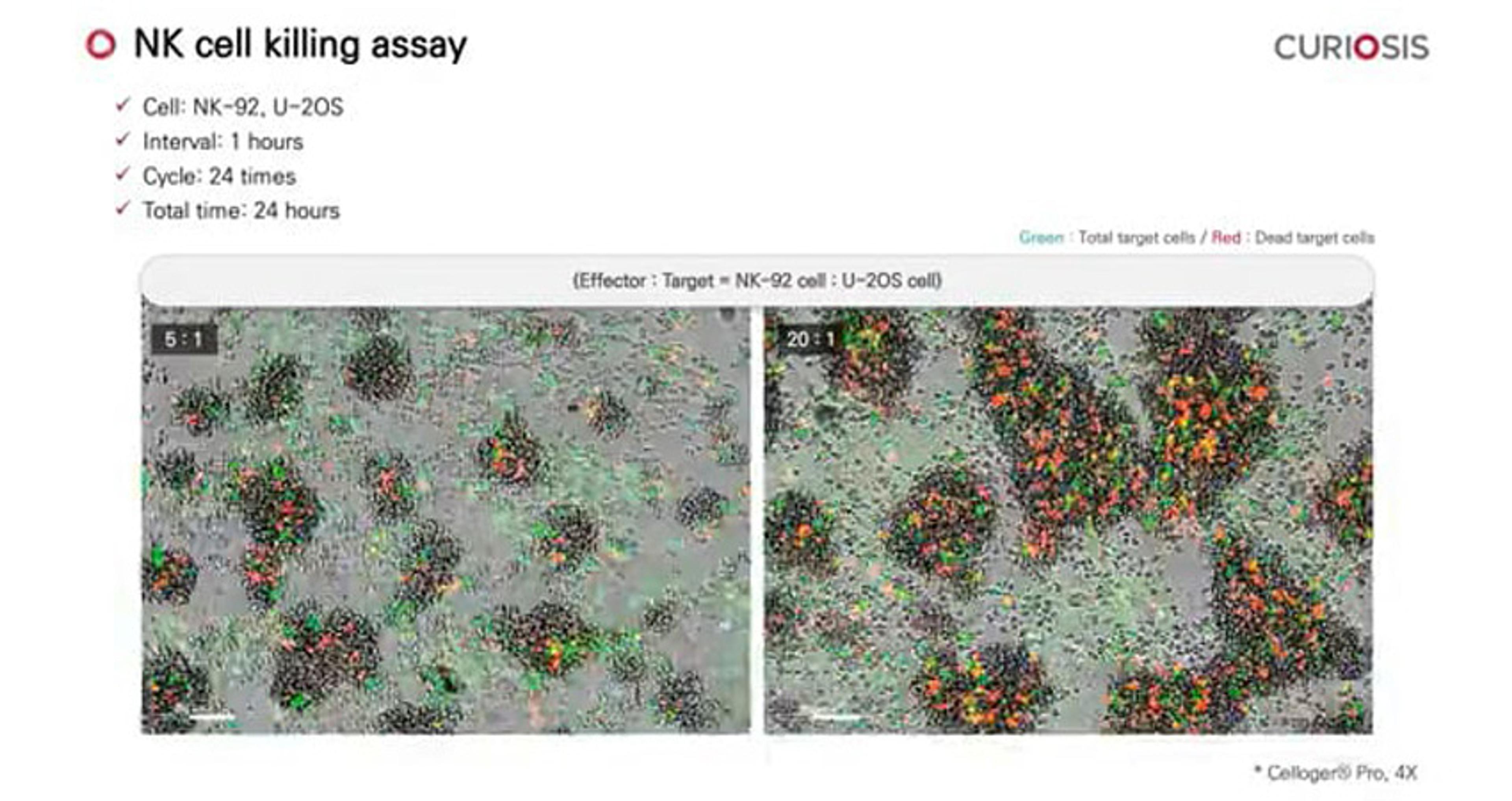
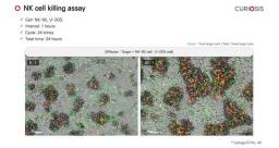
Celloger Pro application: NK cell killing assay
In this video, CURIOSIS demonstrates how the Celloger® Pro can be used to studying NK cell interactions with target cells. To observe these interactions, U-2OS cells (targets) were stained with CellTracker Green CMFDA (green fluorescence) and co-cultured with NK-92 cells (effectors) at effector-to-target ratios of 5:1 and 20:1. Time-lapse imaging began immediately after staining with 4 µM EthD-1 (red fluorescence) and was performed every hour for 24 hours using a Celloger® Pro with a 4X objective lens.
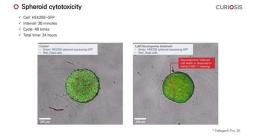
Celloger Pro application: Spheroid cytotoxicity
In this video, CURIOSIS demonstrates how the Celloger® Pro can be used to observe the drug effect on spheroids in real-time. Spheroids were made from HEK293 cells that stably express GFP. Staurosporine and EthD-1 dead cell staining dye were applied, and the images were captured at 30-minute intervals over 24 hours using a Celloger® Pro with a 2X objective.
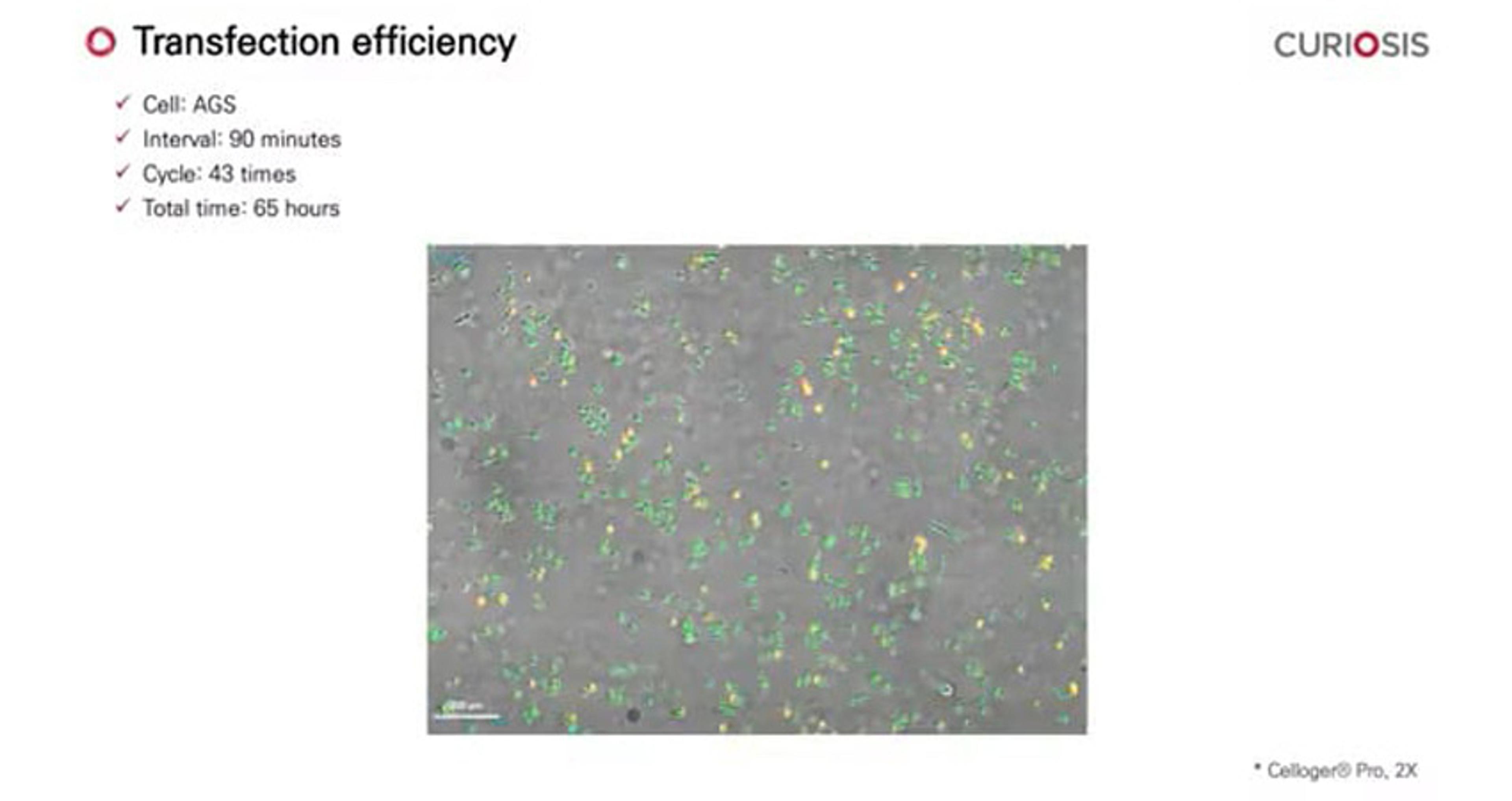
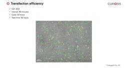
Celloger Pro application: Transfection efficiency
In this video, CURIOSIS demonstrates how the Celloger® Pro can be used to monitor transfection efficiency in real-time. In AGS cell line, cell type was stained with CMFDA dye (green fluorescence) and then transfected with the tdTomato-Lifeact gene (red fluorescence). Seven hours post-transfection, time-lapse imaging was performed every 90 minutes for 65 hours using the Celloger® Pro with a 2X objective.
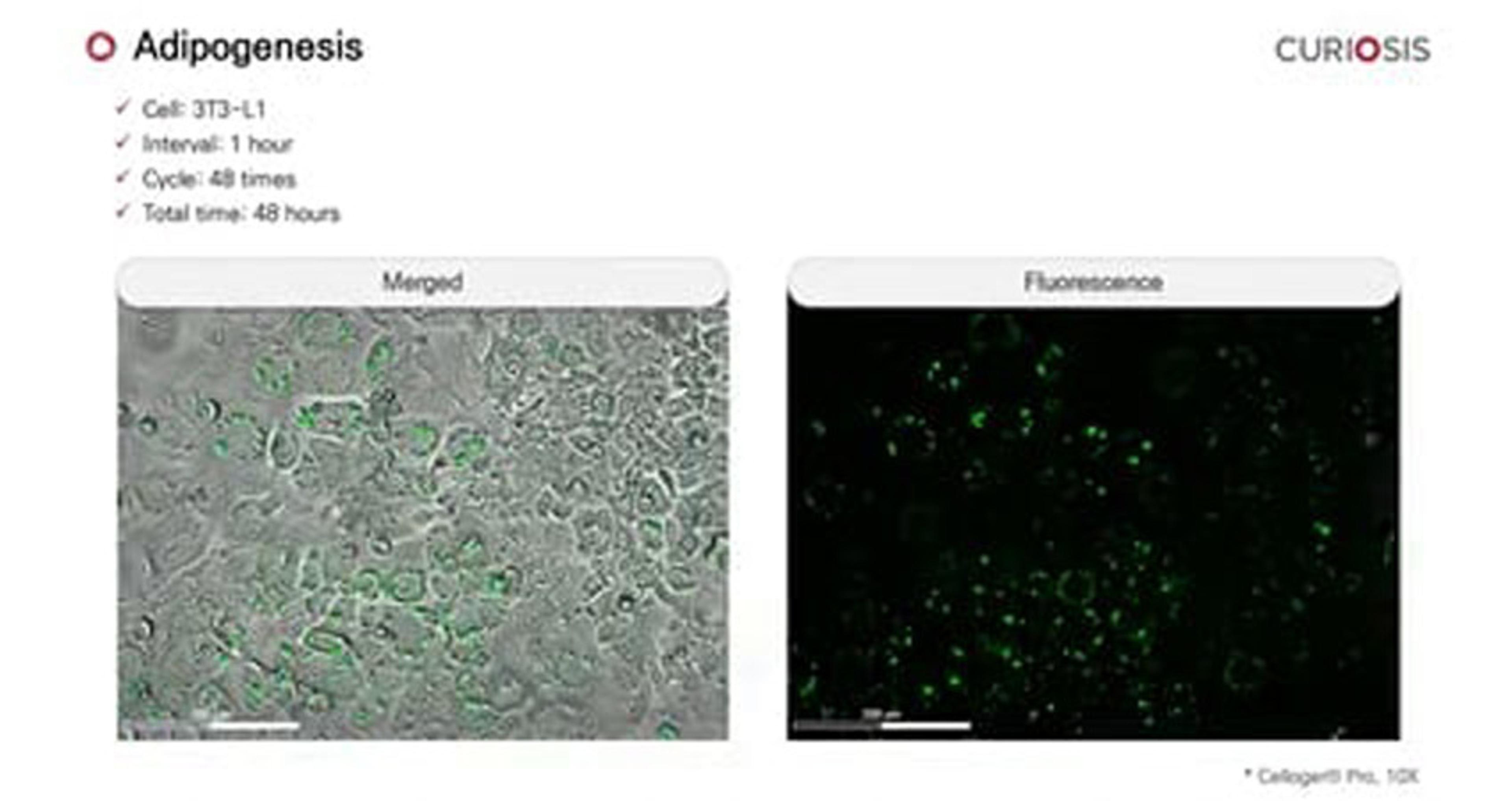
Celloger Pro application: Adipogenesis
In this video, CURIOSIS demonstrates how the Celloger® Pro can be used to monitor the adipocyte differentiation process. To show this, 3T3-L1 cells were stained with LipiDye, and time-lapse images were taken at 1-hour intervals over 48 hours using a Celloger® Pro with a 10X objective. As the cells differentiate, an increase in the distribution of lipid droplets exhibiting green fluorescence can be observed over time.
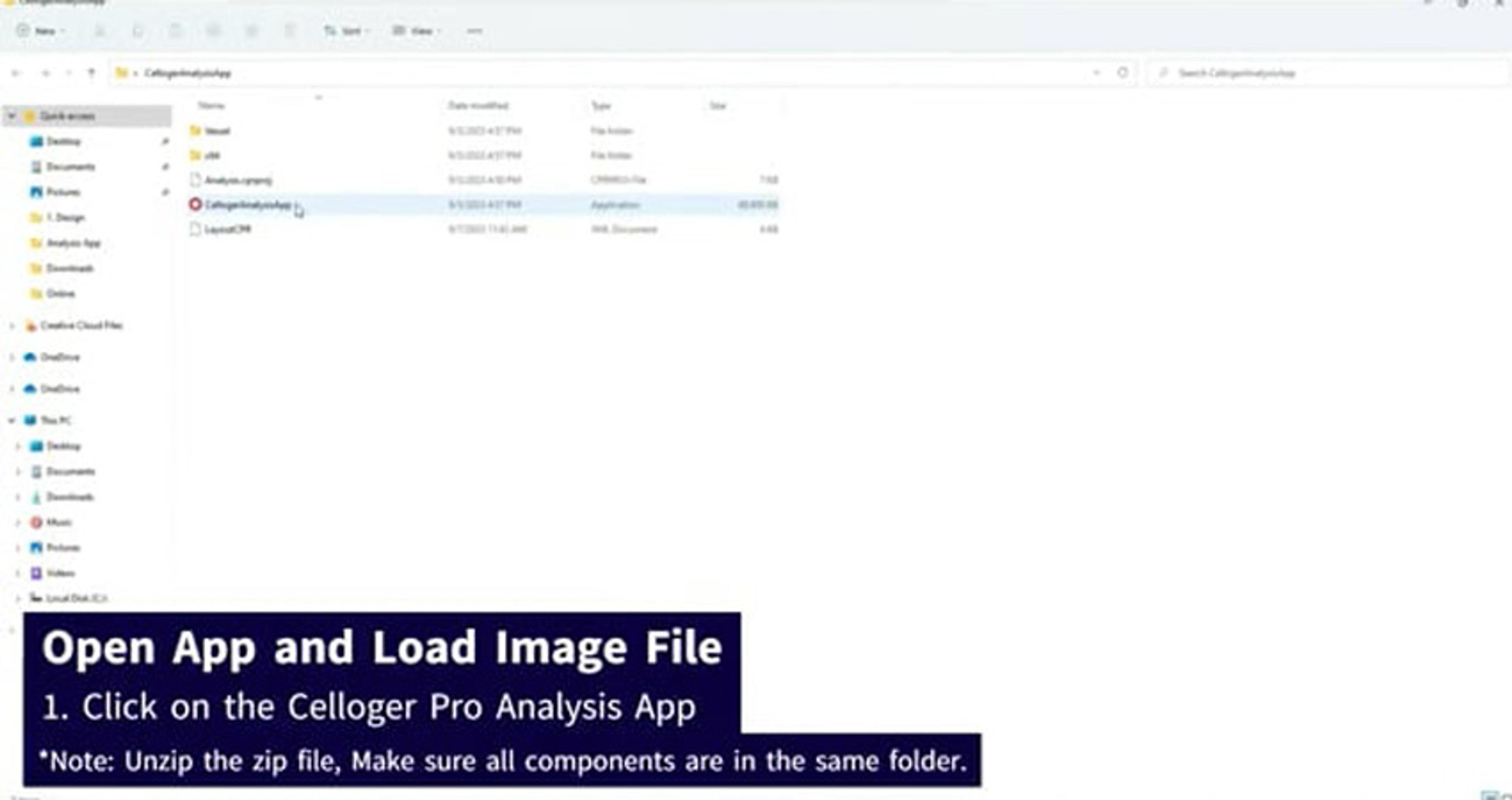
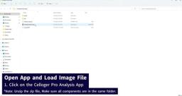
How to use analysis app of Celloger Pro
In this video, CURIOSIS demonstrates the Celloger® Pro, an innovative live cell imaging system that redefines research capabilities, and how to utilize its analysis app.
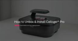
How to unbox and install Celloger Pro
In this video, CURIOSIS demonstrates the Celloger® Pro, an innovative live cell imaging system that redefines research capabilities. Its advanced features allow for seamless observation and tracking of cellular dynamics without disrupting the natural growth environment.
Celloger Pro, automated live cell imaging system
In this video, CURIOSIS demonstrates the Celloger® Pro, a live cell imaging solution for scientific exploration. The system's dual fluorescence and bright-field microscopy enable simultaneous visualization of multiple markers, while the multi-point time-lapse imaging captures dynamic cellular events across different locations. Celloger Pro’s user-friendly interface and intuitive tools make image acquisition and analysis a seamless experience. The system enables real-time cell monitoring inside the incubator, and seamless observation and tracking of cellular dynamics without disrupting the natural growth environment. Additionally, its ability to capture high-resolution images and to effortlessly create videos, eliminates the labor-intensive tasks associated with live cell imaging processes.
The role of automated live cell imaging with the Celloger Pro
Expand horizons in life science research and discover the power of real-time cell monitoring