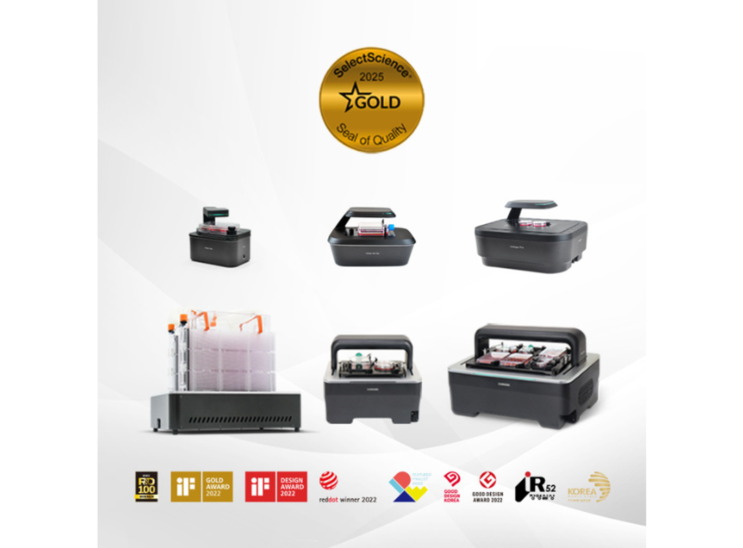
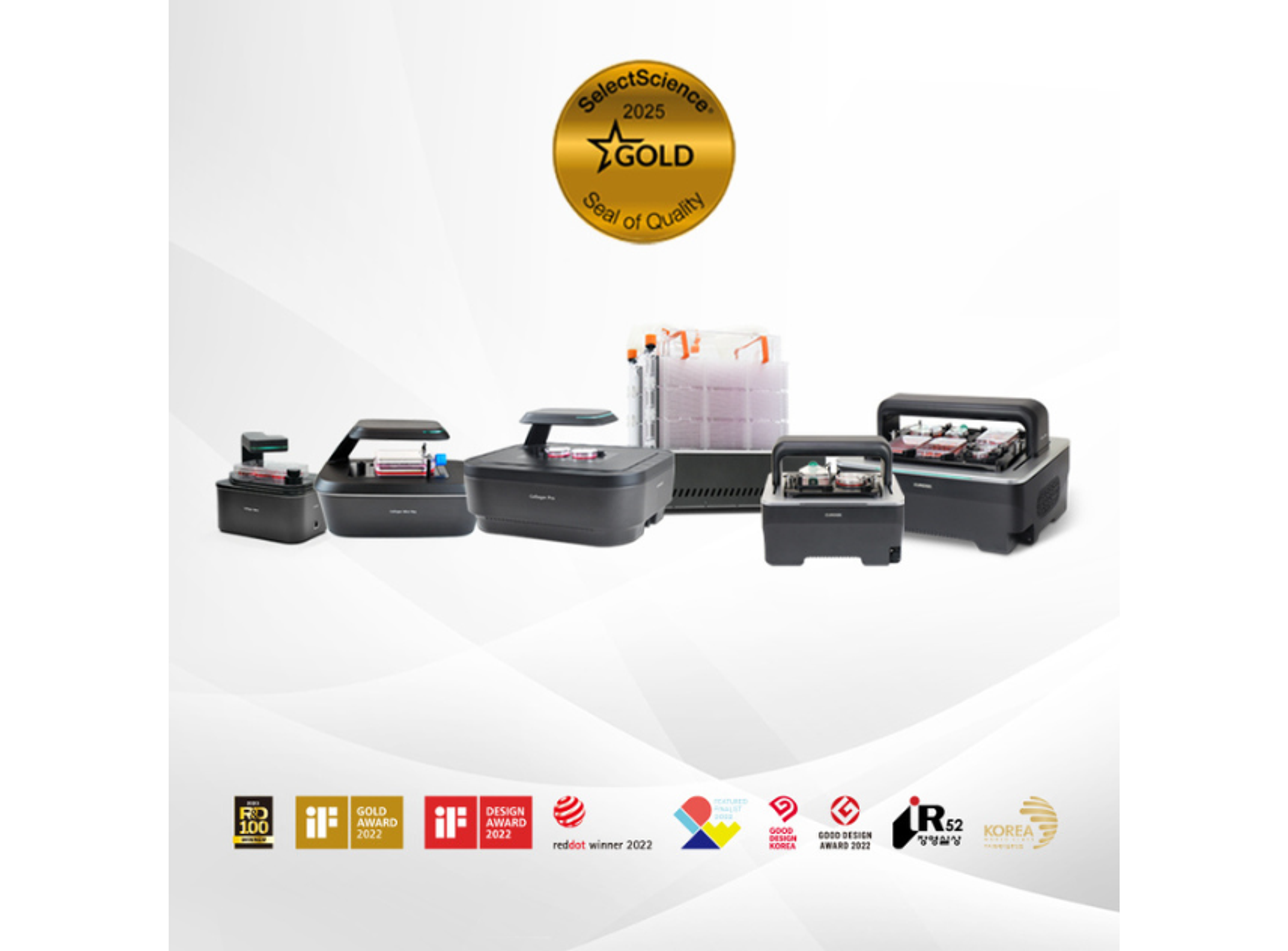
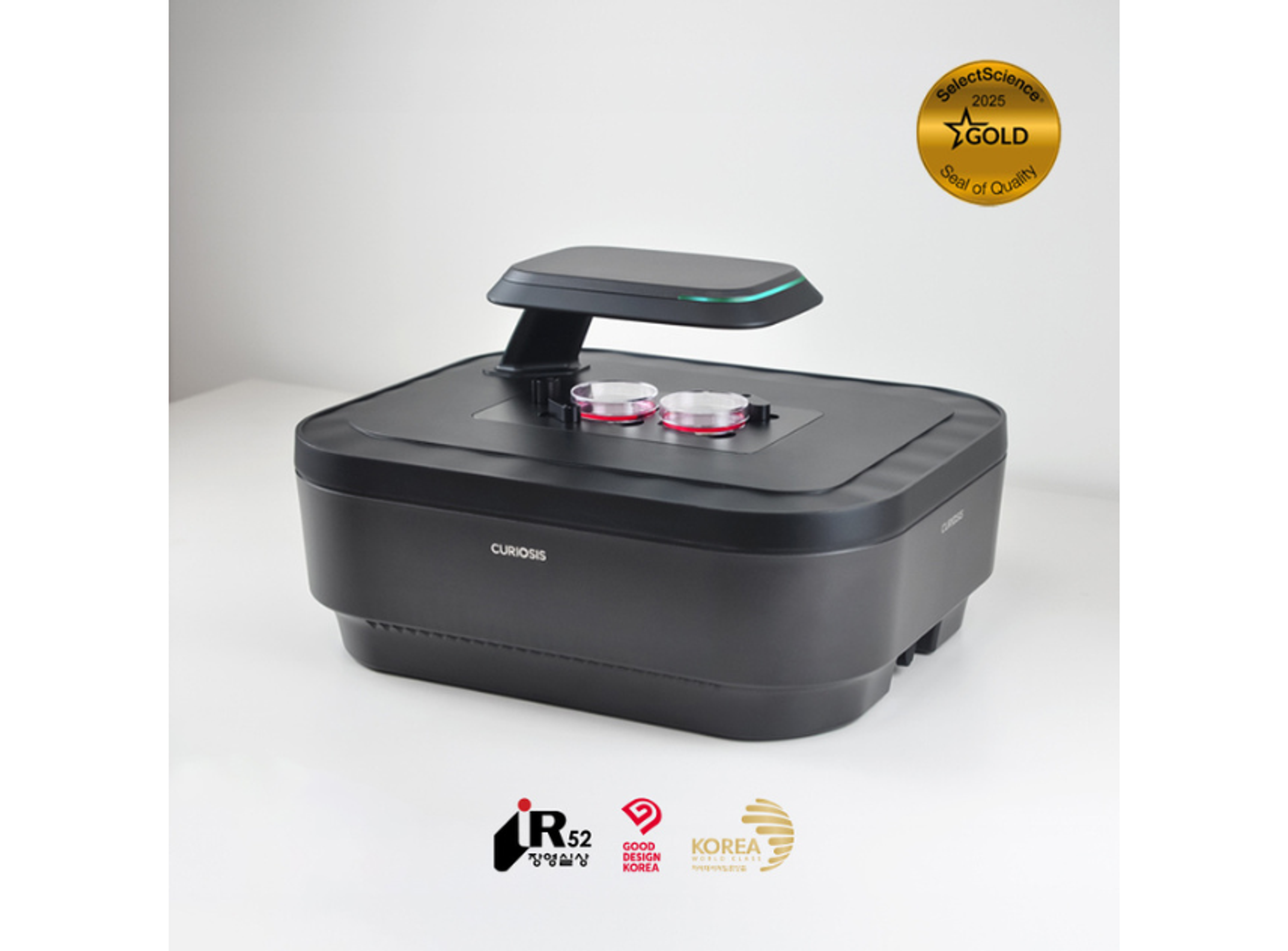
Celloger®, Automated Live Cell Imaging System from Curiosis
Capture the moments of dynamic cellular processes, improving your research with comprehensive insights


Receive your quote directly from the manufacturer.
We were able to easily image and quantify 3D models derived from liver cancer cell lines and human tissue–derived cells.
3D cell model research
Using Celloger® Pro, we were able to easily image and quantify 3D models derived from liver cancer cell lines and human tissue–derived cells. Its stable performance, high-quality imaging, and built-in analysis tools consistently provided the data we needed.
Review Date: 25 Nov 2025 | CURIOSIS
Celloger® Pro made it easy to monitor spheroids under different conditions and track changes in real time.
Cancer biology
Celloger® Pro made it easy to monitor spheroids under different conditions and track changes in their size in real time. After adding fluorescent reagents, we could clearly visualize how the fluorescence signal was distributed within the spheroids. Overall, the real-time fluorescence imaging and analysis features provided exactly the data we needed.
Review Date: 25 Nov 2025 | CURIOSIS
I was especially impressed by the high image quality and clarity of the images.
Cancer biology
I performed an exosome uptake experiment with C2C12 cells and obtained the expected results, and I was especially impressed by the high image quality and clarity of the images.
Review Date: 25 Nov 2025 | CURIOSIS
Using Celloger® Mini Plus for apoptosis experiments made it very easy to monitor cytotoxicity in real time.
Cell death research
Using Celloger® Mini Plus for apoptosis experiments made it very easy to monitor cytotoxicity in real time. It’s simple to use, so I could quickly see how the cells were responding without going through any complicated procedures.
Review Date: 25 Nov 2025 | CURIOSIS
The built-in area measurement and automated wound-healing analysis tools were very convenient.
Cell movement
It was very convenient that the analysis software included area measurement tools and automated functions for wound-healing analysis. I also appreciated that it was compatible not only with standard well plates but also with other types of culture vessels.
Review Date: 24 Nov 2025 | CURIOSIS
The Celloger scan and analysis software was easy to use, and the image quality was very good.
Vascular cell aging and disease research
The Celloger scan and analysis software was easy to use, and the image quality was very good.
Review Date: 24 Nov 2025 | CURIOSIS
The Celloger® Pro system has really boosted my research.
Immunotherapy research
The Celloger® Pro system has really boosted my research. Instead of spending time on tedious imaging work myself, I can rely on the system to handle it automatically. The image quality has been excellent, and the software is very intuitive and easy to work with.
Review Date: 24 Nov 2025 | CURIOSIS
Using the Celloger® Pro for NK cell–mediated cytotoxicity experiments has been very helpful.
Immunotherapy research
Using the Celloger® Pro for NK cell–mediated cytotoxicity experiments has been very helpful. By using two fluorescent markers, I was able to clearly visualize the interactions between NK cells and target cells, and also assess the extent of NK cell–mediated killing. This made it much easier to understand the dynamics of the assay in real time.
Review Date: 24 Nov 2025 | CURIOSIS
Celloger® Mini Plus allowed reliable, stable monitoring of suspension cell growth over time.
Suspension cell growth monitoring
Celloger® Mini Plus allowed reliable, stable monitoring of suspension cell growth over time.
Review Date: 24 Nov 2025 | CURIOSIS
Data acquisition for both wound-healing assays and cell proliferation analysis went very smoothly, with no issues.
Proteostasis network
Data acquisition for both wound-healing assays and cell proliferation analysis went very smoothly, with no issues.
Review Date: 24 Nov 2025 | CURIOSIS
Key features of Celloger®
- - Real-time cell monitoring inside an incubator
- - Compatible with different vessel types
- - Time-lapse imaging capability
- - User-friendly functions included in the software package
Key applications of Celloger®
- - Cell proliferation
- - Wound healing assay
- - Co-culture monitoring
- - Spheroid cell death assay
- - Actin dynamics assay
- - Mitochondrial membrane potential
- - ROS detection
- - Phagocytosis monitoring
- - Zebrafish observation
Brochures
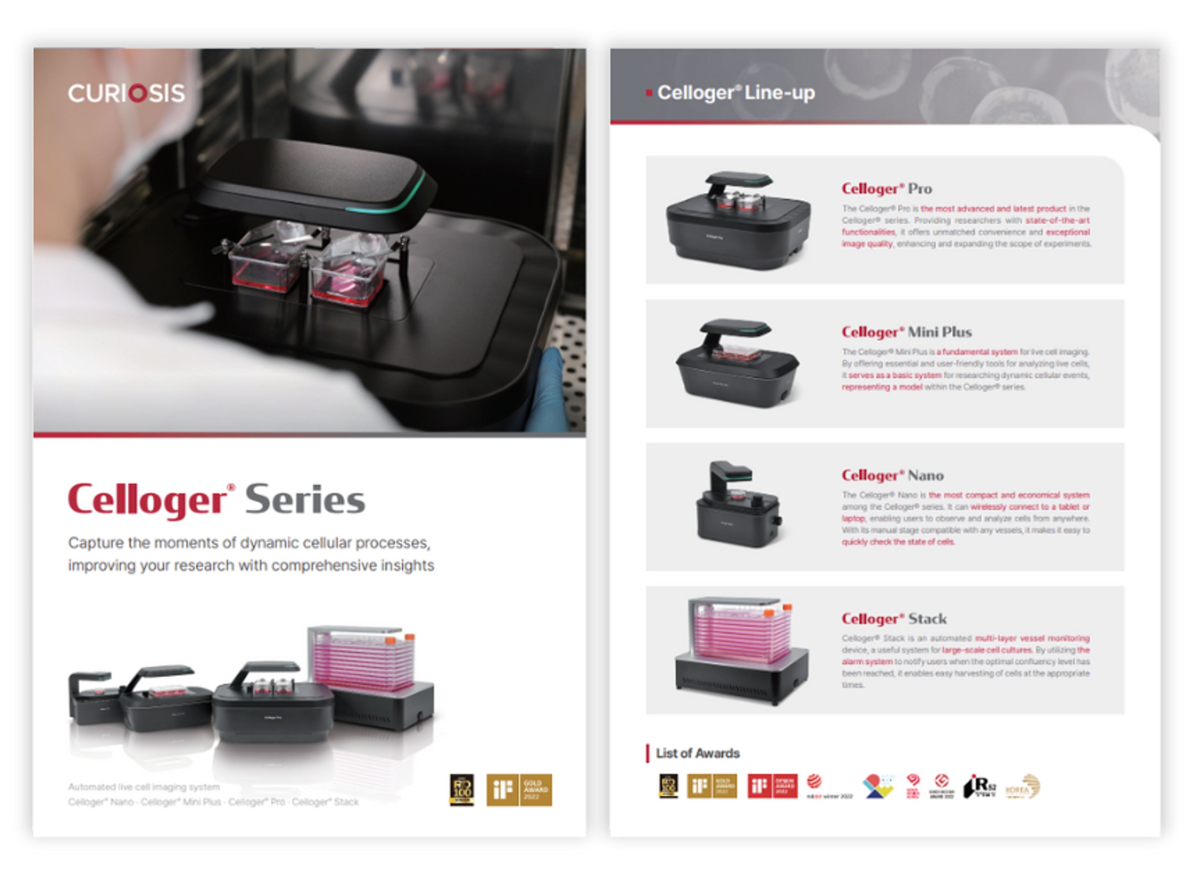
Celloger Series: Automated live cell imaging system
In this product brochure, CURIOSIS presents Celloger®, a range of innovative systems tailored for live cell imaging and monitoring. Celloger® Pro, the latest addition, has advanced functionalities and exceptional image quality, expanding experiment possibilities. The Celloger® Mini Plus provides essential tools for live cell analysis, while the compact Celloger® Nano offers wireless connectivity for remote cell observation. For large-scale cultures, Celloger® Stack automates vessel monitoring, ensuring timely cell harvesting. With versatile applications and user-friendly features, Celloger systems enable real-time cell monitoring inside incubators whilst facilitating time-lapse imaging.
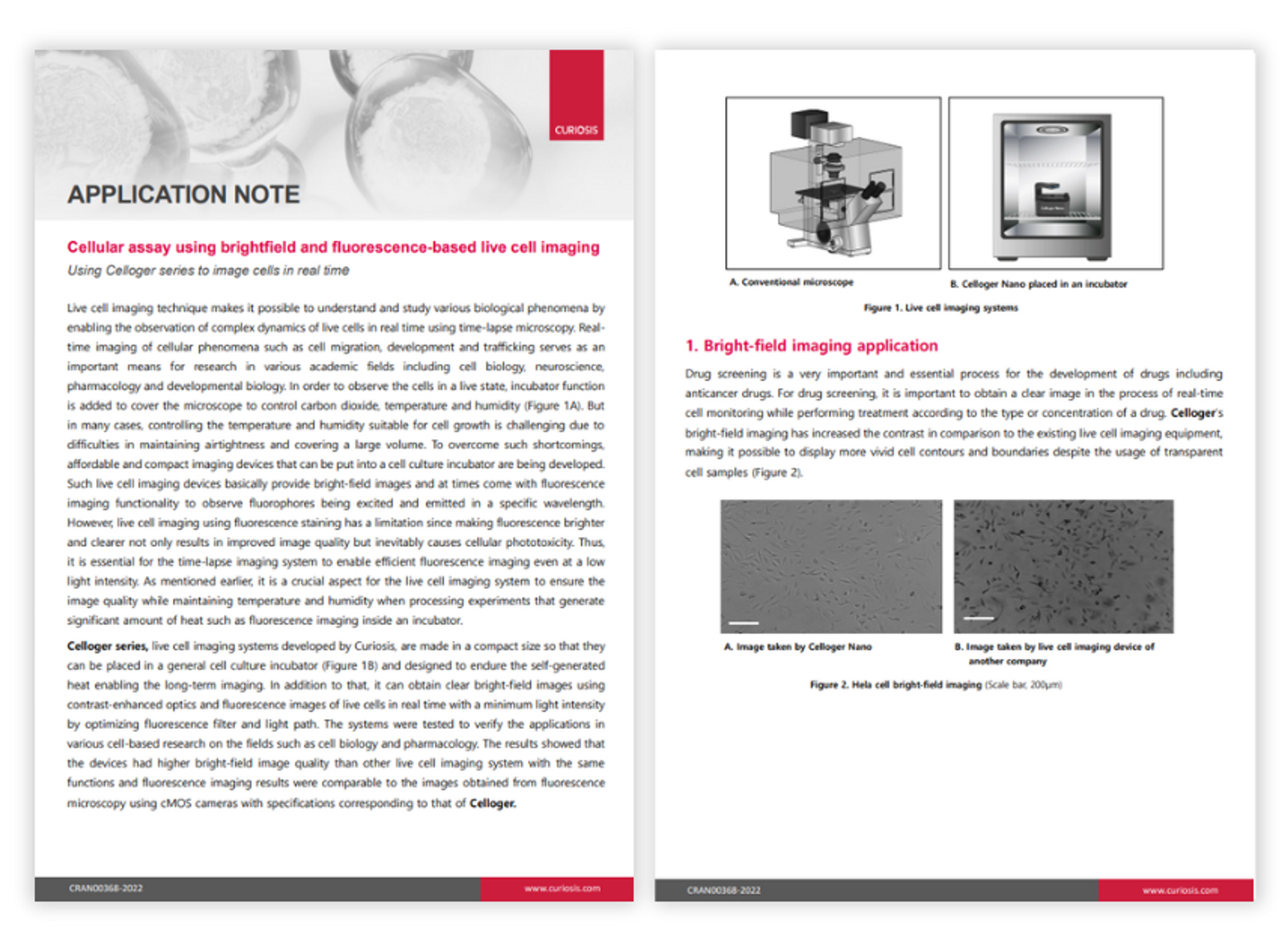
Cellular assay using brightfield and fluorescence-based live cell imaging
In this application note, Curiosis describes the applications of the Celloger series live cell imaging systems, made in a compact size so that they can be placed in a general cell culture incubator and designed to endure the self-generated heat enabling long-term imaging.
Rewriting the rules of muscle aging: Real-time analyses of myogenic progenitor metabolism
Aging is the primary risk factor for numerous life-limiting diseases, including cardiovascular disease, cancer, metabolic disorders, and neurodegenerative conditions. One significant age-related condition is sarcopenia, a progressive decline in muscle mass and function that comes with aging. Sarcopenia not only affects physical abilities but also has a profound impact on overall health and lifespan. Understanding and addressing this decline in muscle health is essential to advancing healthy aging and extending longevity.
Gain insights into cutting-edge research on muscle stem cells (MuSCs), which play a key role in muscle regeneration and growth. The Celloger® live cell imaging system enables real-time observation of differences between young and aged MuSCs. Explore an innovative in vitro parabiosis model built on a 3D microfluidic platform. This system shows how interactions between young and aged vascular and cellular environments can rejuvenate the function of aged MuSCs, offering exciting potential for therapeutic strategies to address musculoskeletal aging.

Introducing the latest Seal of Quality Award recipients
Lab products are recognized for excellence as rated by researchers and lab professionals

Latest Seal of Quality Awards announced
Find out which products have earned a new Seal of Quality, including the rare Diamond Seal of Quality

Our first Seal of Quality recipients of 2024
Discover which products have earned a coveted Seal of Quality award
Product Overview
Links
Products Model Information
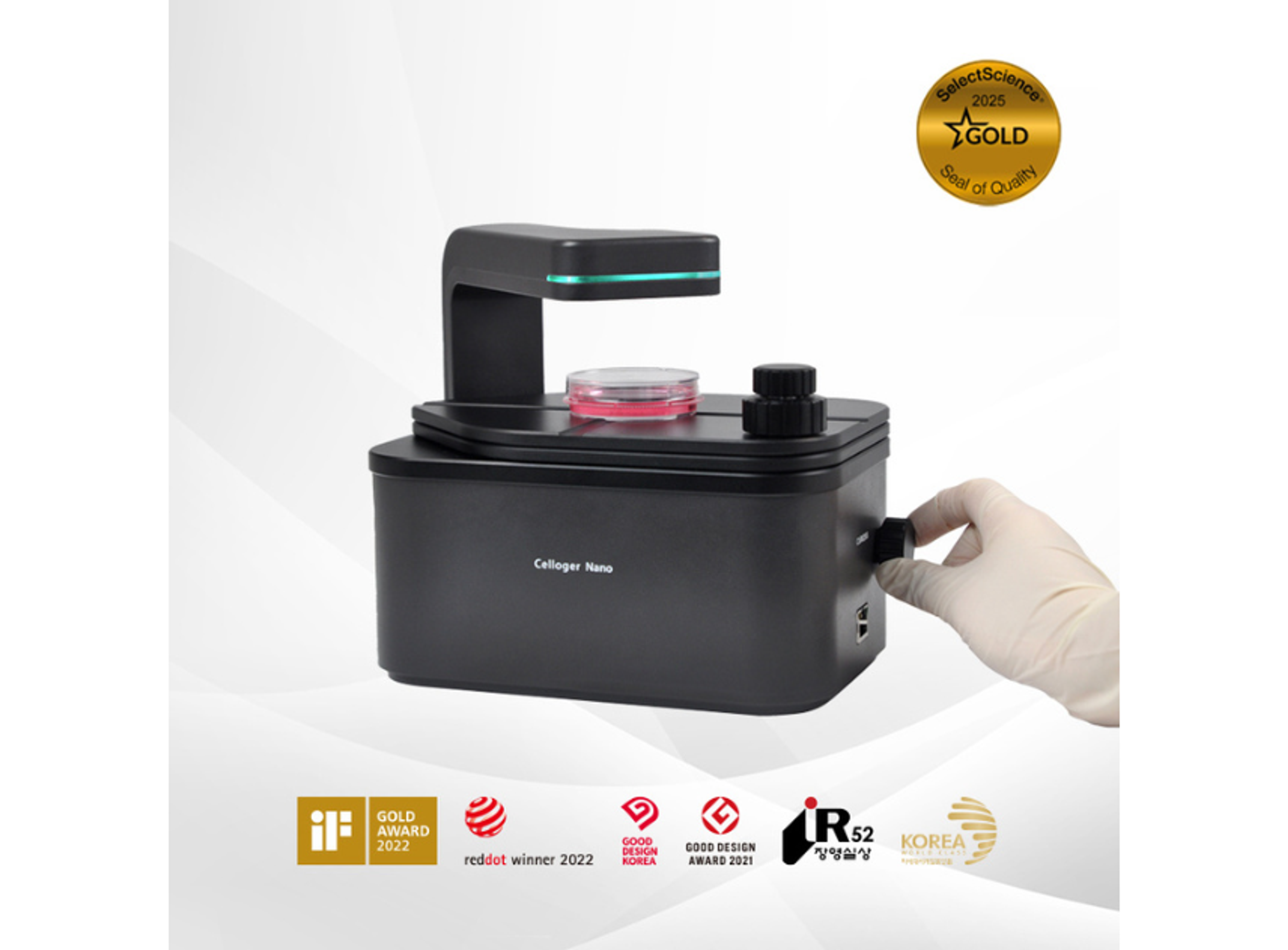
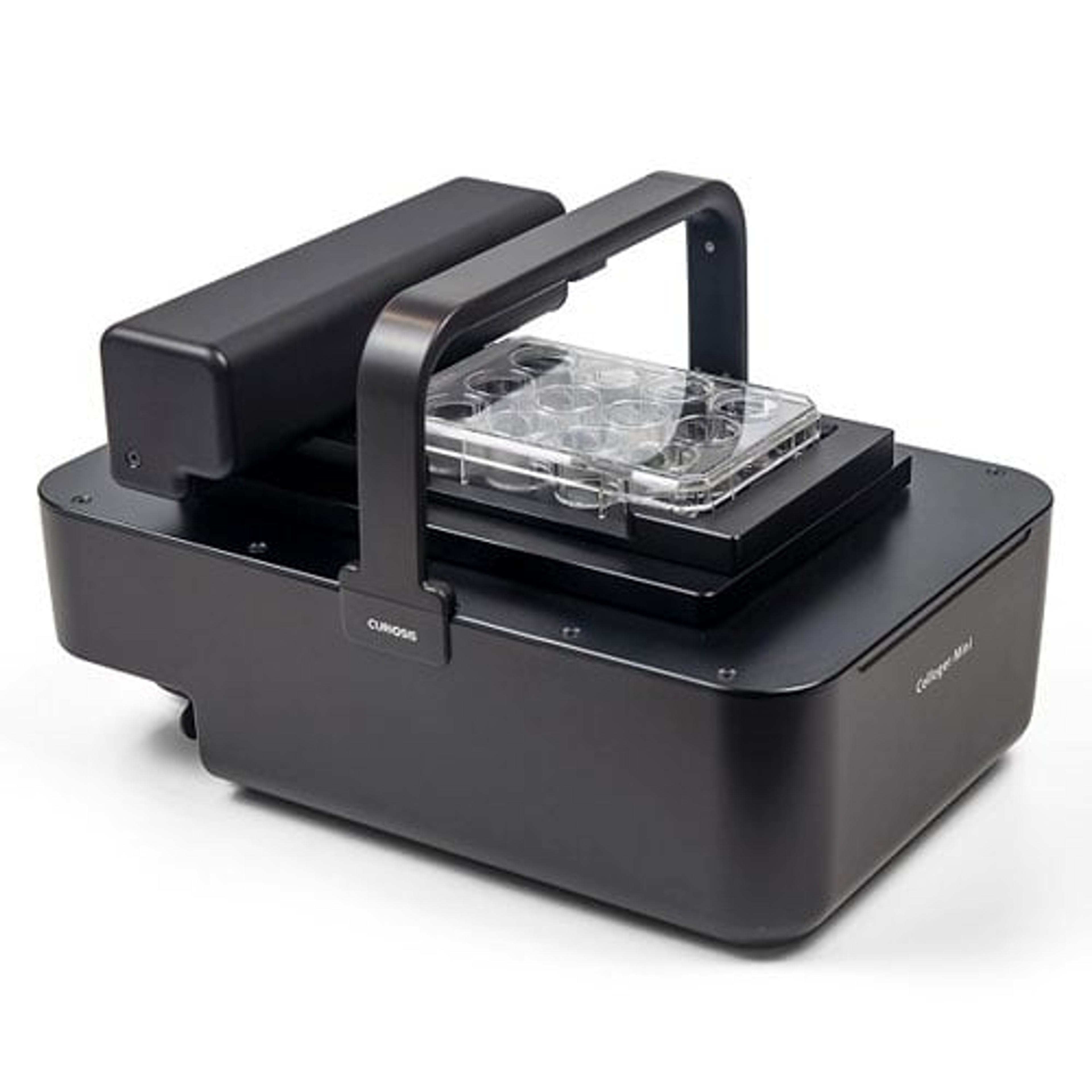
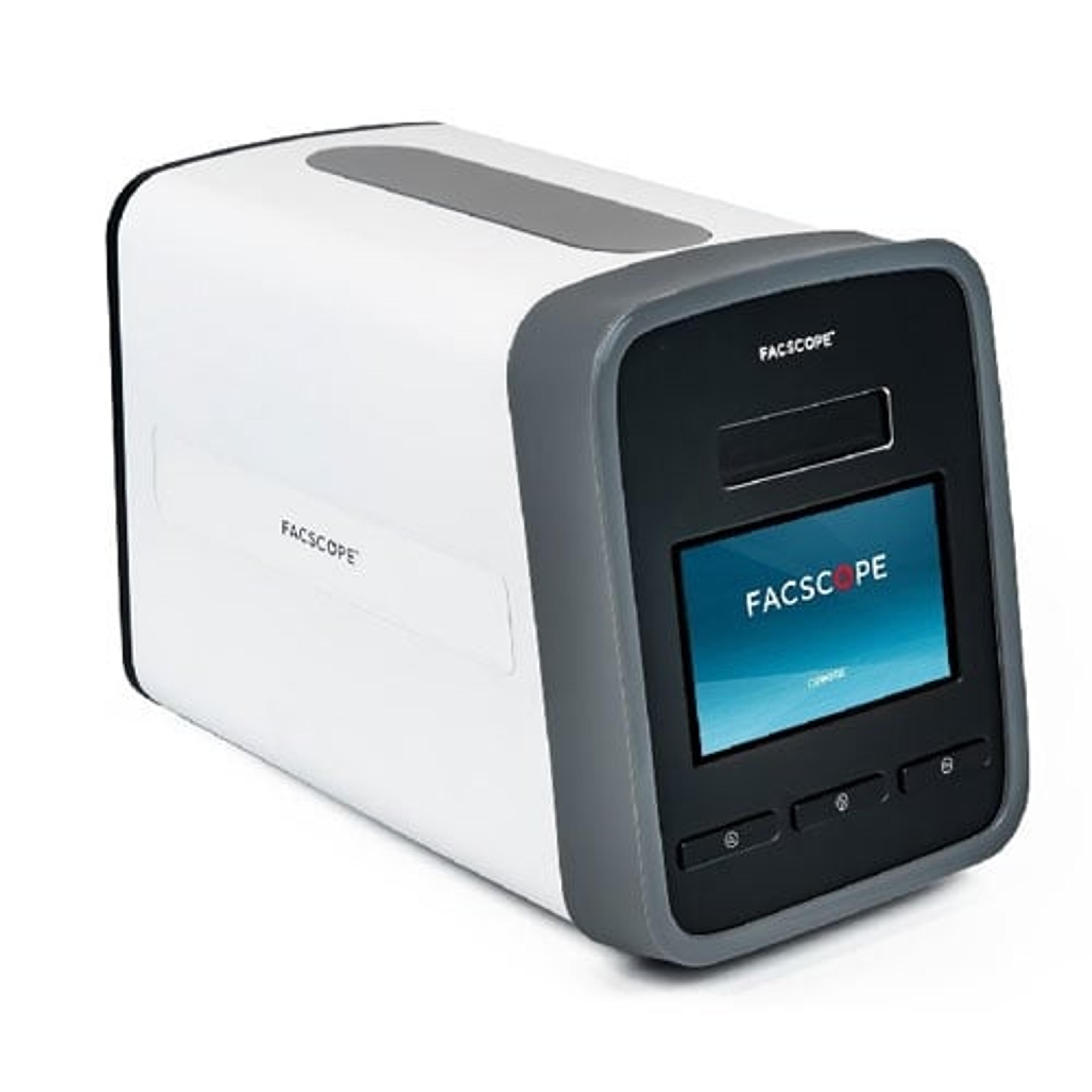
Celloger® Nano, Benchtop Digital Microscope from Curiosis
CURIOSISCelloger® Nano is a benchtop digital microscope equipped with a wireless connection, enabling you to check the state of your cells in real-time from any location within your laboratory. With all the necessary functions to check the cells, you can quickly assess the condition of your cells.
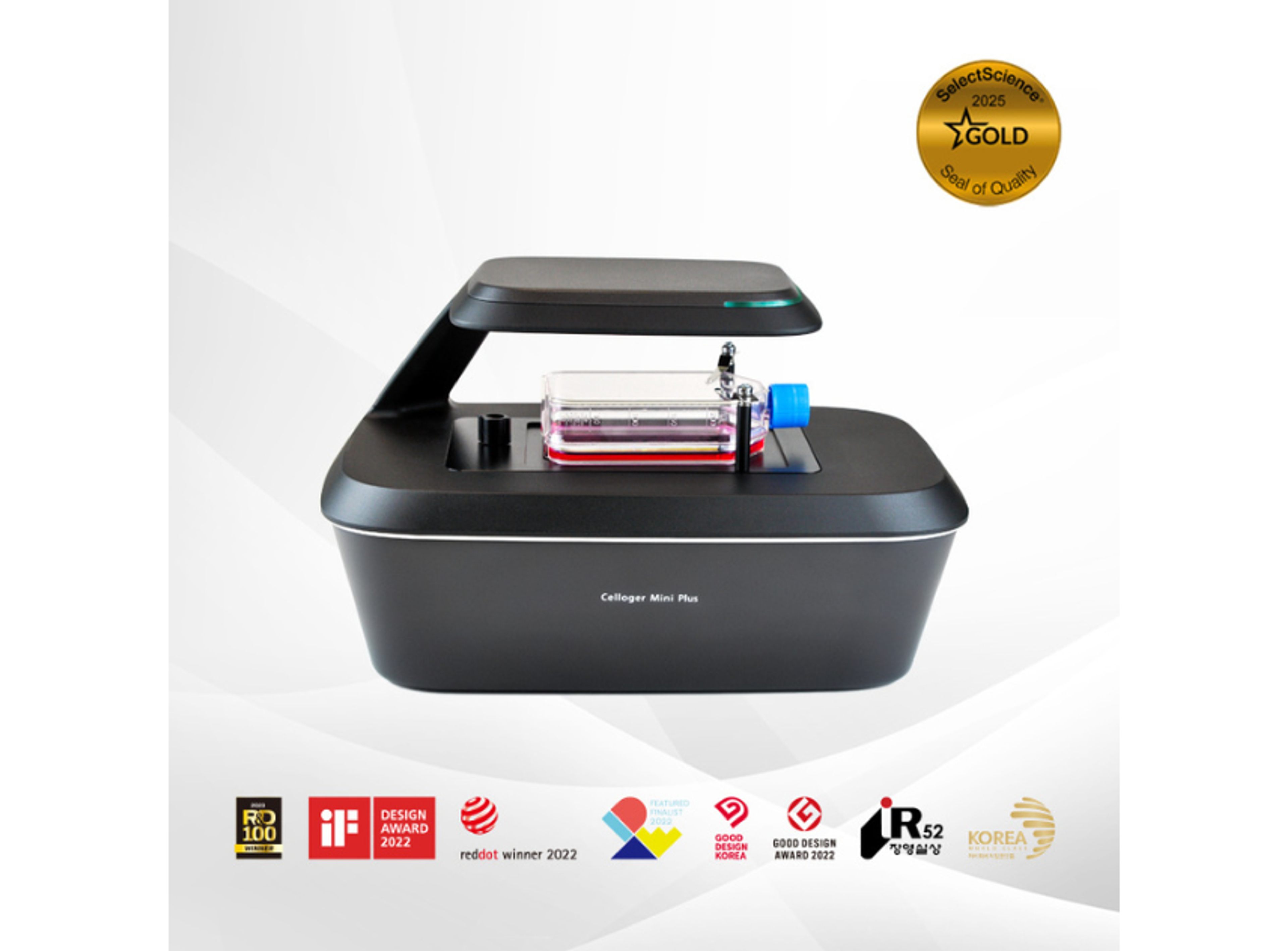
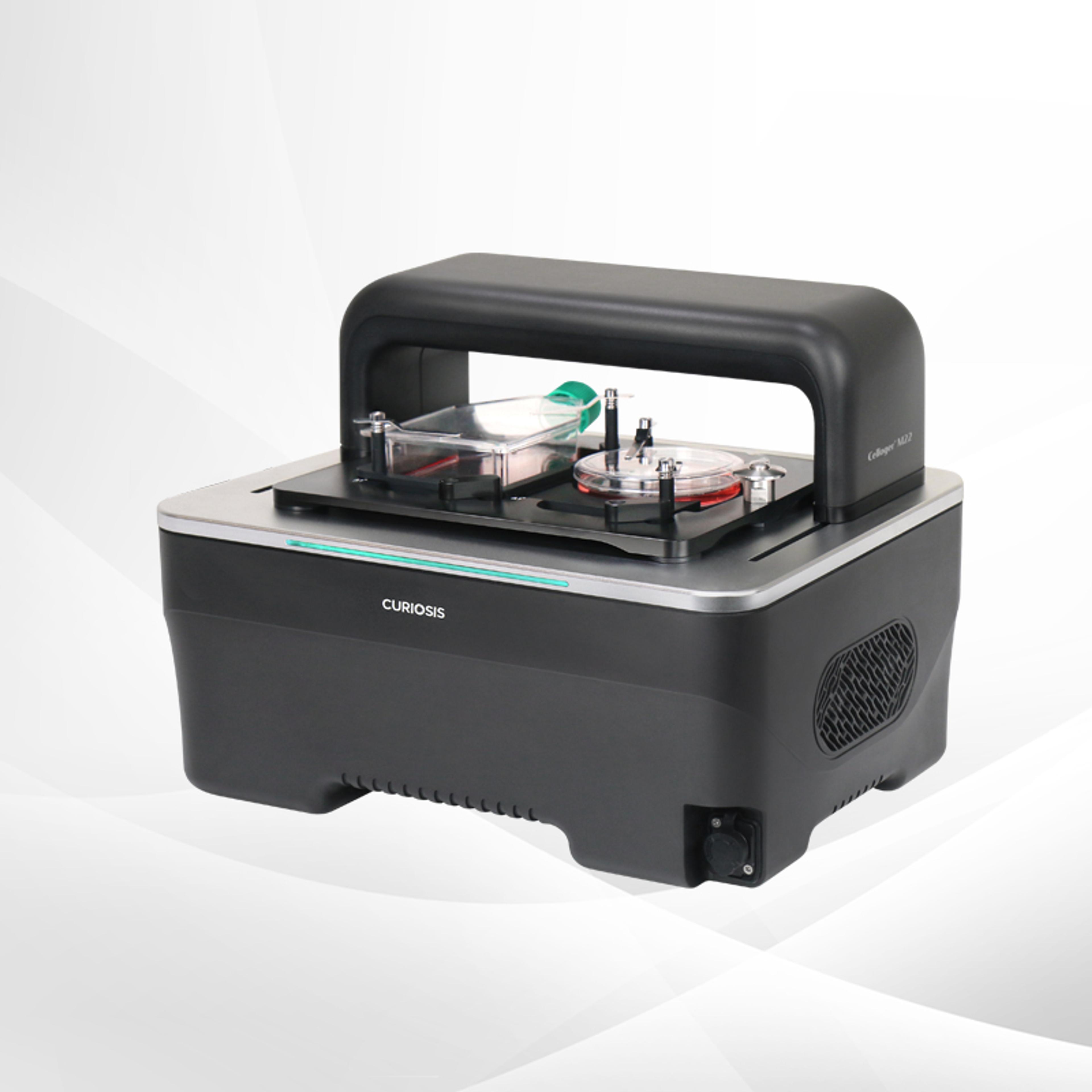
Celloger® Mini Plus, Automated Live Cell Imaging System from Curiosis
CURIOSISCelloger® Mini Plus is an automated live cell imaging system with fluorescence and brightfield microscopy. Celloger® Mini Plus makes it faster and easier to accumulate outstanding research results tailored to your research protocol.
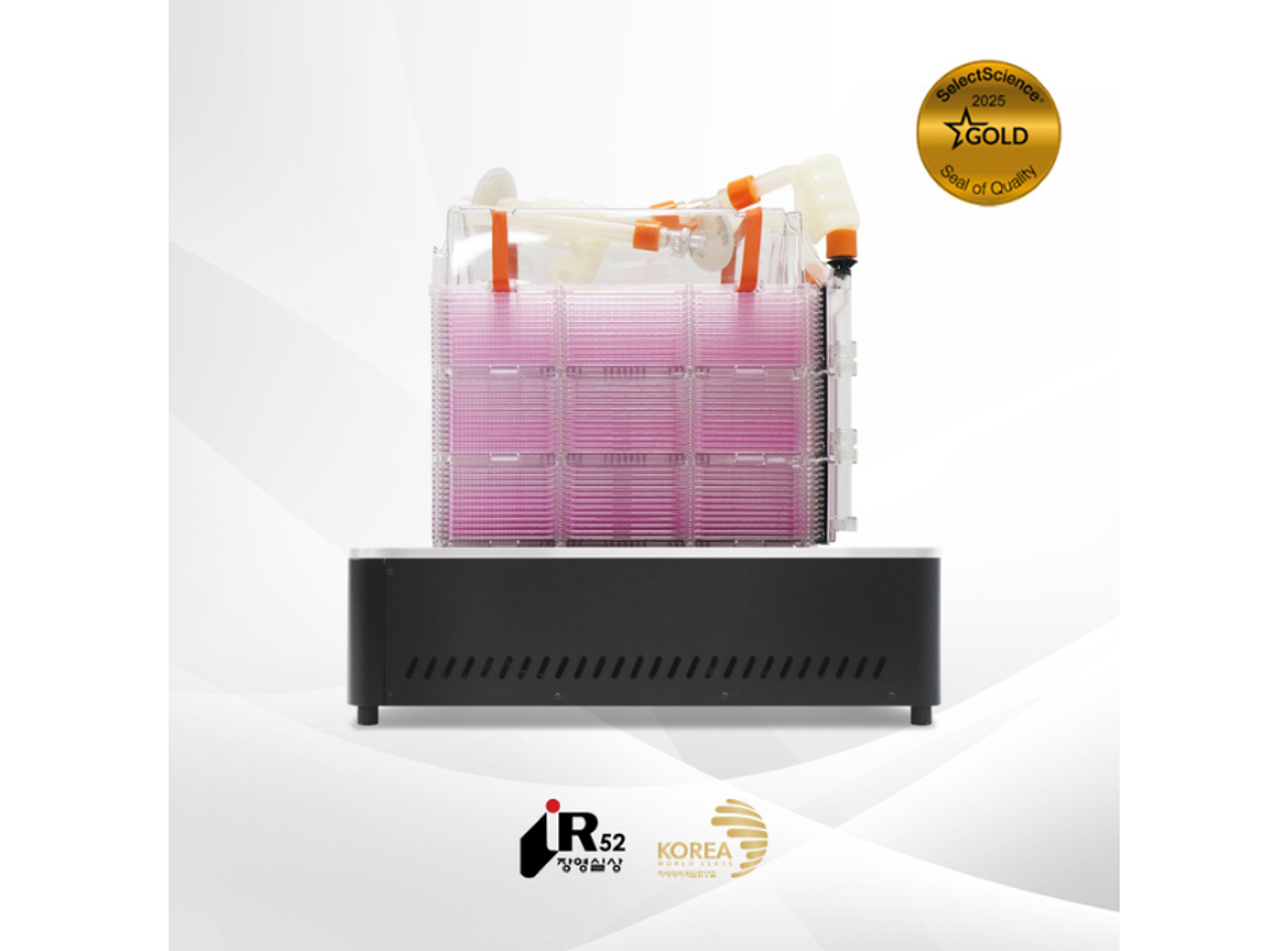
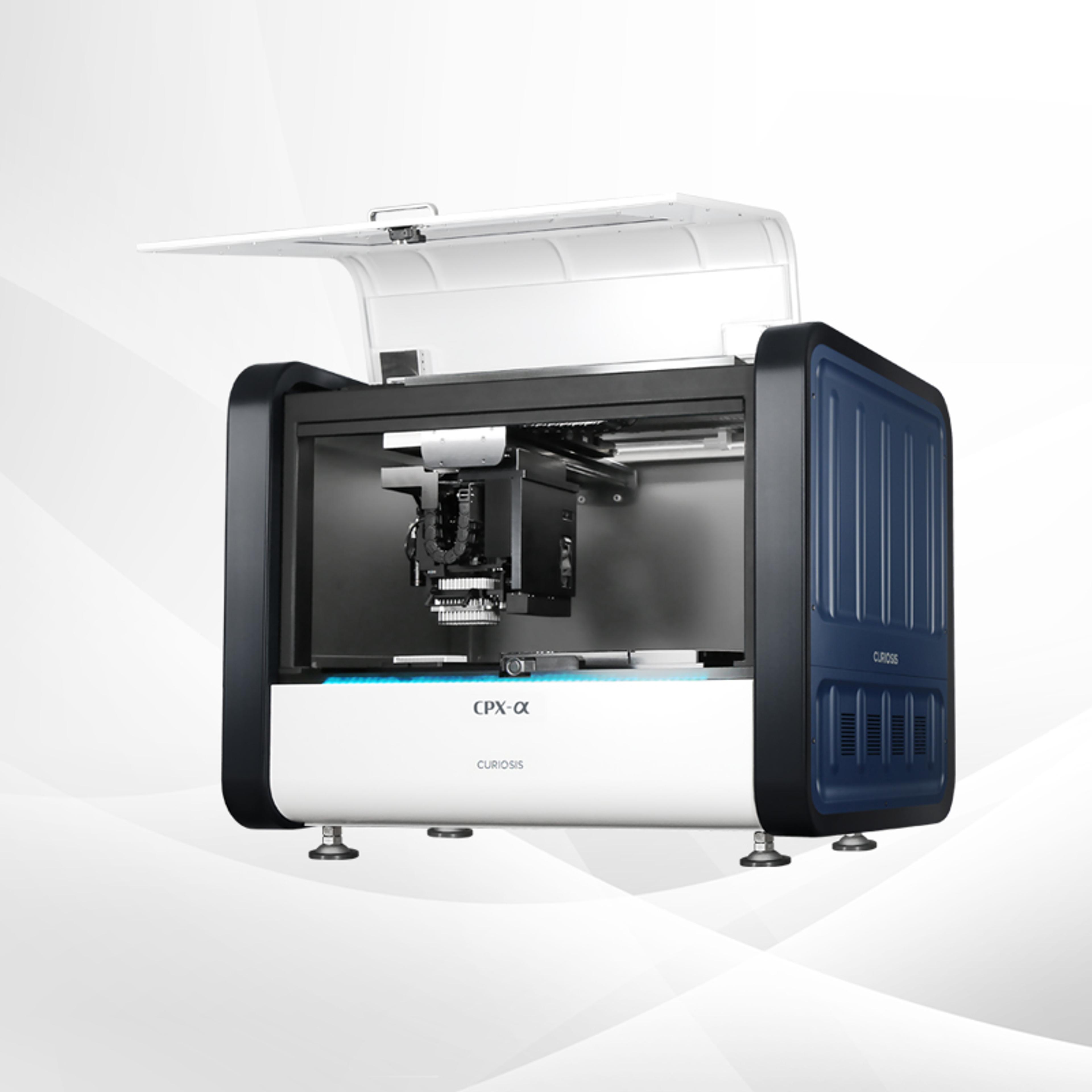
Celloger® Stack, Automated Multi-layer Vessel Monitoring System from Curiosis
CURIOSISCelloger® Stack is an automated multi-layer vessel monitoring system that enables real-time imaging of cell samples while they are cultured in an incubator.
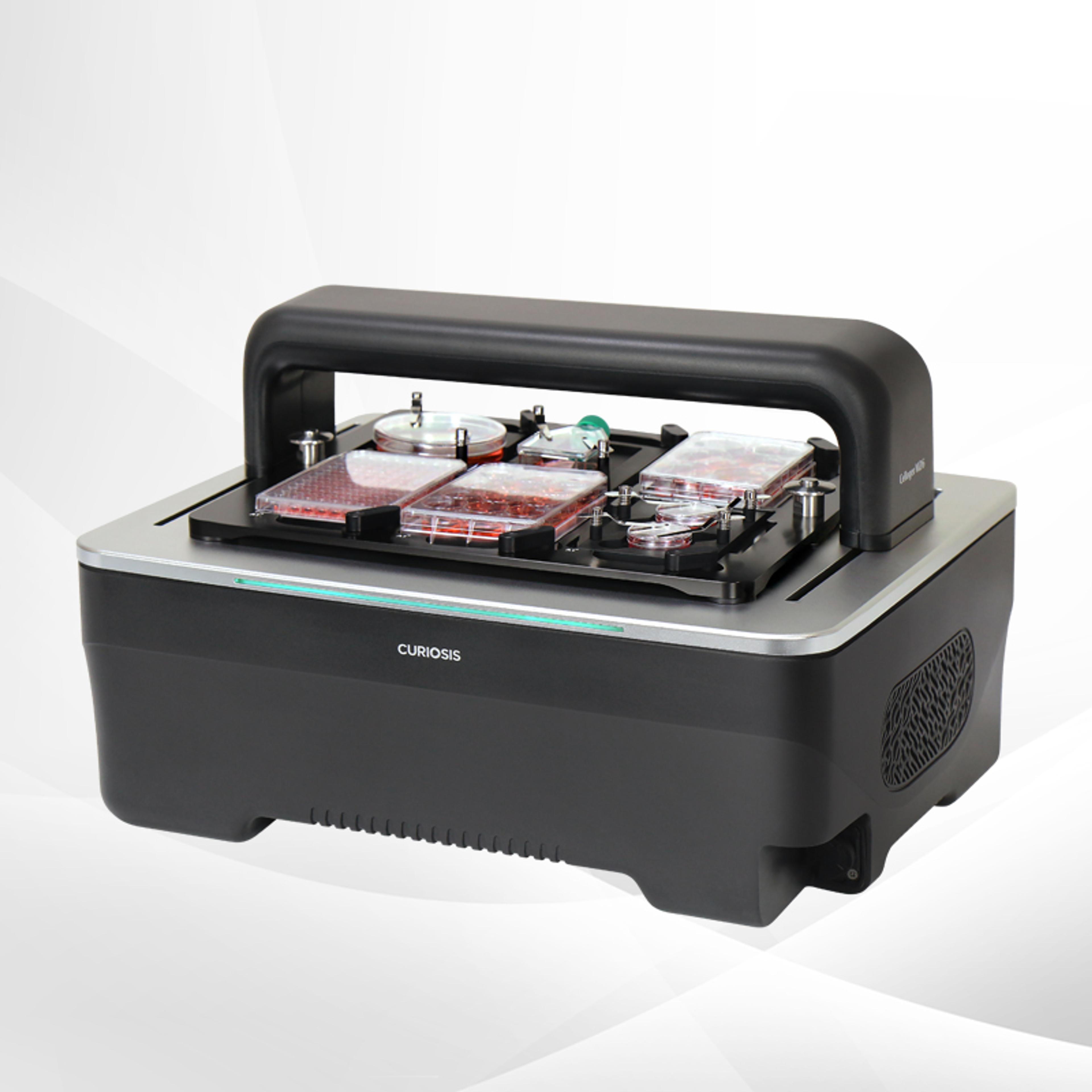
Celloger® Pro, Automated Live Cell Imaging System from Curiosis
CURIOSISWith its streamlined workflow and versatile capabilities, Celloger® Pro is a valuable tool for researchers seeking efficient, accurate, and cost-effective cellular analysis.