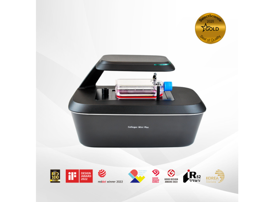
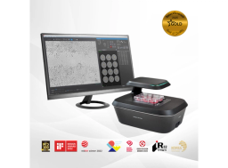
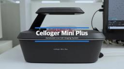
Celloger® Mini Plus, Automated Live Cell Imaging System from Curiosis
Celloger® Mini Plus is an automated live cell imaging system with fluorescence and brightfield microscopy. Celloger® Mini Plus makes it faster and easier to accumulate outstanding research results tailored to your research protocol.


Receive your quote directly from the manufacturer.
Using Celloger® Mini Plus for apoptosis experiments made it very easy to monitor cytotoxicity in real time.
Cell death research
Using Celloger® Mini Plus for apoptosis experiments made it very easy to monitor cytotoxicity in real time. It’s simple to use, so I could quickly see how the cells were responding without going through any complicated procedures.
Review Date: 25 Nov 2025 | CURIOSIS
Celloger® Mini Plus allowed reliable, stable monitoring of suspension cell growth over time.
Suspension cell growth monitoring
Celloger® Mini Plus allowed reliable, stable monitoring of suspension cell growth over time.
Review Date: 24 Nov 2025 | CURIOSIS
Data acquisition for both wound-healing assays and cell proliferation analysis went very smoothly, with no issues.
Proteostasis network
Data acquisition for both wound-healing assays and cell proliferation analysis went very smoothly, with no issues.
Review Date: 24 Nov 2025 | CURIOSIS
Overall, the system was very user-friendly and made it easy to get my work done without any stress.
Spheroid monitoring
Overall, the system was very user-friendly and made it easy to get my work done without any stress. In particular, the coordinate copy/paste, centering, autofocus, and shooting-direction settings (row, column, one-way, or round-trip) were all very convenient, and the holder was easy to attach and detach while still feeling stable during use. I was also pleased with the clear 5-megapixel images. After image acquisition, the function that automatically combines individual images into a single view based on the multi-well layout was extremely useful. Being able to capture images without opening the incubator was a big advantage, as it minimized stress on both the spheroids and the user, and CO₂ consumption remained low. I didn’t notice any vibrations during operation—the system was quiet, and the spheroids didn’t move at all. Thanks to the short imaging time, I could also retake images quickly whenever needed, which made the overall workflow very efficient.
Review Date: 24 Nov 2025 | CURIOSIS
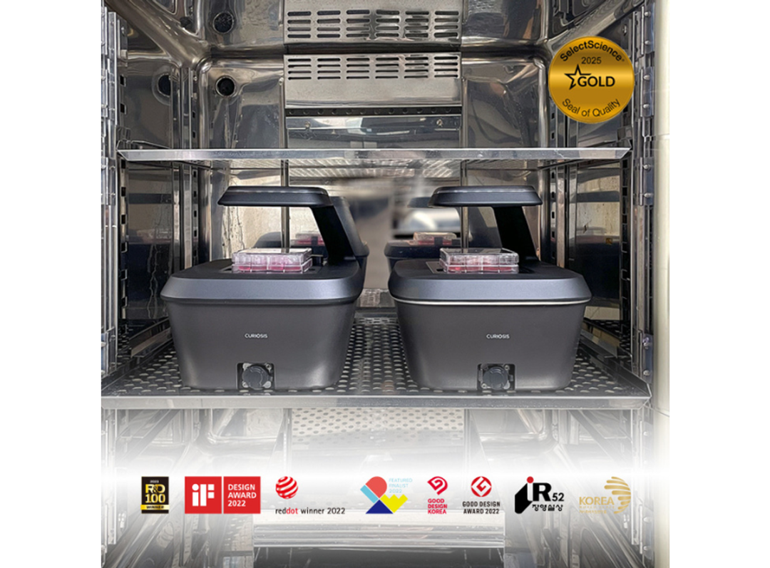
The instrument was compact and easy to install, so it fit well even in the tight space inside the incubator.
Cancer biology
The instrument was compact and easy to install, so it fit well even in the tight space inside the incubator. It operated stably and was simple to maintain. It was also useful that the analysis software could generate results directly, without needing any separate analysis tools.
Review Date: 24 Nov 2025 | CURIOSIS
The ability to set multiple imaging positions within a single well was very helpful for increasing replicate numbers.
Cancer biology
The ability to set multiple imaging positions within a single well was very helpful for increasing replicate numbers. It was also a big advantage to monitor live cells in real time directly inside the incubator without disturbing the culture. By defining several points per well and capturing them in one acquisition, it was easy to collect more replicates efficiently.
Review Date: 24 Nov 2025 | CURIOSIS
The analysis software offered convenient built-in tools for area measurement and automated wound-healing analysis.
Cancer biology
The analysis software offered convenient built-in tools for area measurement and automated wound-healing analysis. Because the captured images could be analyzed directly in the software, the overall workflow was much more efficient, and the flexible time-lapse scheduling made it easy to tailor experiments to different cell types and conditions.
Review Date: 24 Nov 2025 | CURIOSIS
Z-stacking function was a strong feature, allowing the acquisition of images at multiple focal planes
Biochemistry
For COS7 cell research, the Z-stacking function was a strong feature, allowing the acquisition of images at multiple focal planes and combining them to generate enhanced 2D images with improved depth information. The image stitching function was also valuable, facilitating the analysis of large cell populations by merging multiple images into a single comprehensive view.
Review Date: 16 Apr 2025 | CURIOSIS
Compatibility with various culture vessels was one of the most appreciated features
Biochemistry
In conjunctiva cell research, compatibility with various culture vessels was one of the most appreciated features. The system supported flasks, dishes, and well plates, allowing for flexible experimental setups. The user-friendly software made it easy to track cell confluency, monitor growth curves, and perform data analysis. The time-lapse video function was especially useful for visualizing dynamic cellular changes over time, making it an excellent tool for cell behavior analysis.
Review Date: 16 Apr 2025 | CURIOSIS
One of the most satisfying aspects of using Celloger® Mini Plus was its real-time monitoring capability
Biochemistry
One of the most satisfying aspects of using Celloger® Mini Plus for pterygium cell research was its real-time monitoring capability. It allowed continuous observation of cell growth and changes without disturbing the culture environment, ensuring stable data acquisition. Additionally, the multi-position imaging function enabled the simultaneous analysis of multiple samples, significantly improving experimental efficiency. The autofocus function was also highly reliable, providing consistently sharp images without losing focus.
Review Date: 16 Apr 2025 | CURIOSIS
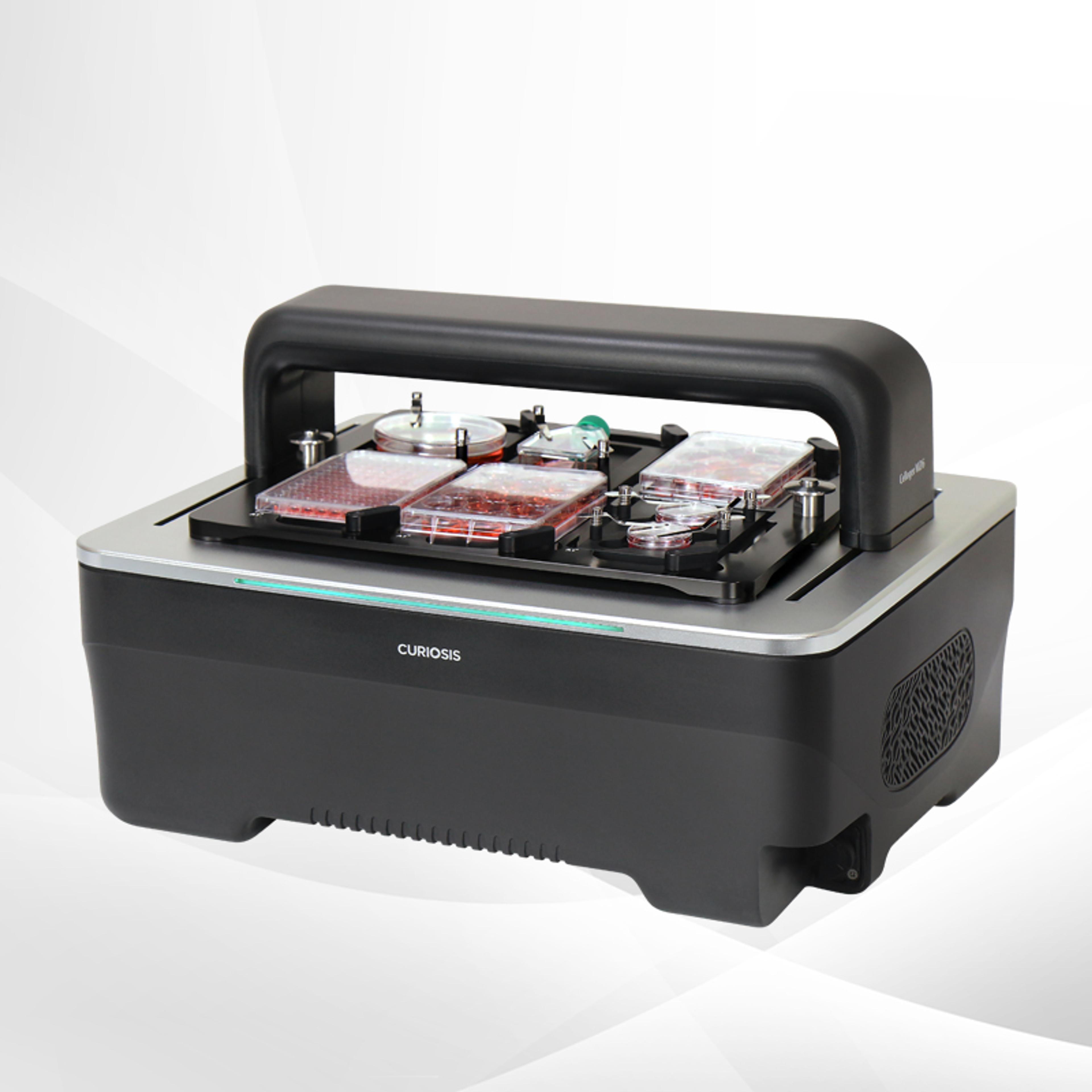
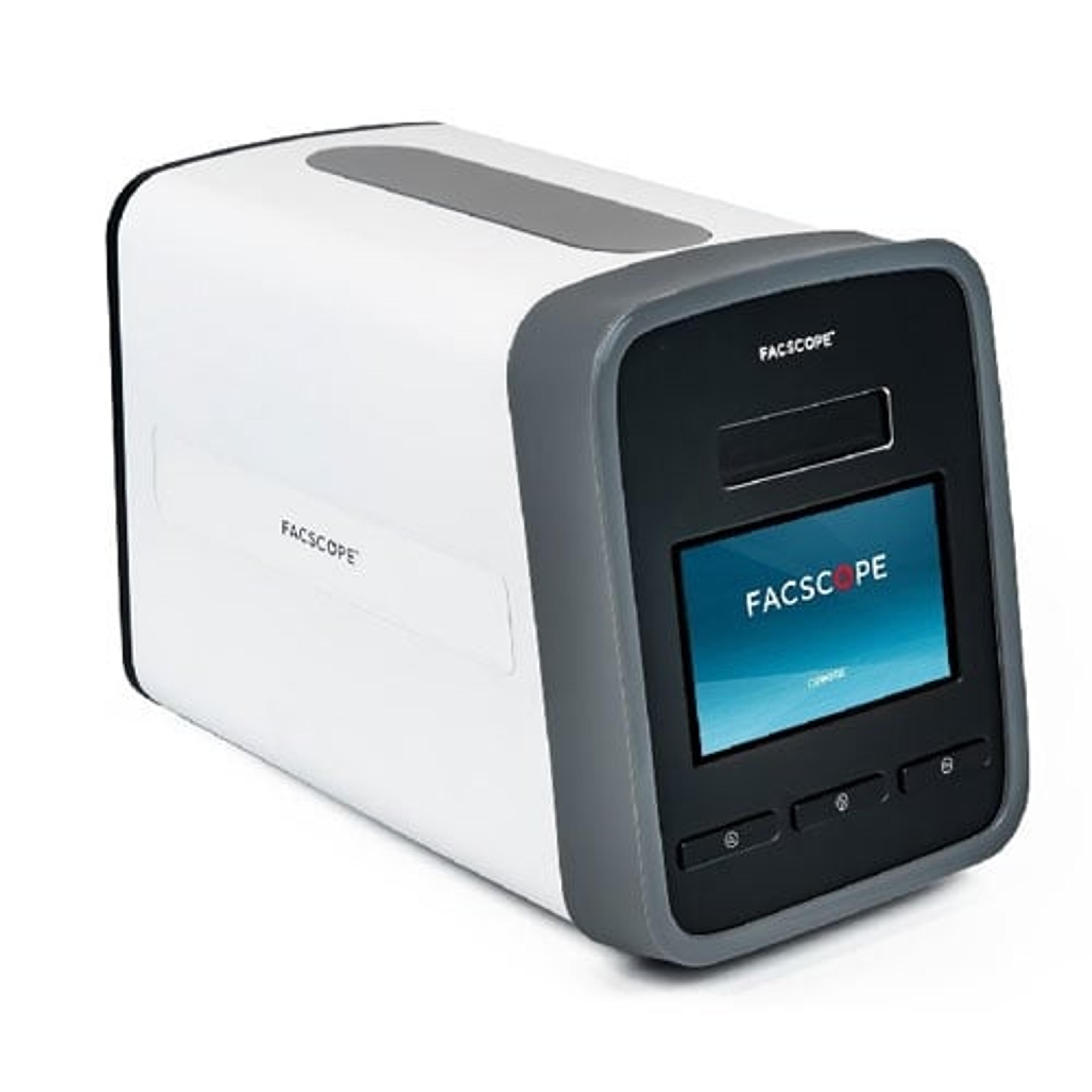
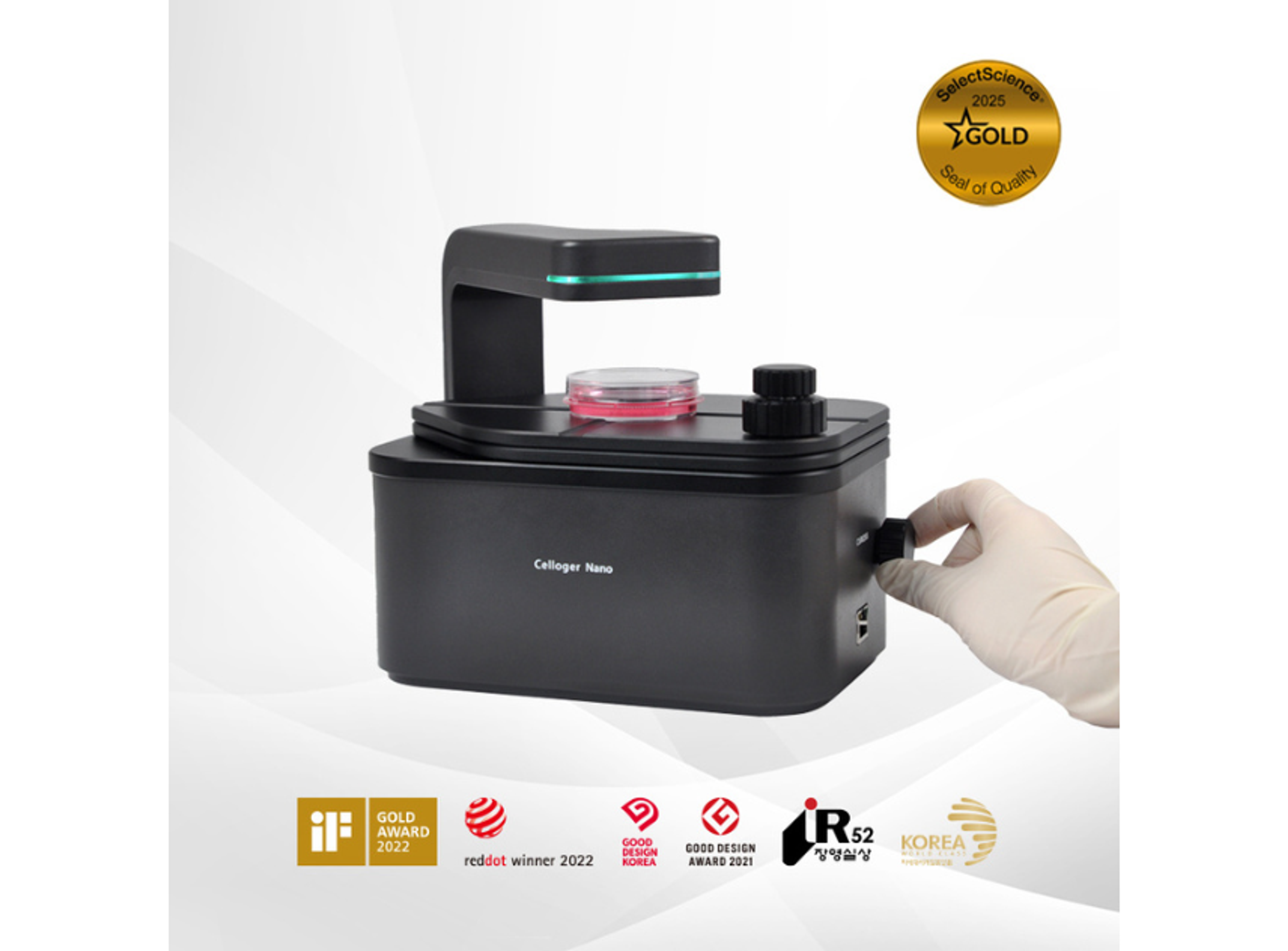
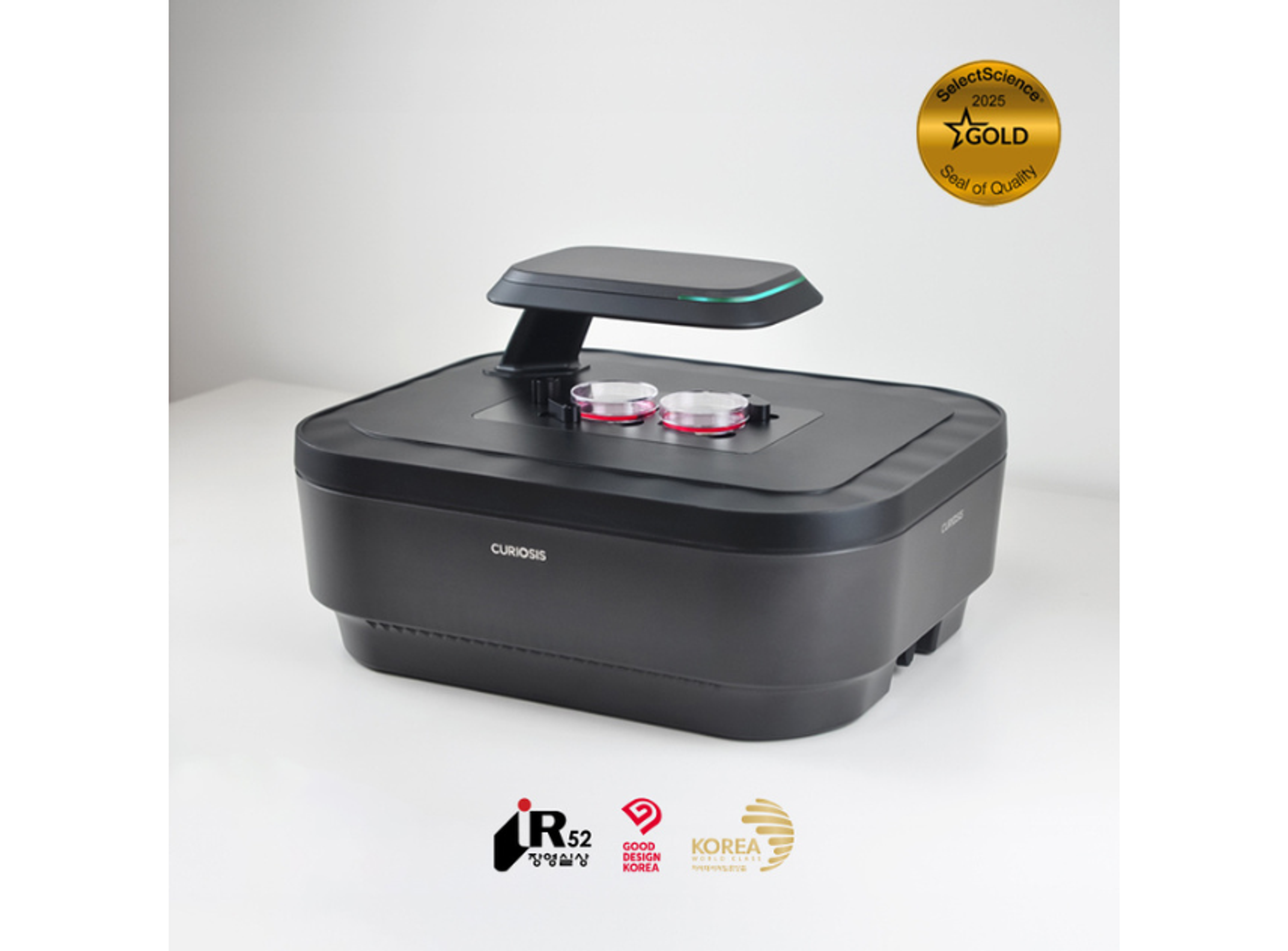
Celloger® Mini Plus is a live cell imaging system based on bright field and fluorescence(green/red) microscopy. The motorized stages move on X, Y, and Z axes that minimize vessel movement, allowing stable cell observation with no shaking. Researchers may obtain stable research results in line with individual research protocols through the use of functions that are optimized for cell research, such as automatic focusing, real-time multi-position imaging, etc.
Key features
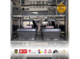
- Compact size that easily fits into standard CO₂ incubators
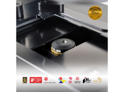
- Fully motorized multi-position imaging up to 96 well plate
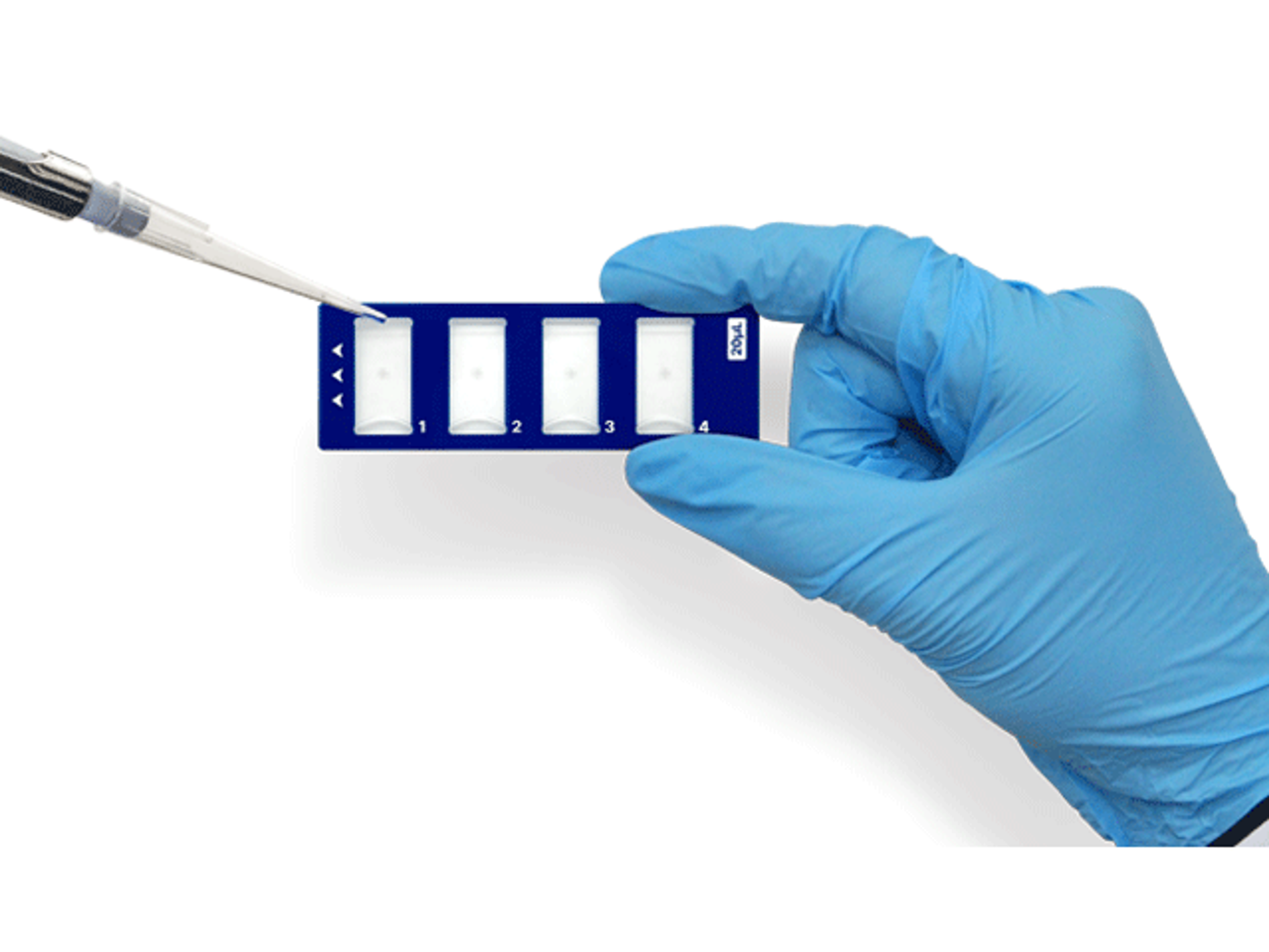
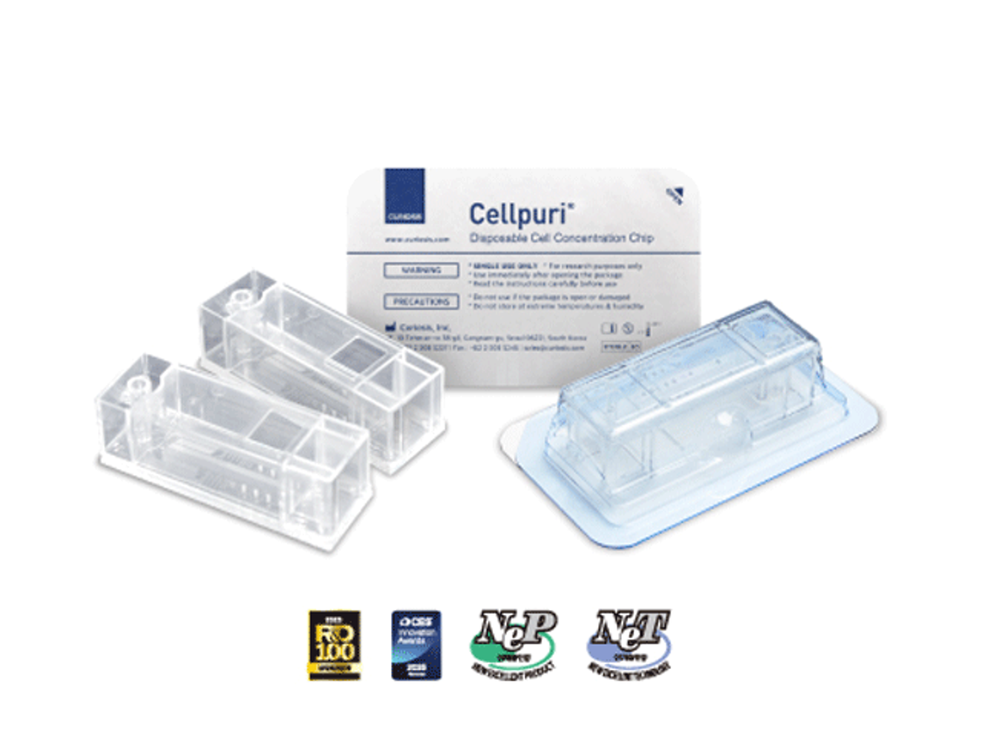
- Compatible with various cell and tissue culture vessel types
- Multiple focal planes can be captured through Z-stack imaging
- Intuitive UI/UX and easy to acquire confluency data
- Increased focus speed and reproducibility with reliable autofocusing function
- Image stitching possible to enable analysis of larger volume and sections
Applications
- Wound healing assay
- Cell migration
- Cell morphology
- Cell confluency
- Cell proliferation
- Cytotoxicity assay
- Transfection efficiency assessment
- Coculture monitoring
- Multipoint cell monitoring
Specification
- Dimension: 226 x 358 x 215mm
- Weight: 5.6kg / 12.3lb
- Objective: 4X / 10X
- Imaging modes: Brightfield, Fluorescence (Green/Red)
- Fluorescence: Green - Excitation (470/40x), Emission (510lp) / Red - Excitation (525/30x), Emission (570lp)
- Light source: LED
- Stage: Motorized XYZ
Brochures
Celloger Mini Plus brochure
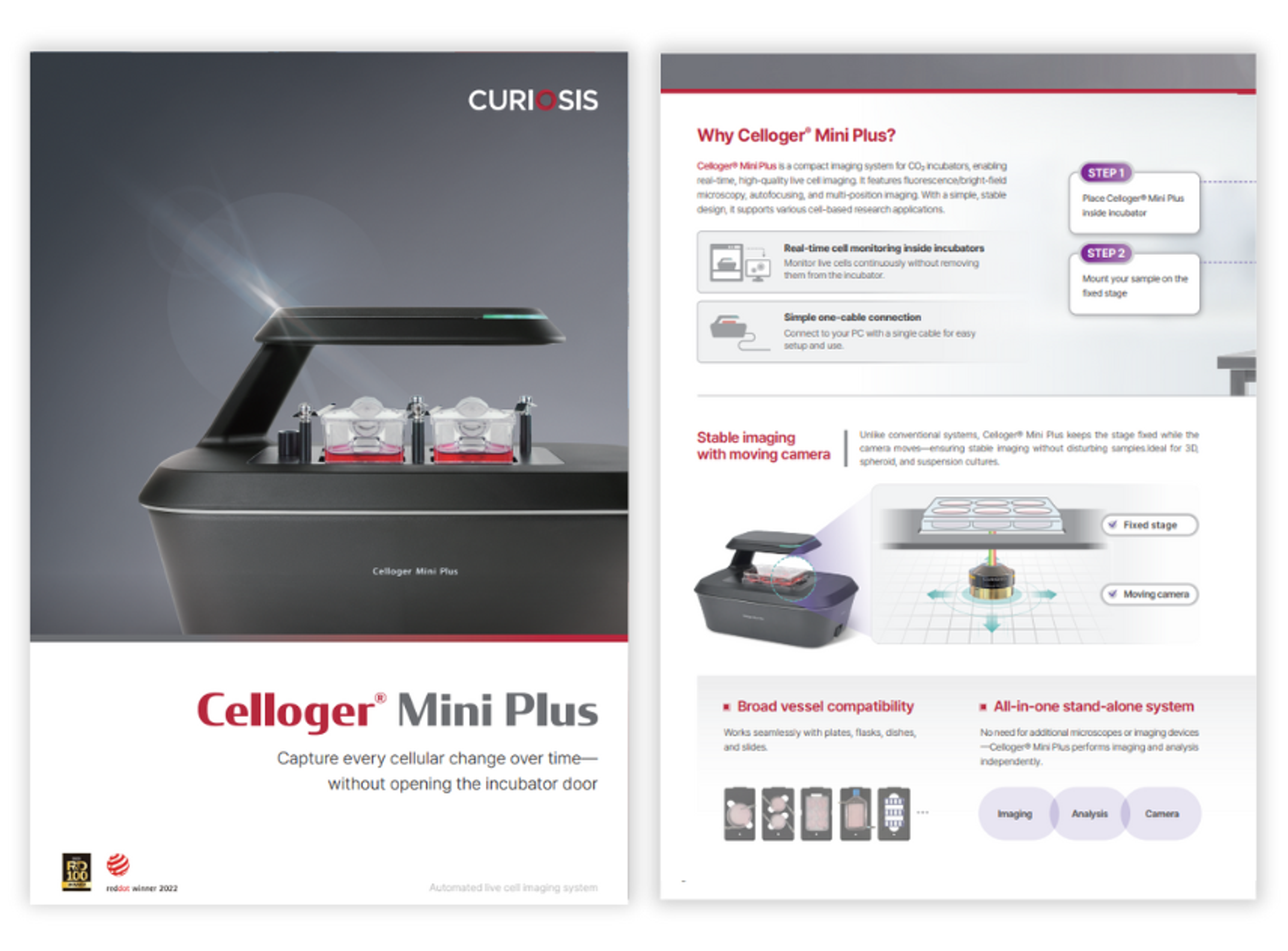
In this product brochure, CURIOSIS demonstrates the Celloger® Mini Plus, compact imaging system for CO₂ incubators, enabling real-time, high-quality live cell imaging. It features fluorescence/bright-field microscopy, autofocusing, and multi-position imaging. With a simple, stable design, it supports various cell-based research applications.
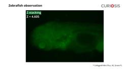
Zebrafish observation: Utilizing Z-stacking function with Celloger Mini Plus
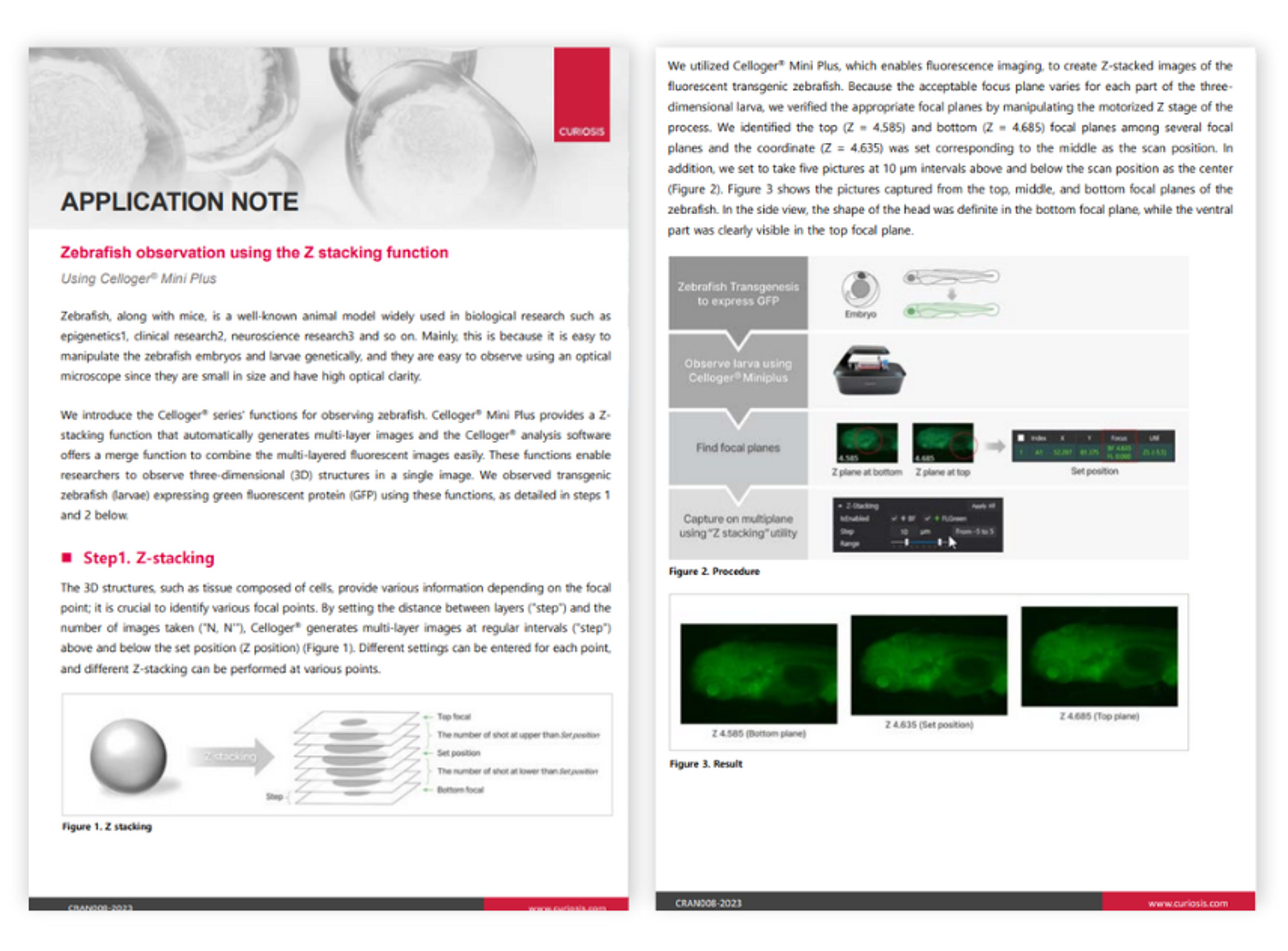
In this application note, CURIOSIS highlights the versatility of zebrafish as a widely used animal model in biological research. The Celloger® series introduces specialized functions for zebrafish observation, including the Celloger Mini Plus with its Z-stacking capability. This function automatically generates multi-layer images, complemented by the merge function in the Celloger analysis software. These features enable researchers to observe three-dimensional structures in a single image. The application note demonstrates the application of these functions by observing transgenic zebrafish larvae expressing green fluorescent protein (GFP).

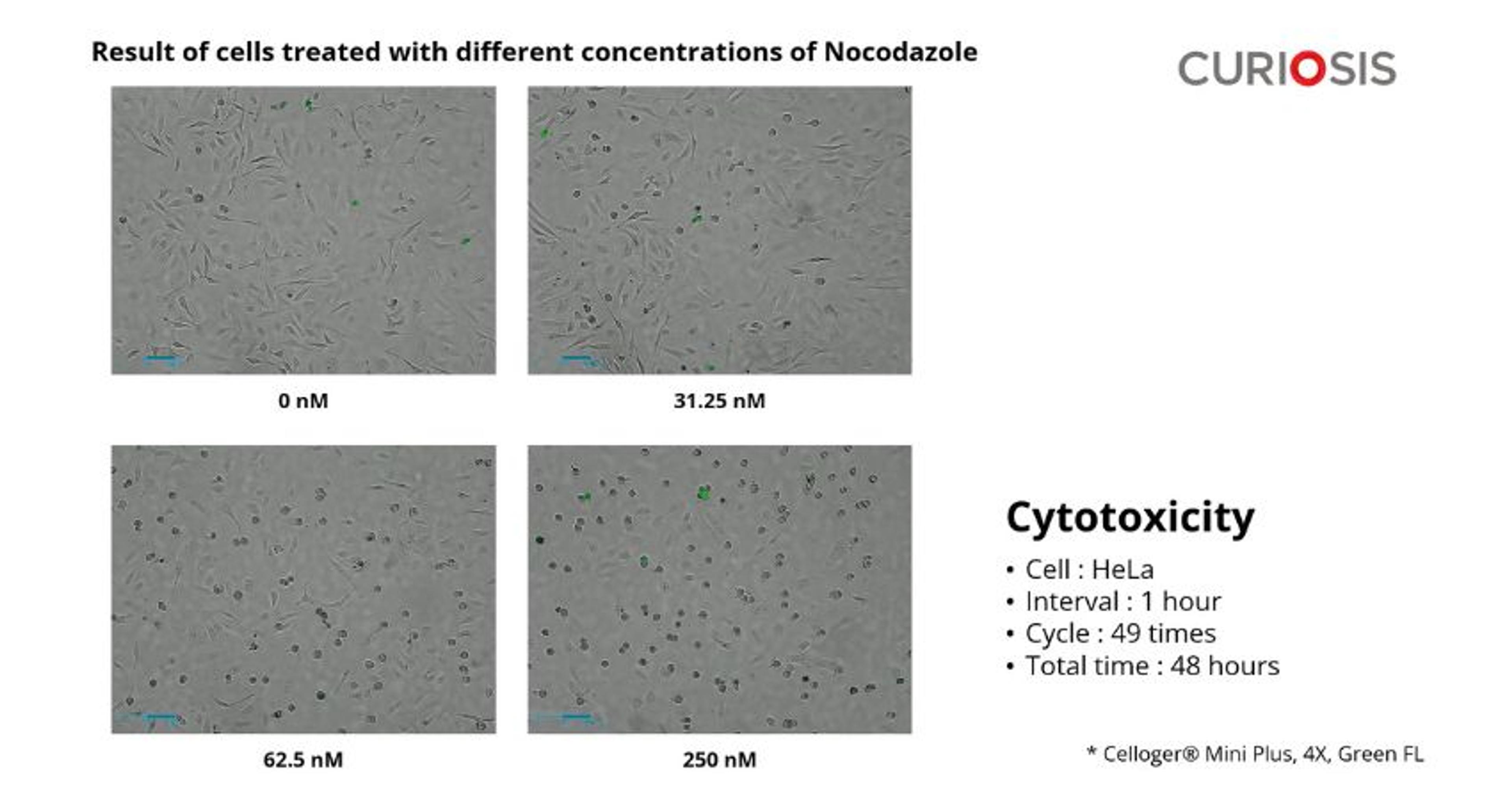
Analysis of nocodazole-induced cytotoxicity using Celloger Mini Plus
In this application note, CURIOSIS aimed to examine the performance of a cytotoxicity assay using real-time imaging. Cells treated with various concentrations of Nocodazole, the anti-cancer drug, were stained with a fluorescent dye during cell death, then monitored with the Celloger® Mini Plus.

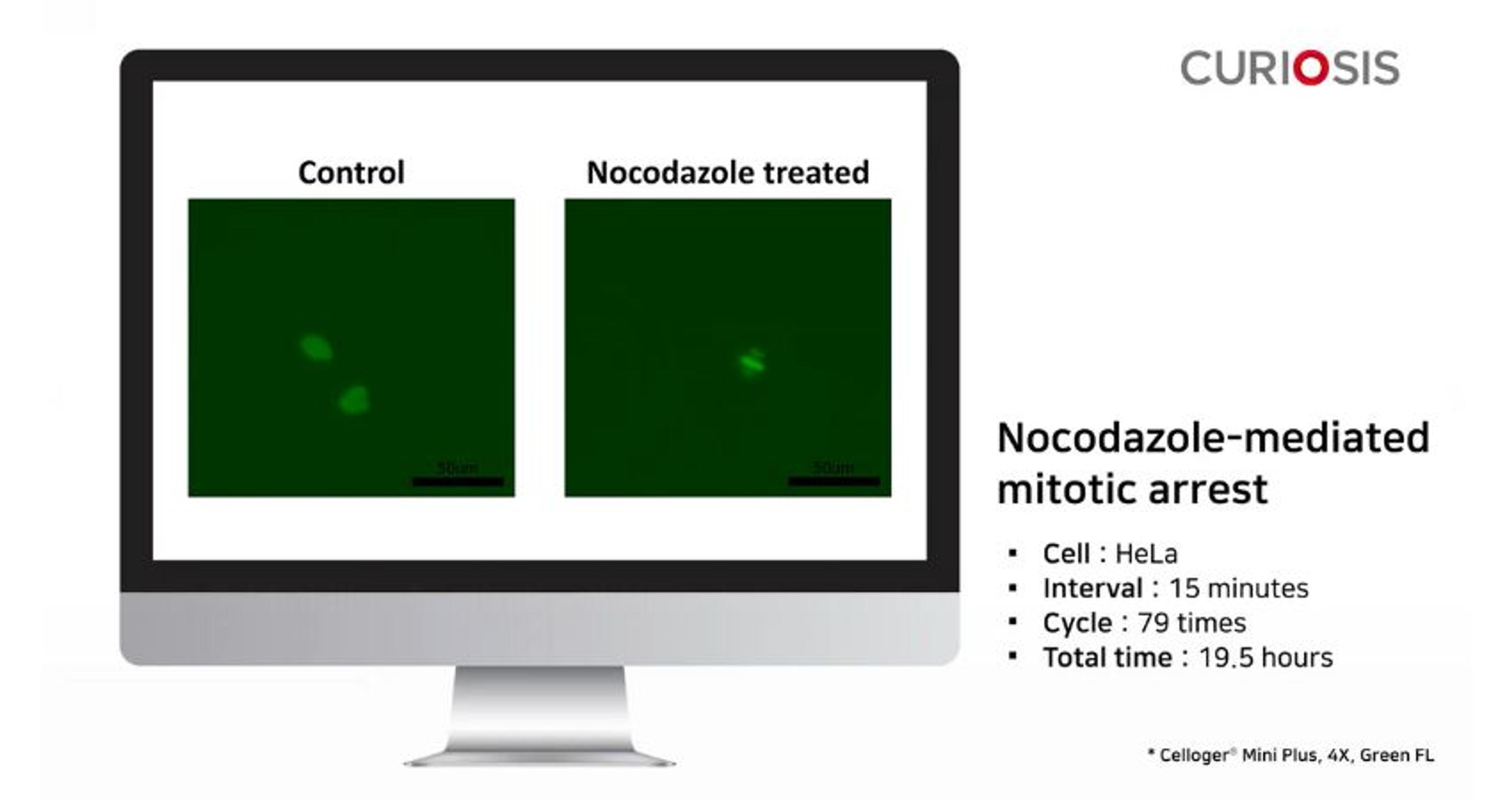
Observation of mitosis using Celloger Mini Plus
In this application note, the anti-mitotic activity of nocodazole against cancer cell line was examined by monitoring the cell division process. The cell division was monitored in real-time after treating the cell with or without nocodazole using the Celloger® Mini Plus.
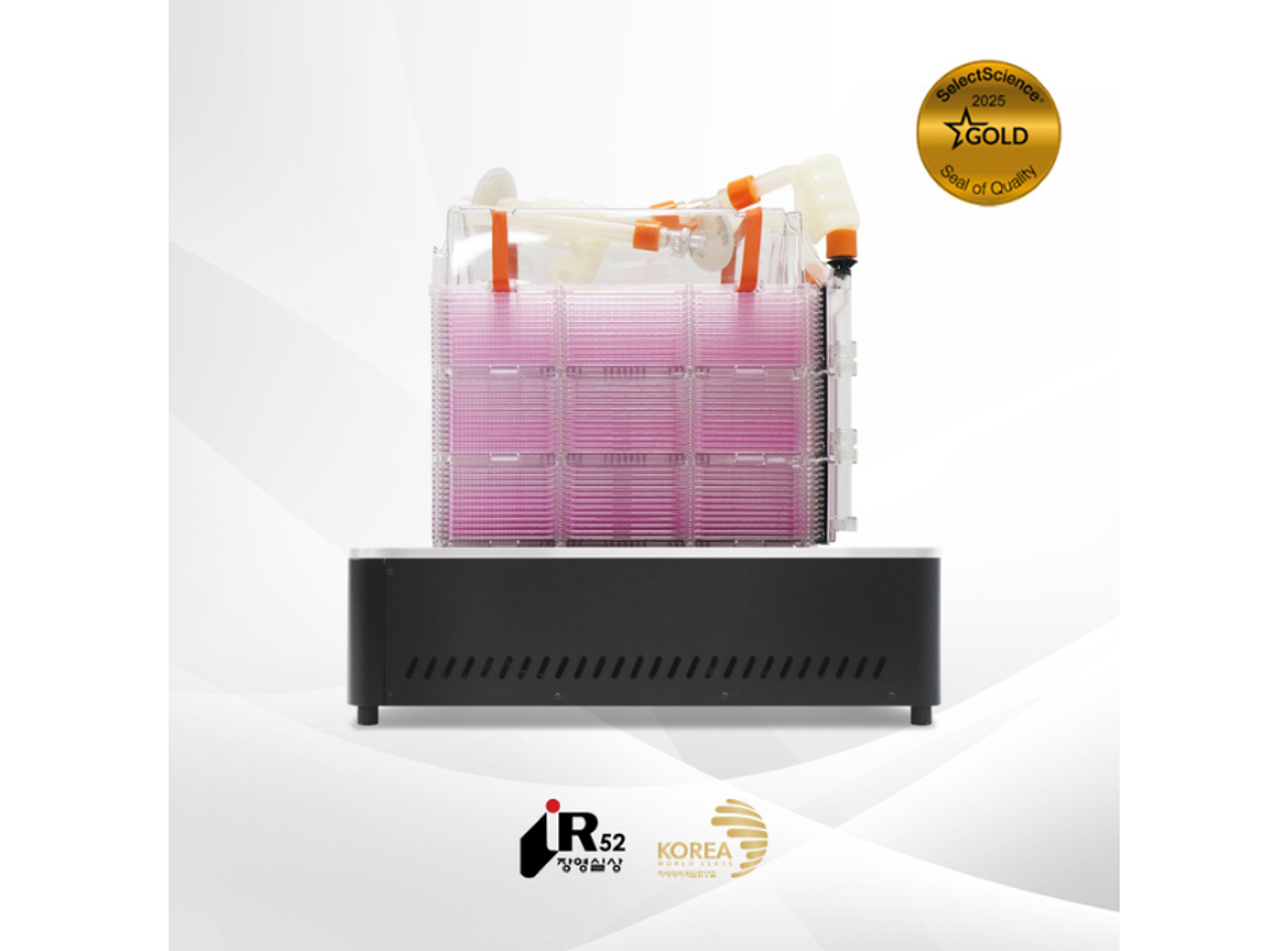
An integrated live cell monitoring system for stable imaging of suspension cells in immunological research
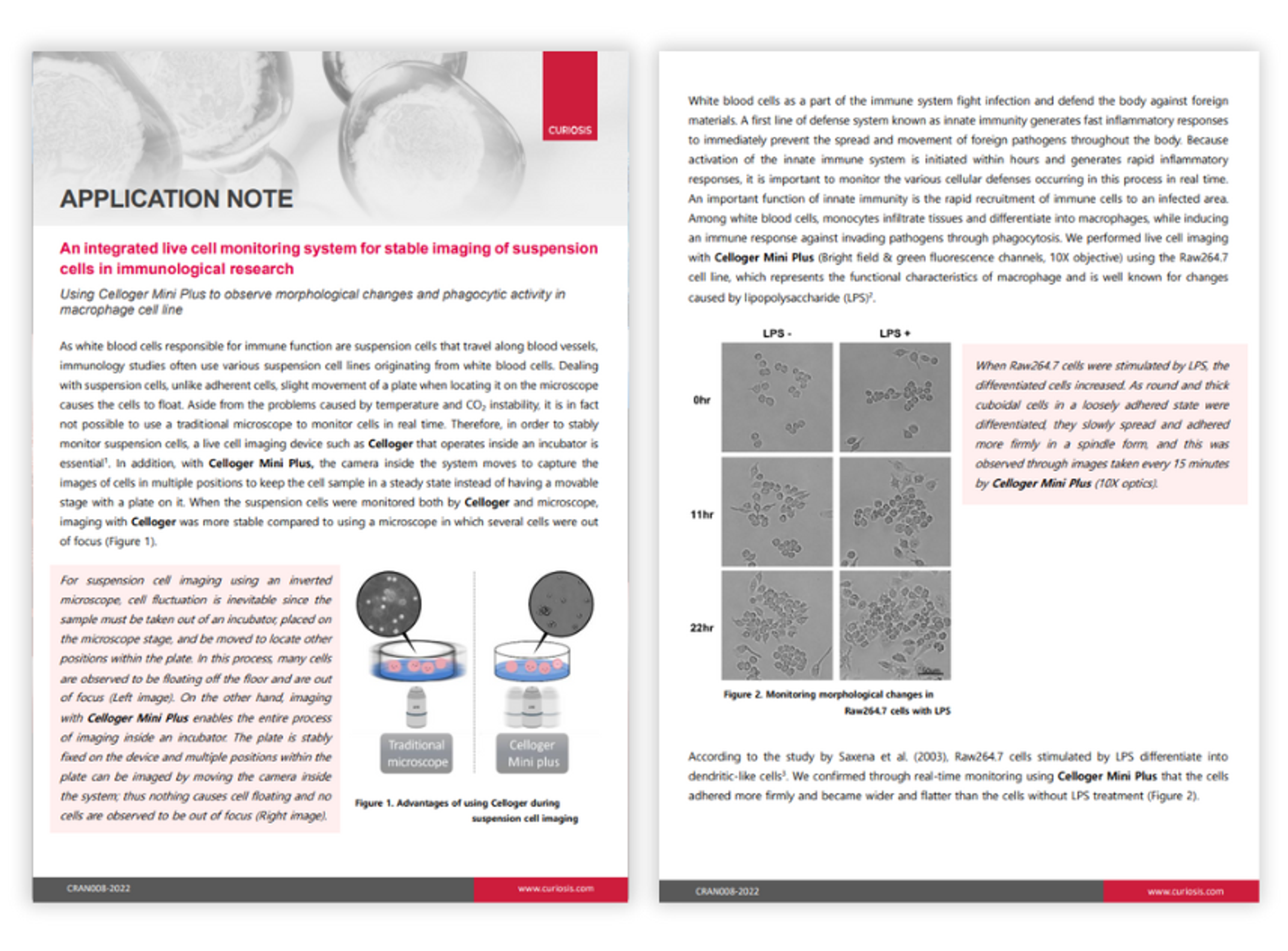
In this application note, CURIOSIS performs live cell imaging with Celloger Mini Plus using the Raw264.7 cell line, which represents the functional characteristics of macrophages. This was in order to monitor the rapid inflammatory responses and cellular defenses occurring in innate immunity in real time.
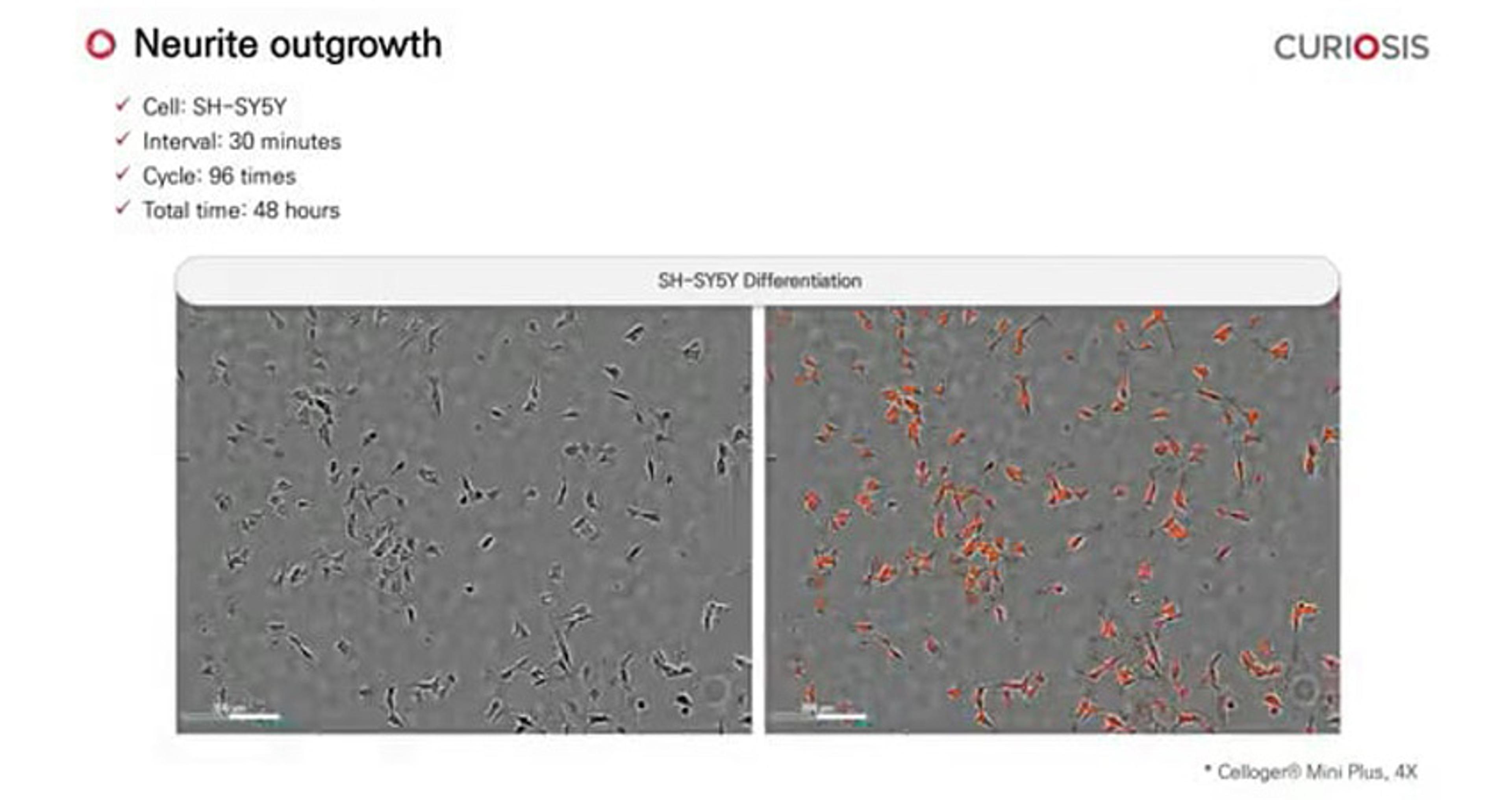
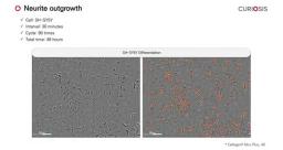
Celloger Mini Plus application: Neurite outgrowth
In this video, CURIOSIS demonstrates the real-time process in which you can monitor cells utilizing the Celloger® Mini Plus. SH-SY5Y cells were stained with PKH26 dye on the third day of differentiation. The following day, the cells were treated with BDNF (brain-derived neurotrophic factor) and imaged every 30 minutes for two days using the Celloger® Mini Plus with a 4X objective.
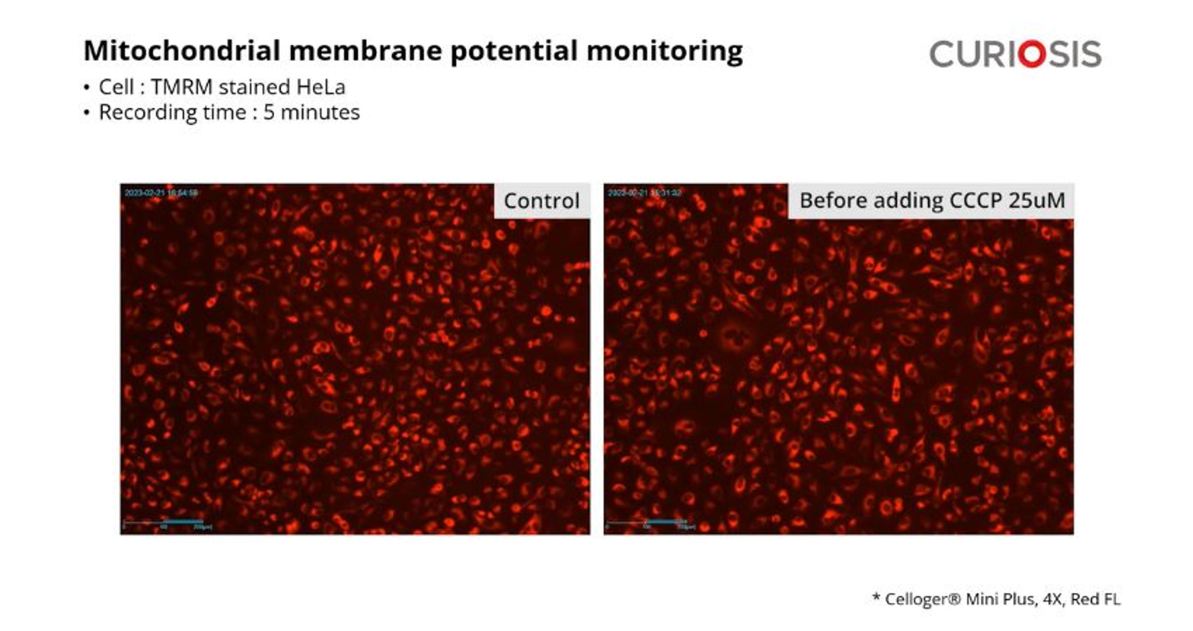
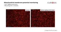
Celloger Mini Plus application: Mitochondrial potential monitoring
In this video, CURIOSIS discusses the vital role of mitochondria in generating cellular energy and supporting biological functions. Monitoring changes in mitochondrial membrane potential (MMP) provides crucial insights into cellular health and mitochondrial function.
HeLa cells treated with CCCP 25 uM had their mitochondrial membrane potential monitored using Celloger® Mini Plus. This automated imaging system promises exceptional fluorescence and bright-field technology for high-quality images and time-lapse videos. It enables real-time cell monitoring and observation of applications like wound healing, apoptosis, reactive oxygen species, and transfection.
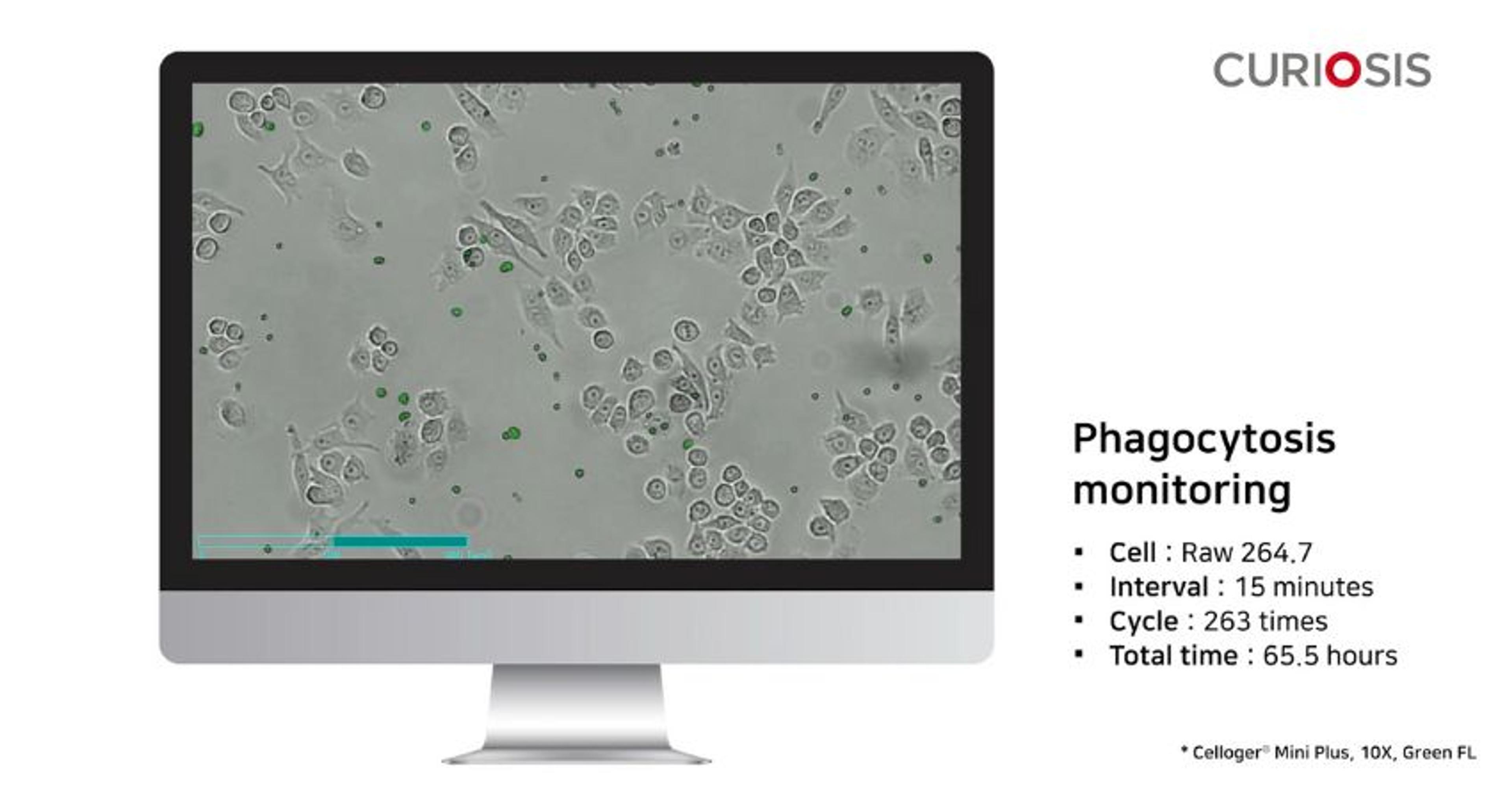
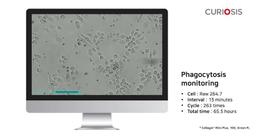
Celloger Mini Plus application: Phagocytosis monitoring
In this video, CURIOSIS demonstrates the performance of fluorescence imaging using fluorescent latex beads engulfed by macrophages. The activated Raw264.7 cells, stimulated by lipopolysaccharides (LPS), effectively engulfed the fluorescent beads. Time-lapse images were captured at 15-minute intervals to observe cell migration towards and uptake of the beads. This process was carried out using the Celloger® Mini Plus, which utilized a 10x objective and green fluorescence.
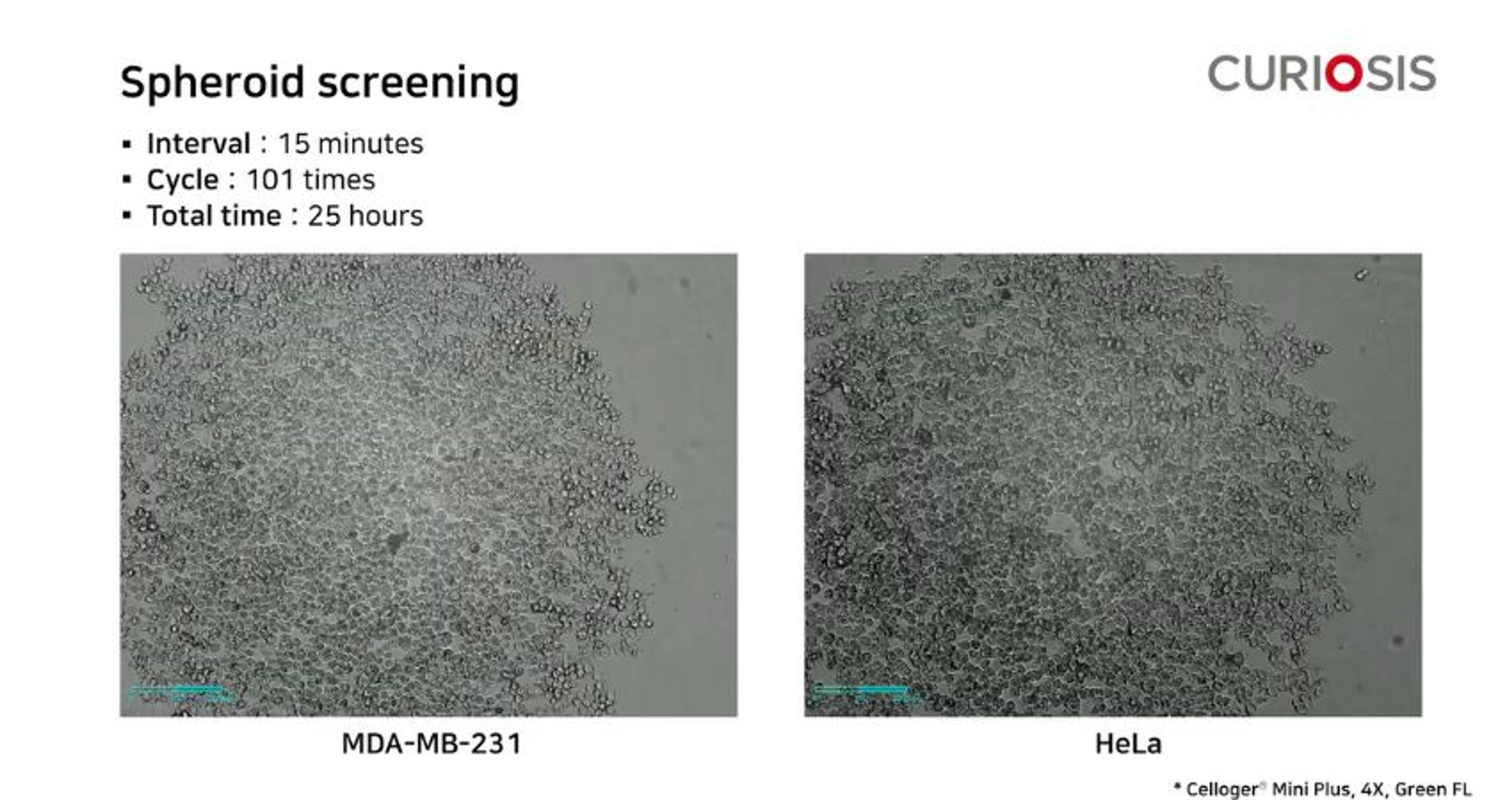
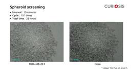
Celloger Mini Plus application: Spheroid screening
In this video, CURIOSIS demonstrates the monitoring of concentration-dependent spheroid formation in two cell lines, MDA-MB-231 and Hela cells, using the Celloger® Mini Plus. With a 4x objective and green fluorescence, the system captured the differences in spheroid size between the two cell lines, despite having the same number of cells forming the spheroids. Hela cells occupied a smaller area and showed more effective spheroid formation, suggesting stronger cell aggregation compared to MDA-MB-231. The cells were seeded at different densities (10,000, 5,000, and 1,000 cells per well) in a round-bottom 96-well plate and cultured for 26 hours.
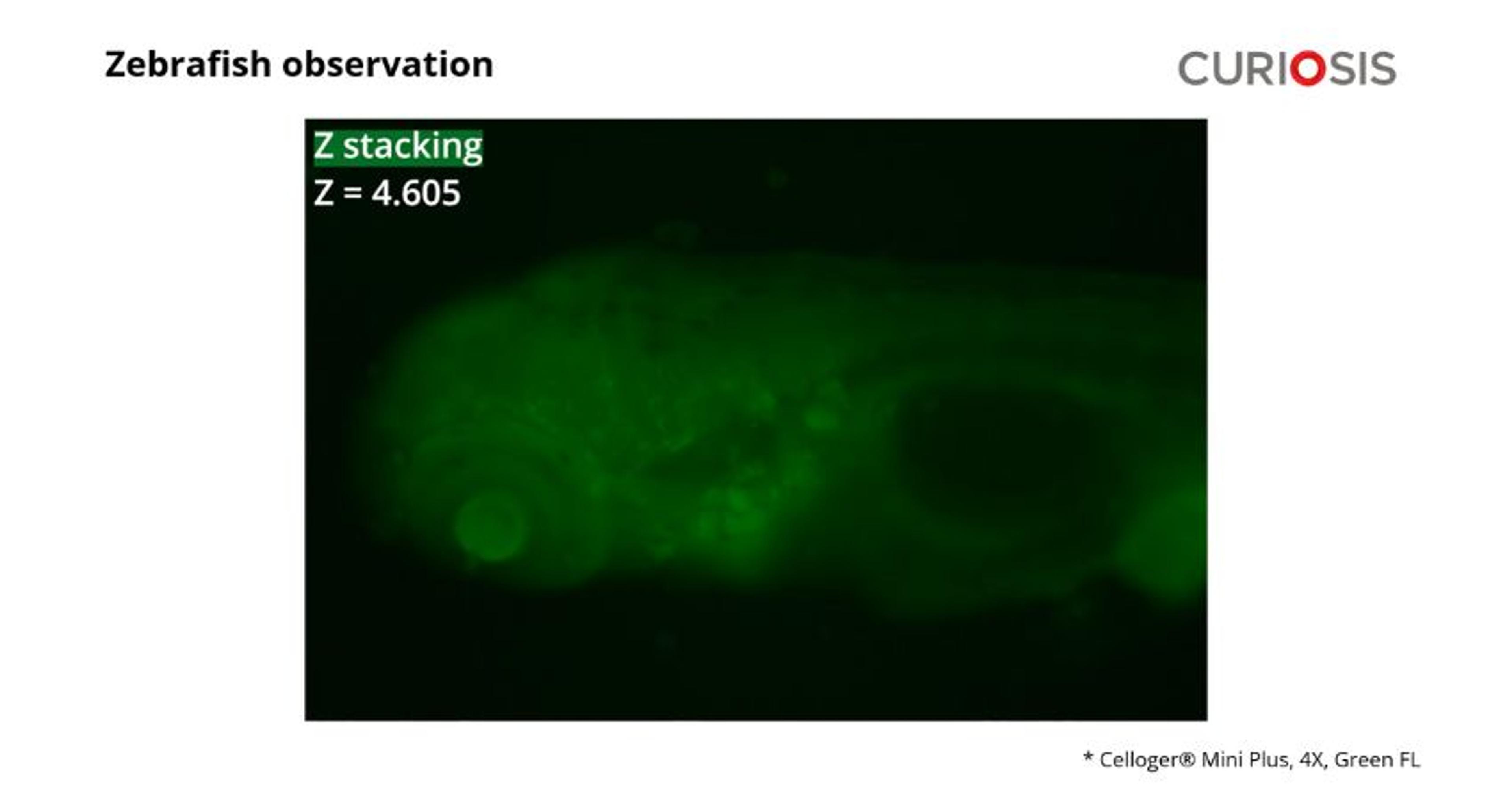
Celloger Mini Plus application: Zebrafish observation
In this video, CURIOSIS demonstrates the use of the Celloger® Mini Plus Z-stacking function to observe a transgenic Zebrafish expressing green fluorescent protein. Zebrafish, a commonly used experimental animal model, possesses a small size and high optical clarity, making it easy to observe using optical microscopy. Additionally, the genetic manipulability of Zebrafish embryos and larvae allows for their utilization in a wide range of studies.
The video showcases the capabilities of the Celloger Mini Plus in monitoring cells in real-time. Equipped with fluorescence and bright-field technology, the system provides high-quality images and time-lapse videos.
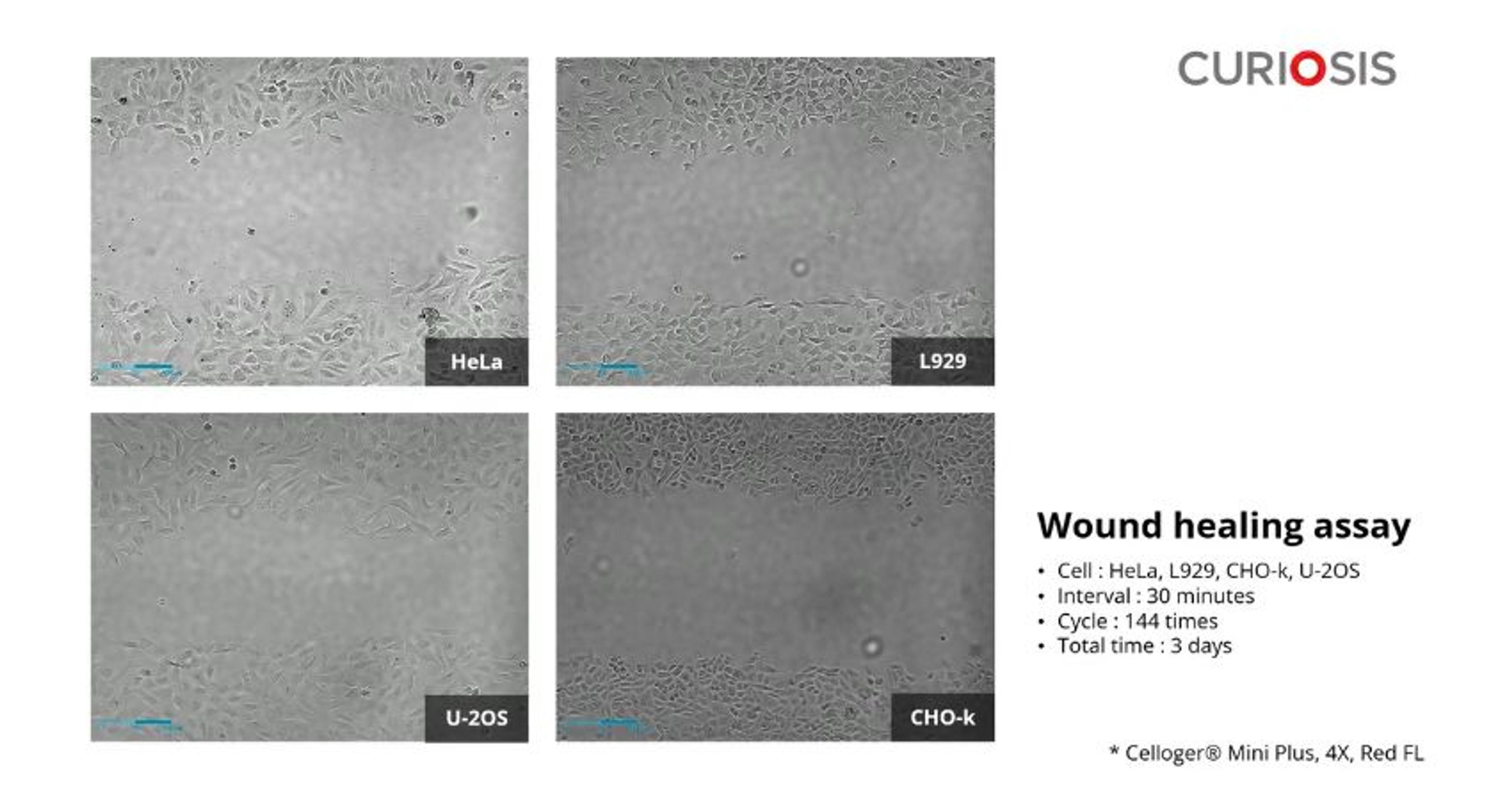
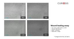
Celloger Mini Plus application: Wound healing assay
In this video, CURIOSIS showcases the Celloger® Mini Plus 4x objective for observing the wound healing process in four cell lines. The wound healing assay is a fast and simple way to assess cell migration, where cells fill in a created scratch or space in the monolayer. The time-lapse video captures the closure of the wounds over three days, with images taken every 30 minutes.
Celloger Mini Plus application: Cytotoxicity
In this video, CURIOSIS focuses on measuring the cytotoxicity of nocodazole by staining dead cells with the green fluorescent CellTox™ dye. The HeLa cells are imaged every hour for a total of 49 hours using the Celloger® Mini Plus, capturing green fluorescence. This advanced automated live cell imaging system offers exceptional fluorescence and bright-field technology, ensuring high-quality images and time-lapse videos. It also provides innovative functions for observing various applications, including wound healing, apoptosis, reactive oxygen species, transfection, and more.
Celloger Mini Plus application: Nocodazole-mediated mitotic arrest
In this video, CURIOSIS examines the anti-mitotic activity of nocodazole against a cancer cell line by monitoring cell division. HeLa cells, transfected with H2B-GFP plasmid, are seeded in a 24-well plate at 4 x 10⁴ cells per well. After allowing the cells to attach overnight, they are incubated with or without 62.5 nM of nocodazole. Real-time imaging is conducted using the Celloger® Mini Plus 4x objective, capturing green fluorescence. Time-lapse images are taken every 15 minutes over a period of 19.5 hours.
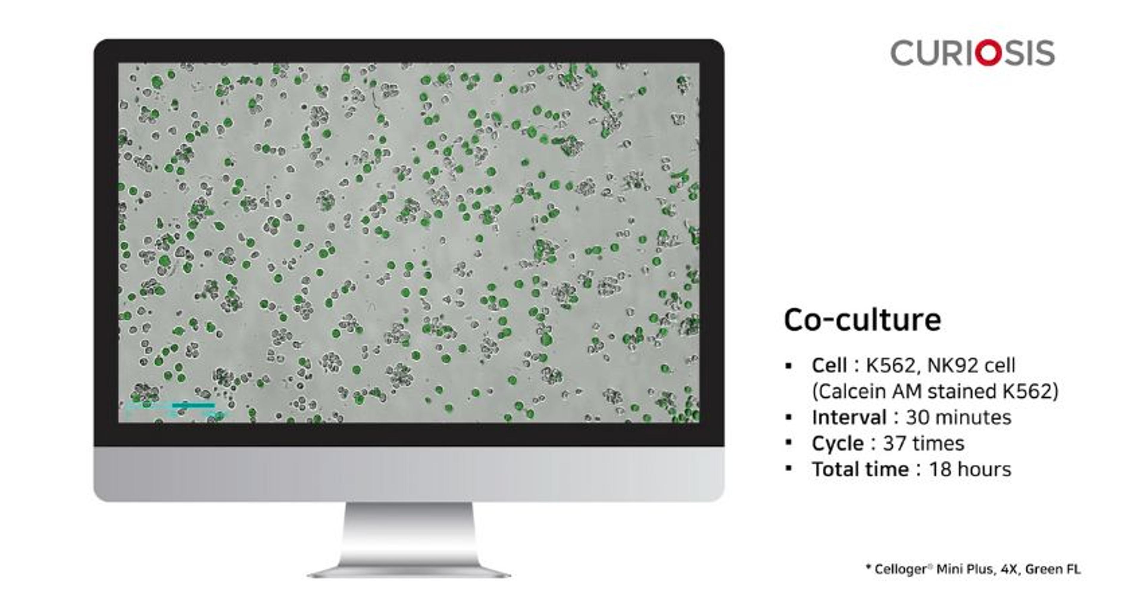
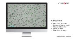
Celloger Mini Plus application: Co-culture
In this video, CURIOSIS showcases the co-culture of K562 and NK92 cells, demonstrating the growth of these two different cell types grown together. The time-lapse video captures the growth process, with images taken every 30 minutes over a total duration of 18 hours. The Celloger® Mini Plus, equipped with a 4x objective and green fluorescence, enables this observation.
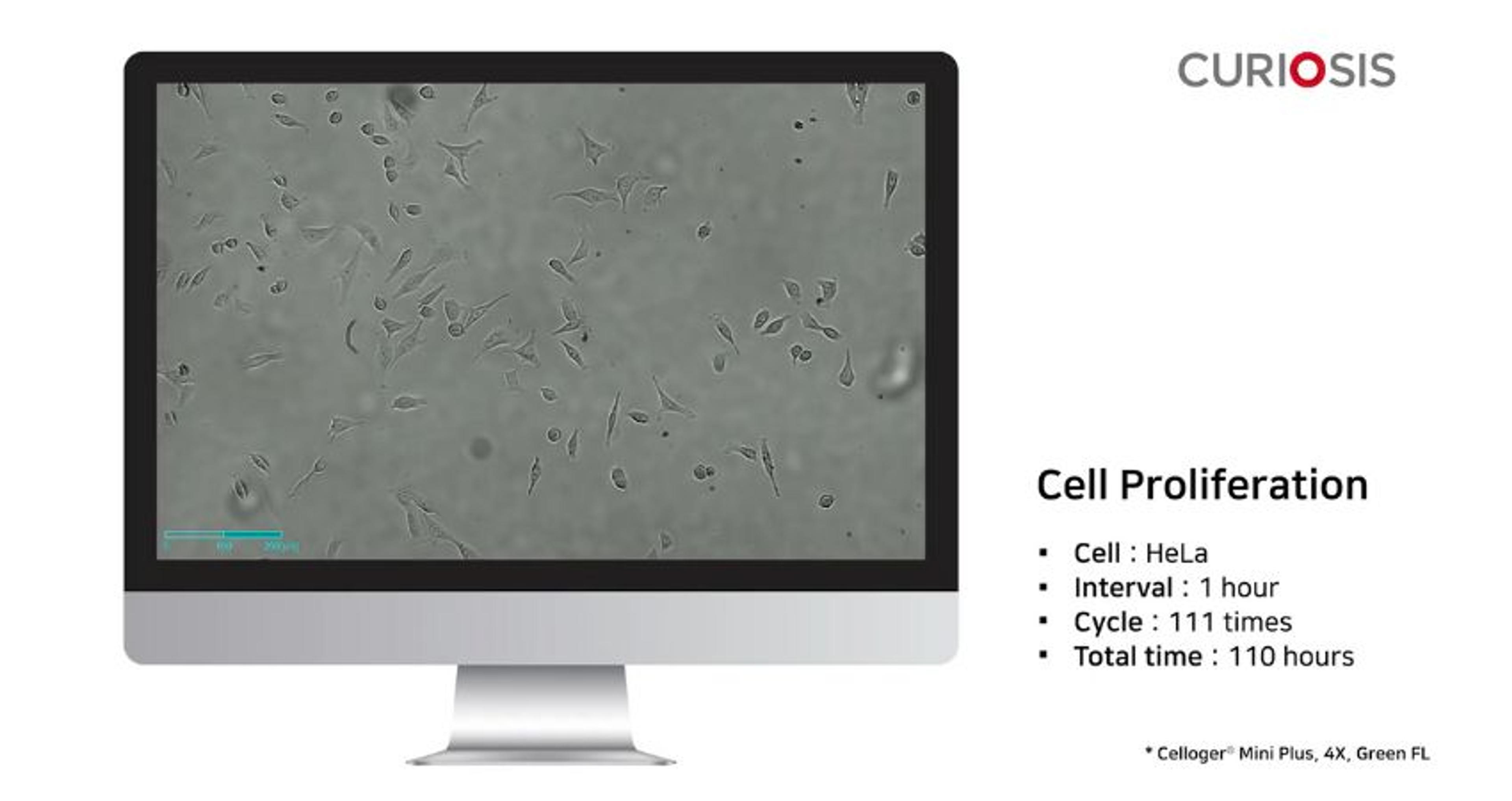
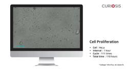
Celloger Mini Plus application: Cell proliferation
In this video, CURIOSIS focuses on cell proliferation as a means to quantify the increasing number of cells over time, confirming their normal growth process. The time-lapse video specifically observes HeLa cell proliferation, capturing images every one hour over a total duration of 110 hours, using the Celloger® Mini Plus with a 4x objective.

Materials science: Key technological advancements to celebrate
Key cell biology lab products reviewed by our scientists
From everyday reagents to state-of-the-art tech, find reviews on a range of cell biology lab products and services
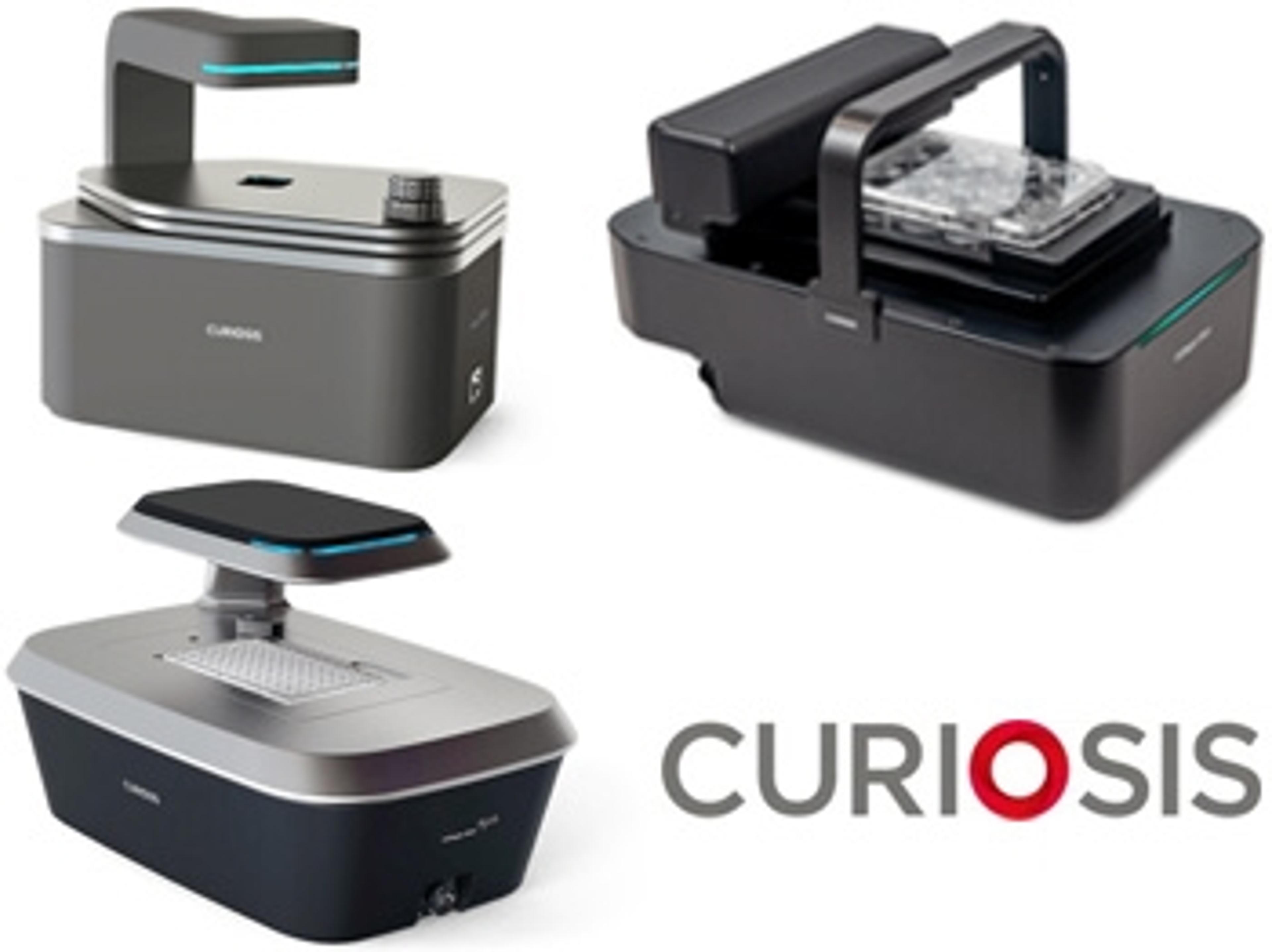
Looking for a compact microscope that works perfectly inside your CO2 incubator?
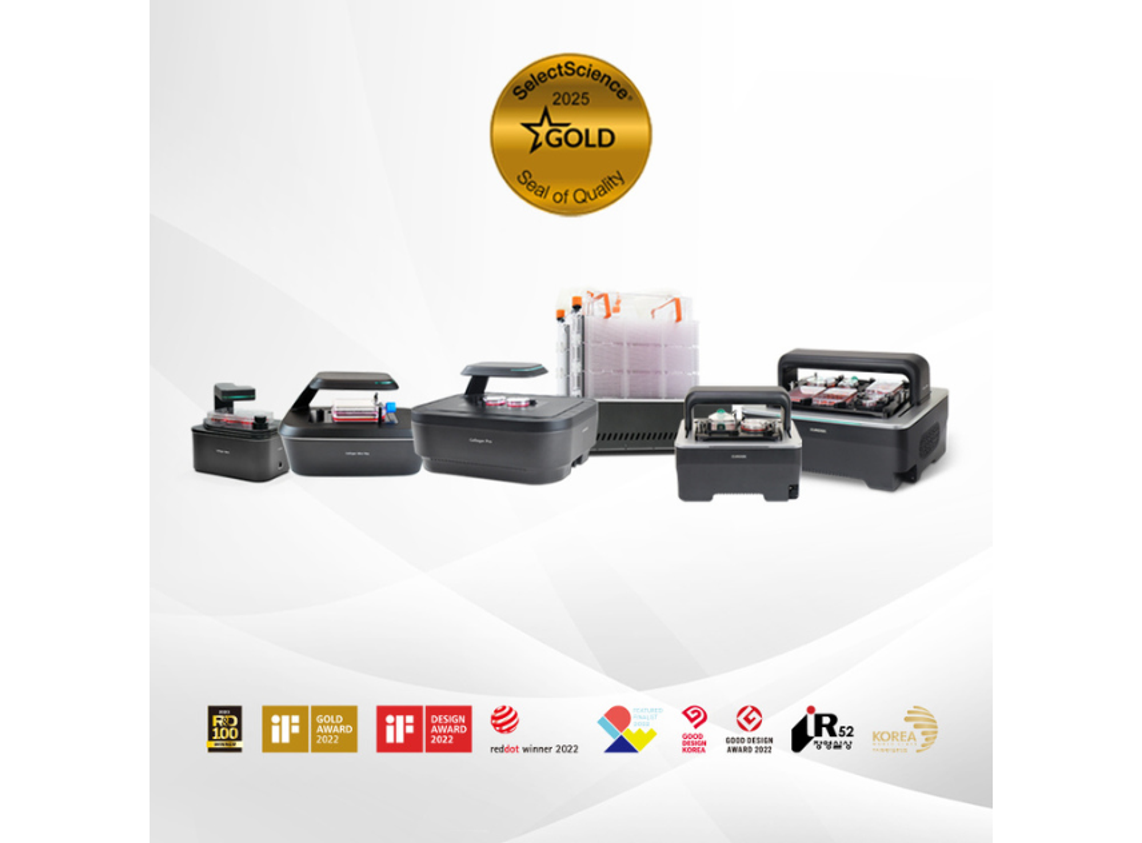
Introducing Curiosis’ automated live-cell imaging systems: The Celloger Series