
Flexible high-throughput live-cell imaging workflows for complex 3D cell models with the Celloger M22 and M26
Automated imaging systems support real-time analysis of spheroids and organoids in scalable drug discovery workflows
28 May 2026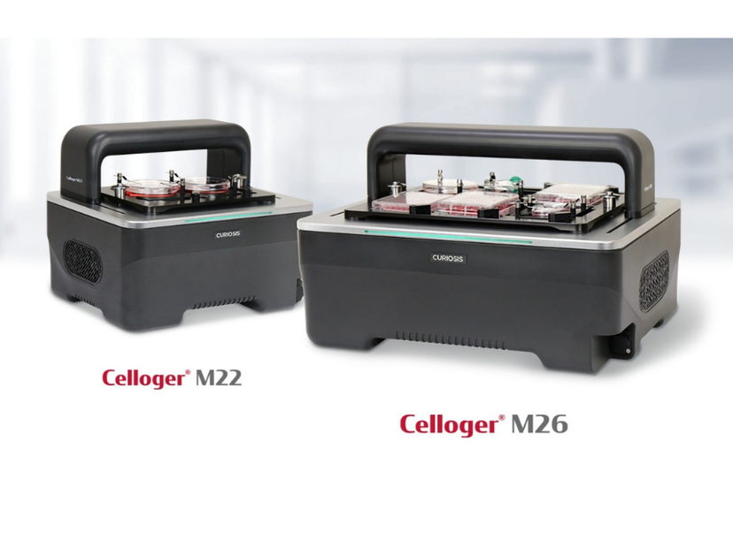
The new Celloger® M22 and Celloger® M26
As the pharmaceutical industry explores alternatives to traditional animal testing, interest in advanced cell-based screening platforms is rapidly increasing. Recent regulatory changes are further accelerating this trend. In particular, the FDA has emphasized the expanded use of alternative preclinical evaluation methods and non-animal testing approaches to support more human-relevant drug discovery workflows.
3D cell models such as spheroids and organoids are becoming increasingly important for evaluating drug efficacy, toxicity, and treatment responses under physiologically relevant conditions. Unlike conventional 2D cultures, these models better replicate the structural complexity and cellular interactions of human tissues. As a result, they are becoming valuable tools in drug screening and translational research.
As the adoption of these advanced in vitro models continues to grow, researchers increasingly need to monitor dynamic cellular responses over extended experimental periods. At the same time, growing numbers of drug candidates and diverse biological samples are driving large-scale preclinical studies.
Because drug responses can vary depending on cell type and sample characteristics, efficiently monitoring large numbers of samples across multiple treatment conditions has become a major challenge in high-throughput screening workflows.
To support these growing research demands and help researchers manage diverse experimental conditions, CURIOSIS has introduced the new Celloger® M22 and Celloger® M26. These next-generation automated live-cell imaging systems are designed for high-throughput, real-time cellular analysis directly inside a standard CO₂ incubator.
Building upon the core strengths of the Celloger® Series, the new systems combine scalable multi-vessel imaging with enhanced optical performance. These capabilities provide an optimized solution for drug discovery and cell-based screening applications.
Key features of Celloger M22 and M26
Stable long-term imaging directly inside the incubator
The Celloger® M22 and M26 are designed to operate directly inside a standard CO₂ incubator, enabling continuous live-cell imaging without repeated sample transfer. While the systems remain inside the incubator, researchers can monitor cellular images live in real time on an external PC.
Designed with low-power operation, the Celloger® M22 and M26 help minimize temperature fluctuations and condensation inside the incubator, supporting reliable imaging during extended live-cell experiments involving large numbers of samples.
High-throughput multi-vessel imaging for scalable workflows
The Celloger® M22 supports simultaneous imaging of up to two vessels, while the Celloger® M26 can image up to six vessels at once. The systems are compatible with various culture vessels including well plates, flasks, dishes, and slides, enabling flexible workflows for large-scale live-cell imaging and high-throughput screening applications.
Individual imaging settings can be applied to each vessel, allowing multiple experiments with different conditions to be performed simultaneously on a single system. This flexibility helps researchers efficiently manage increasing numbers of samples and experimental conditions.
Enhanced image quality for complex 3D cell models
The Celloger® M22 and M26 are designed with a fixed vessel stage while the optical system moves beneath the sample during image acquisition. This design minimizes sample disturbance and supports consistent imaging of sensitive 3D cell models such as spheroids and organoids across multiple positions over time. In addition, the synchronized illumination system moves together with the camera to maintain optical alignment throughout imaging, supporting uniform brightfield image acquisition across multiple imaging areas.
Flexible imaging with interchangeable objective lenses
The Celloger® M22 and M26 support user-interchangeable 4X, 10X, and 20X objective lenses, allowing researchers to optimize imaging conditions based on sample type and experimental purpose. From large-area screening to detailed cellular observation, the systems provide flexible imaging workflows for a wide range of research applications.
User-friendly software and flexible analysis workflows
The Celloger® M22 and M26 provide intuitive image acquisition and analysis software designed to simplify live-cell imaging workflows. Functions including time-lapse imaging, Z-stacking, image stitching, fluorescence analysis, and 3D cell morphology analysis support efficient monitoring and analysis across a wide range of experimental applications.
In addition, exported image datasets are compatible with various third-party analysis platforms, allowing researchers to integrate Celloger® image data into existing workflows and perform further downstream analysis using their preferred software tools.
Want the latest science news straight to your inbox? Become a SelectScience member for free today>>
Related products
Request Quote for All Products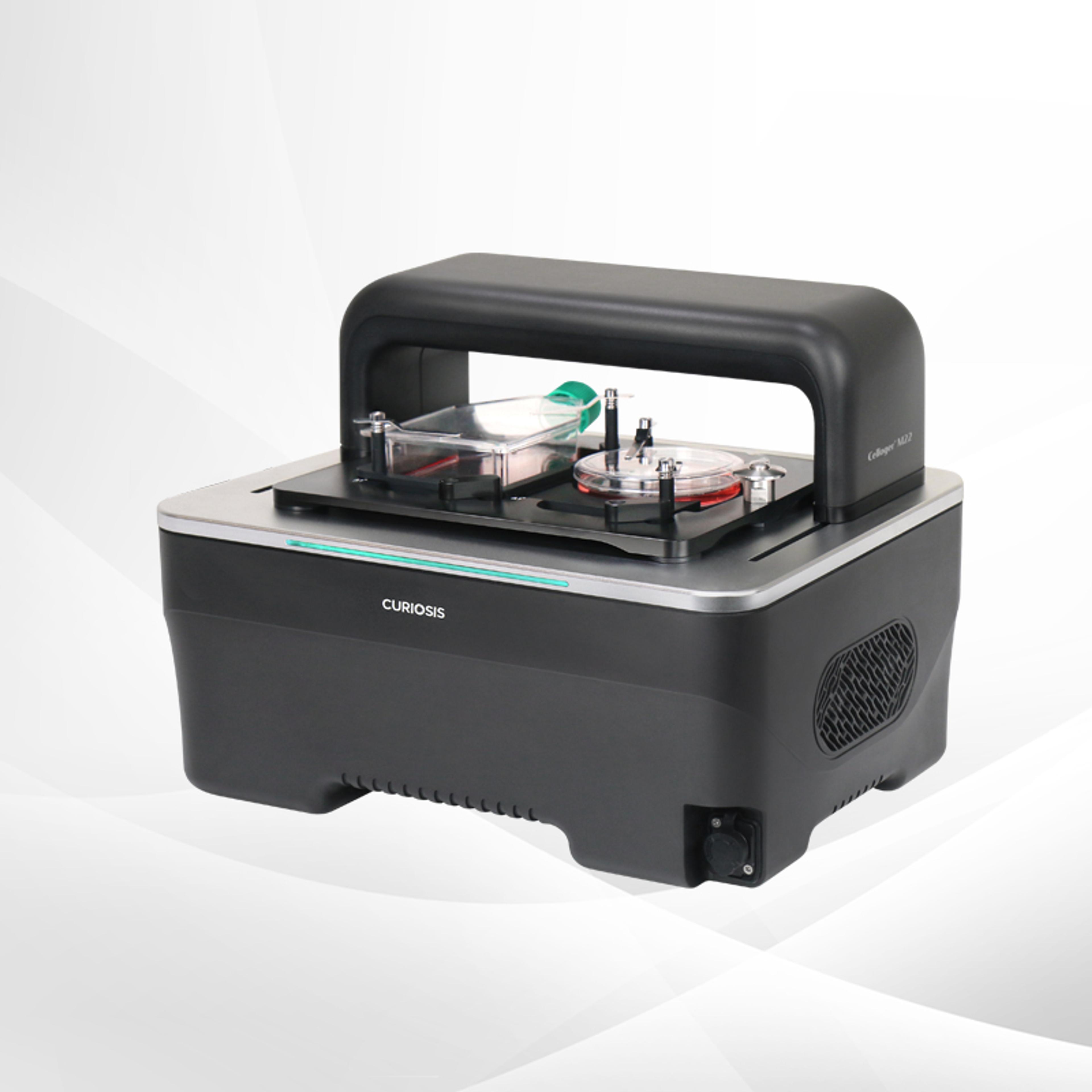
Celloger® M22, Dual-vessel Live Cell Imaging System from Curiosis
CURIOSISCelloger® M22 is a dual-vessel live cell imaging system for simultaneous monitoring inside the incubator. With synchronized imaging under identical conditions, it reduces variability and delivers consistent results for parallel experimental workflows.
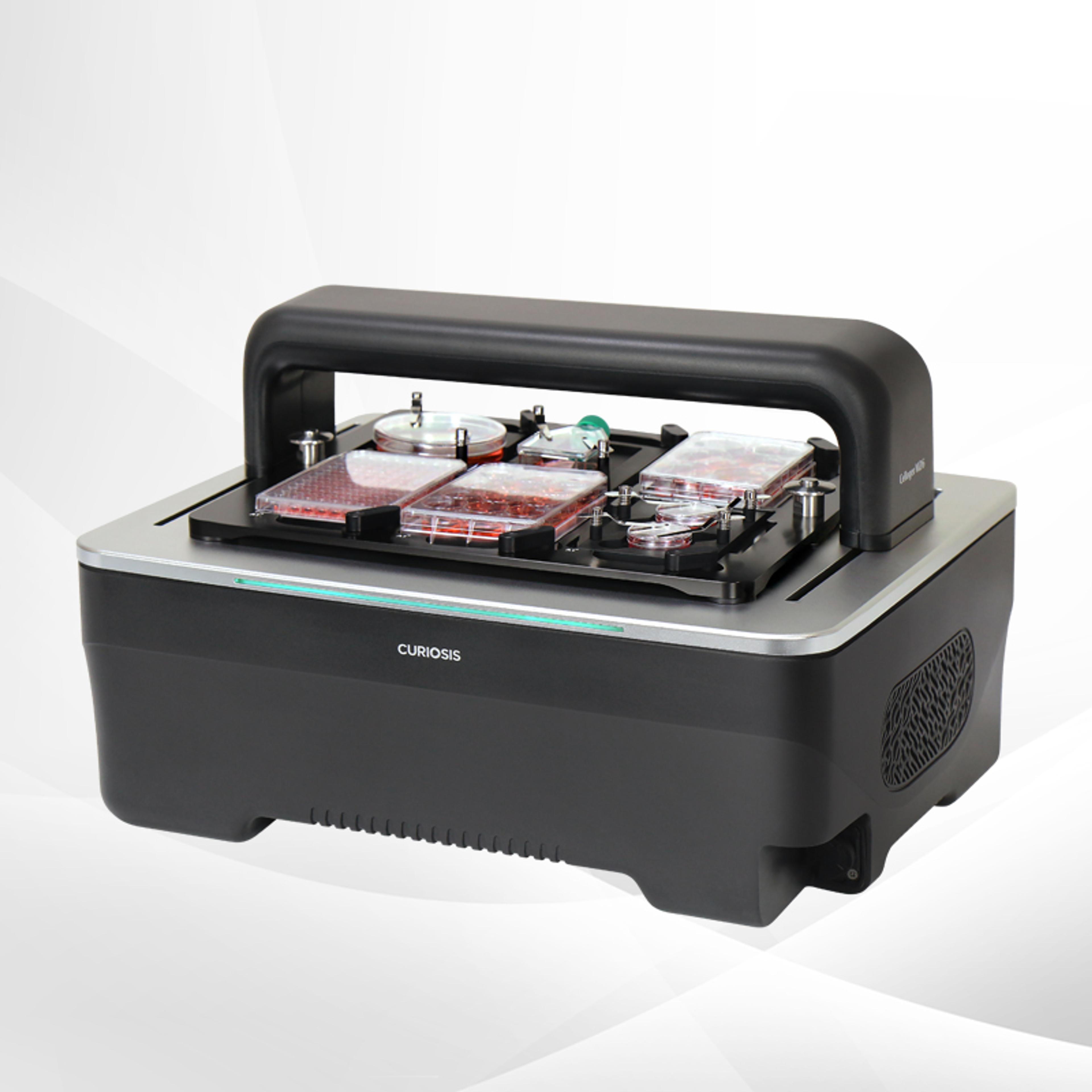
Celloger® M26, High-throughput Live Cell Imaging System from Curiosis
CURIOSISCelloger® M26 is a high-throughput live cell imaging system for real-time monitoring inside the incubator. It offers synchronized illumination, ultra high-resolution imaging, interchangeable lenses, and brightfield/fluorescence modes, enabling simultaneous imaging of up to six plates for large-scale cellular analysis.
Links
Tags
Frequently asked questions
Show frequently asked questions
How do Celloger M22 and M26 support high-throughput drug discovery using 3D cell models such as spheroids and organoids?
Celloger® M22 and M26 are next-generation automated live-cell imaging systems designed for high-throughput, real-time cellular analysis directly inside a standard CO₂ incubator. They support scalable multi-vessel imaging, with Celloger® M22 imaging up to two vessels and Celloger® M26 imaging up to six vessels simultaneously.
Both systems are compatible with well plates, flasks, dishes, and slides, enabling large-scale live-cell imaging and high-throughput screening workflows. Their fixed vessel stage and moving optical system minimize sample disturbance, providing consistent, high-quality imaging of complex 3D cell models such as spheroids and organoids for drug efficacy, toxicity, and treatment response studies.
What key features make Celloger M22 and M26 suitable alternatives to traditional animal testing in pharmaceutical and drug discovery workflows?
Celloger® M22 and M26 address the growing demand for advanced in vitro models driven by regulatory emphasis on non-animal testing and more human-relevant preclinical evaluation methods. Operating directly inside a standard CO₂ incubator, they enable stable, long-term live-cell imaging without repeated sample transfer, minimizing temperature fluctuations and condensation.
These systems support time-lapse imaging, Z-stacking, image stitching, fluorescence analysis, and 3D cell morphology analysis, allowing researchers to monitor dynamic cellular responses over extended periods. By combining high-throughput multi-vessel imaging with advanced analysis tools, Celloger® M22 and M26 provide an optimized platform for cell-based screening and translational research that complements and reduces reliance on traditional animal testing.
How do the imaging and software capabilities of Celloger M22 and M26 enhance preclinical screening and translational research?
Celloger® M22 and M26 feature a moving optical system with synchronized illumination that maintains optical alignment, supporting uniform brightfield imaging across multiple areas and time points. User-interchangeable 4X, 10X, and 20X objective lenses enable flexible imaging from large-area screening to detailed cellular observation, accommodating diverse sample types and experimental purposes.
Their intuitive software simplifies live-cell imaging workflows with functions such as time-lapse imaging, Z-stacking, image stitching, fluorescence analysis, and 3D cell morphology analysis. Exported image datasets are compatible with various third-party analysis platforms, allowing seamless integration into existing preclinical and translational research workflows and facilitating large-scale, data-rich drug screening studies.