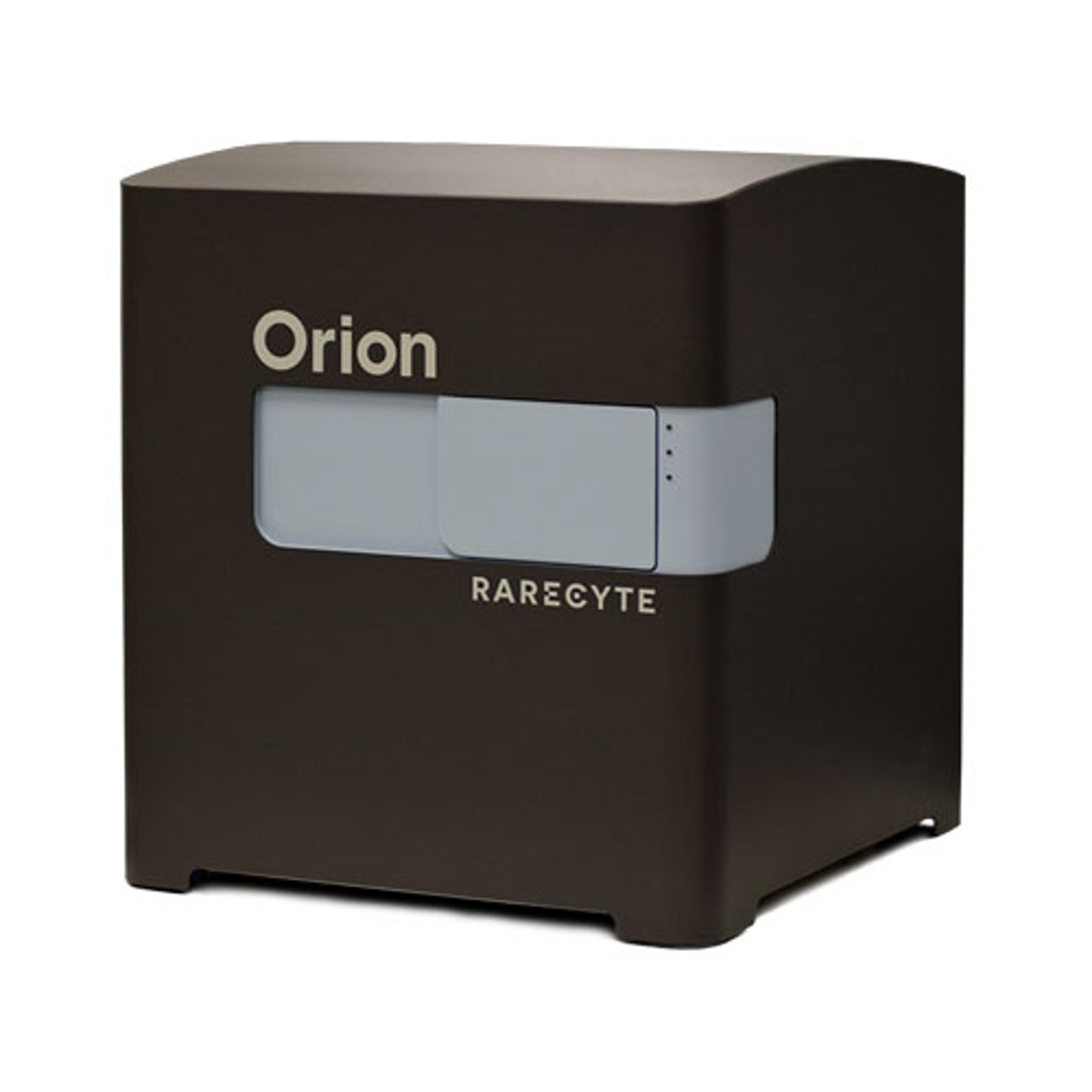
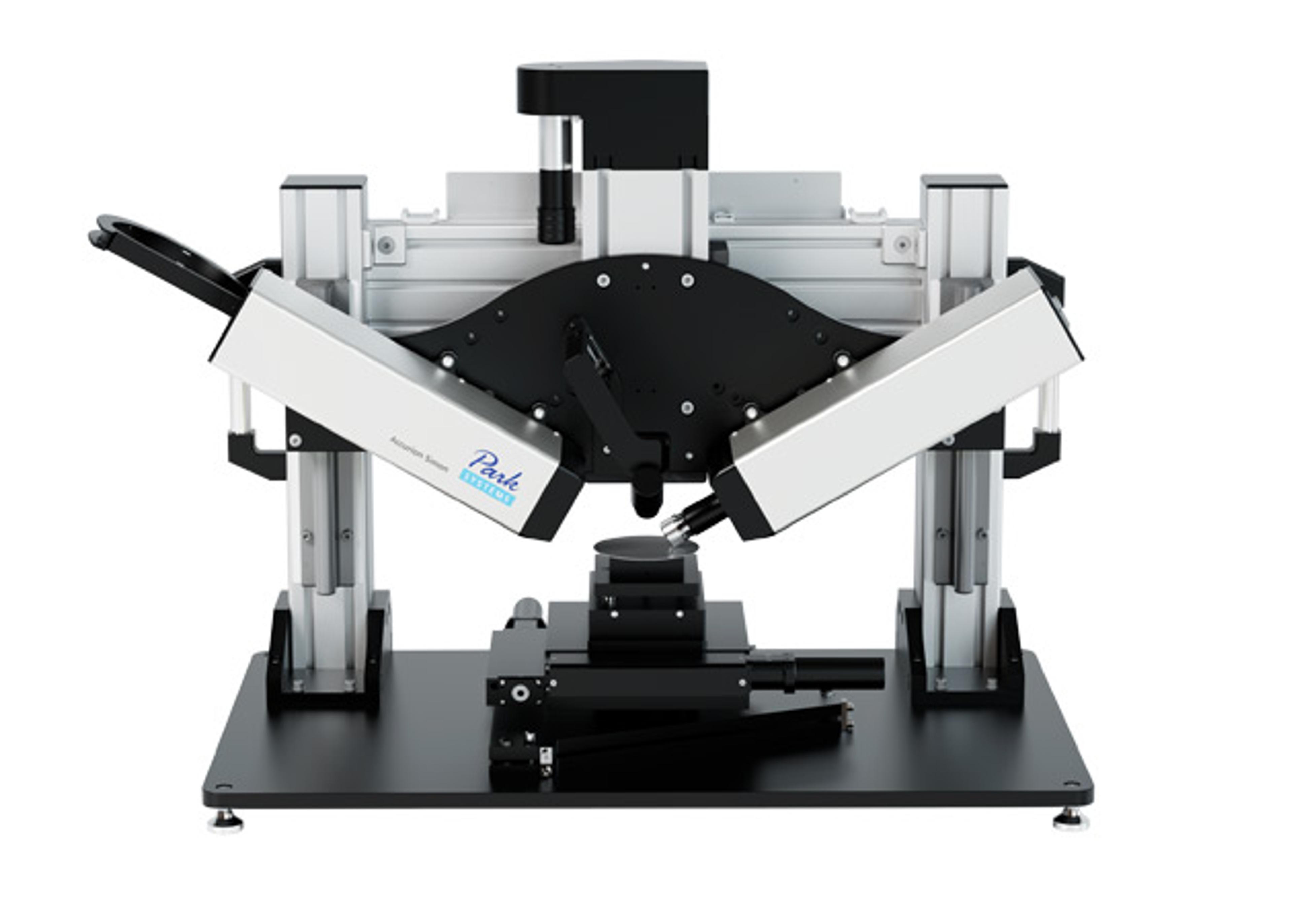
Orion Multiplex Imaging Instrument
The Orion Platform delivers spatial biology and cellular phenotyping at a speed and resolution needed for translation from research to the clinic. It utilizes single round immunofluorescence staining and imaging of standard FFPE or fresh frozen tissue to deliver subcellular quantitation of biomarker targets across a whole slide in only a few hours. Combining speed and resolution with the additional benefits of industry-stan…

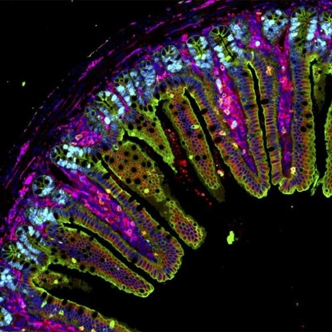
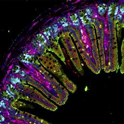
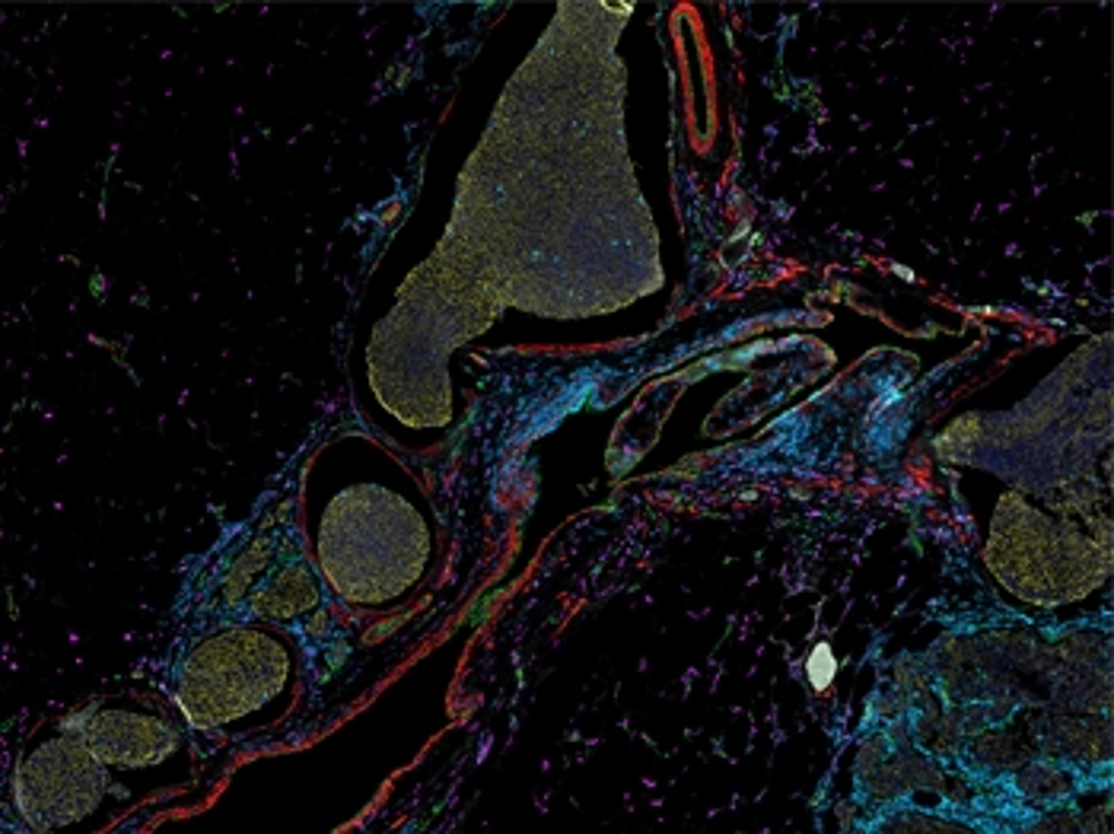
IF imaging of mouse ileum
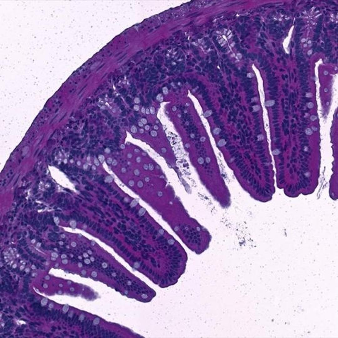
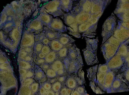
Same section imaging of mouse ileum


The supplier does not provide quotations for this product through SelectScience. You can search for similar products in our Product Directory.
The Orion Platform delivers spatial biology and cellular phenotyping at a speed and resolution needed for translation from research to the clinic. It utilizes single round immunofluorescence staining and imaging of standard FFPE or fresh frozen tissue to deliver subcellular quantitation of biomarker targets across a whole slide in only a few hours.
Combining speed and resolution with the additional benefits of industry-standard H&E and IHC modes, comprehensive phenotypic profiling of patient cohorts can be generated for characterization of tissue architecture, tumor heterogeneity, and the immune response.
Use your own antibodies, run panels from an extensive biomarker list, or engage RareCyte services for custom panels and programs.
Brochures

Product brochure: Orion Multiplex Imaging System for Spatial Biology
In this product brochure, discover the Orion™ novel technology and services platform offering fast path to whole slide, high-plex imaging. Combining speed and resolution, the Orion platform enables comprehensive phenotypic profiling and characterization of tissue architecture, tumor heterogeneity, and the immune response for whole sections in hours.
Specification sheet: Orion Multiplex Imaging System
In this product brochure, explore the specifications of the Orion™ platform which is designed to unlock highly multiplexed spatial biology by providing biomarker depth and flexibility with the convenience of a rapid, single-cycle process.
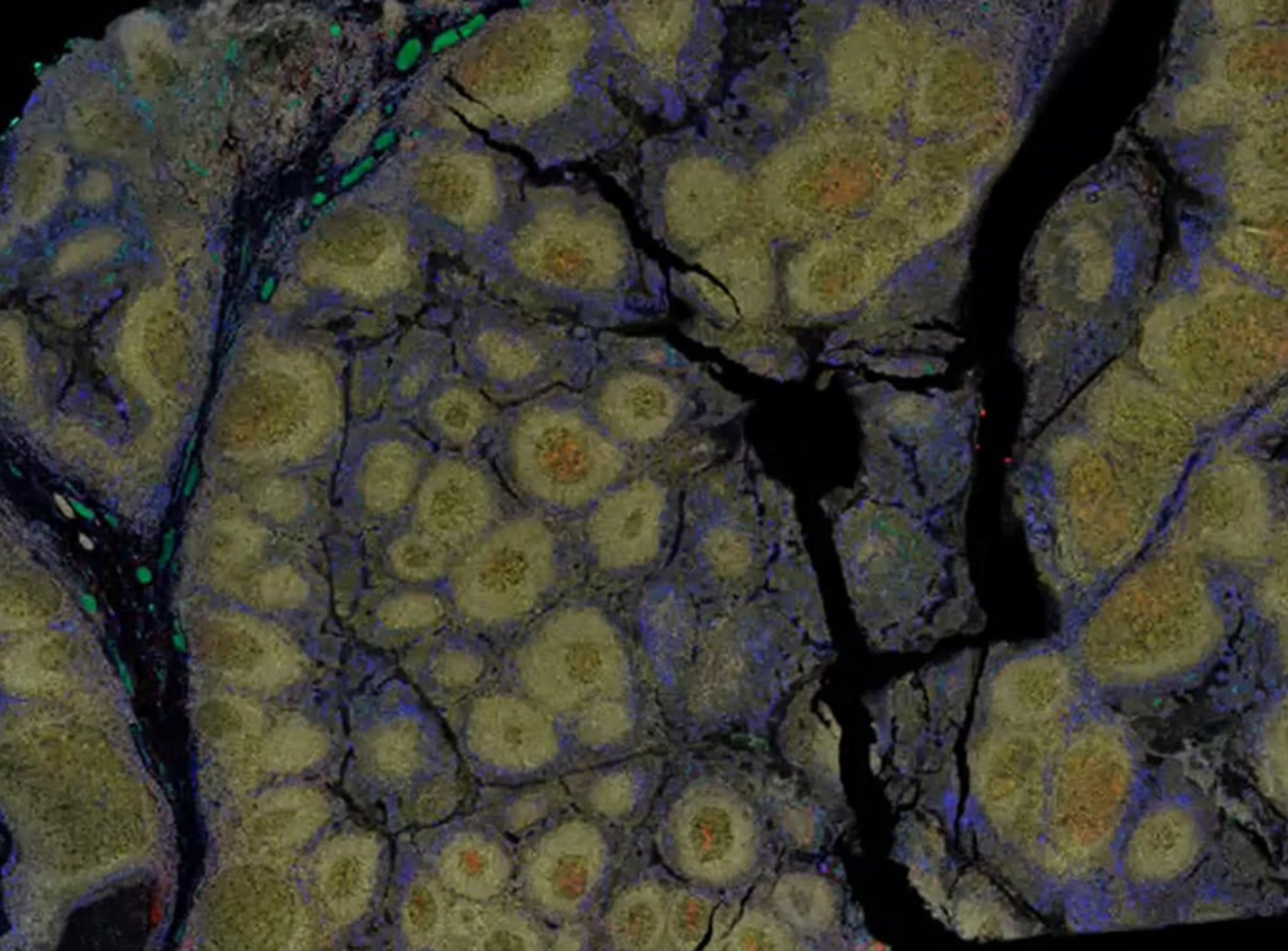
14-plex sample imaging of liver cancer modalities using one-step staining and imaging
RareCyte presents a study of a tumor microenvironment, using the Orion™ imaging system. The formalin-fixed paraffin-embedded (FFPE) liver section was stained using a 14-plex immunofluorescence (IF) panel in one staining round, followed by whole-slide imaging with the Orion instrument, allowing for the traditional pathology analysis to be complimented by same-cell phenotyping. The liver section exhibits extensive infiltration by metastatic moderately-differentiated adenocarcinoma with high Ki-67 proliferation index and central dirty necrosis morphologically consistent with primary tumor from the colon.
Multiplex data utilizing a single-step staining and imaging workflow to investigate multiple sample types
Understanding the tumor microenvironment has proven particularly important for oncology and immune oncology studies. Resolving tissue complexity across a statistically relevant number of patient biopsies at a cellular level has historically been challenged by image resolution, the number of targets that can be simultaneously assessed and sample throughput. These barriers have been broken with the generation of high plex, whole-slide immunofluorescence (IF) imaging data in a single cycle using the Orion™ platform to deliver multiplex biomarker quantitation in just hours. RareCyte gives some specific examples of how the Orion one-step stain-and-scan approach has been used to rapidly generate high-quality, subcellular quantitative IF data for multiple biomarkers across whole tissue sections. Plus, RareCyte describes how this, combined with traditional H&E on the same platform, can be used to provide accretive insights across the same cells and tissue microenvironments from the same biopsy sample.
Quantitative analysis of colorectal adenocarcinoma images
Understanding the tumor microenvironment is particularly important for oncology studies. RareCyte demonstrates how the Orion™ spatial biology platform has been used to investigate a sample of invasive colorectal adenocarcinoma using whole slide, single-step high-plex staining and imaging at single-cell resolution followed by quantitative analysis. The data reveal a distinction between normal colonic epithelium, well-differentiated adenocarcinoma with immune cell collection, and an infiltrating border of the carcinoma. These data also highlight the importance of sufficient plex, resolution and whole slide context to derive reliable spatial biomarkers of potential prognostic value.
Orion Platform imaging for tumor-immune interactions
Deeper understanding of the intricacies between tumor microenvironments and immune cell infiltrates is critical for creation of next generation therapies. In this scientific poster, RareCyte presents a single-step 15-plex fluorescence immunohistochemistry (IHC) protocol using the Orion platform to analyze tumor microenvironments and immune cell phenotypes across multiple tumor types. The poster highlights a study aimed to enhance biomarker analysis by utilizing formalin-fixed paraffin-embedded (FFPE) tumor microarrays. The results demonstrate that the Orion platform can efficiently detect 15 distinct biomarkers on a single slide, surpassing traditional methods limited to 6 markers. The method offers significant workflow advantages, reducing tissue and signal loss, and potentially leading to better diagnostic and therapeutic outcomes by providing deeper insights into immune surveillance and mechanisms of resistance in various cancers.
Orion tissue investigation: Tonsil lymphoid hyperplasia
Here, RareCyte presents its Orion tissue investigation into tonsil lymphoid hyperplasia.
Orion tissue investigation: Colorectal carcinoma
Here, RareCyte presents a whole-slide tissue section of an invasive colorectal adenocarcinoma stained with a 17-plex Immuno-oncology biomarker panel and imaged with the Orion system in a single staining and scanning process.
Accelerating Data and Throughput in Spatial Imaging: A Core Facility Perspective
Core facilities are often handling diverse samples and juggling multiple projects simultaneously. When choosing a spatial imaging platform, Dr. Zbigniew Mikulski, Director of the Microscopy and Histology Core Facility at La Jolla Institute for Immunology, needed a solution that balanced high-quality data, cost-effectiveness, and throughput to accommodate multiple users and high volume. Seamless integration into the core lab’s workflow was key due to the volume the facility generates, up to 15,000 histology slides annually.
In this webinar, Dr. Mikulski will share best practices from working on over 18 different projects, and how the Orion platform for spatial biology facilitated a fast, low run cost solution that works with FFPE samples. Dr. Mikulski will also discuss practices that other labs might adopt to further improve workflow efficiency and cost-effectiveness.
Key learning objectives
- Gain tips for integrating multiplexing into a busy histology laboratory
- Learn how to easily add custom biomarkers to your panel
- Understand the importance of sample quality for proper immunostaining results
Who should attend?
- Academic researchers
- Pharmaceutical companies
- Core laboratories
Certificate of attendance
All webinar participants can request a certificate of attendance, including a learning outcomes summary, for continuing education purposes.
Open source software for multiplex staining & image analysis
Dr. Sarah McArdle presents on open source software for quantitative image analysis, focusing on multiplex staining for high dimensional analysis. She explains the RareCyte Orion microscope and its image acquisition process. The discussion covers the advantages of open source software, challenges in analyzing large datasets, and the importance of validating staining quality.

Advanced spatial biology sheds new light on therapeutic research
See how high-resolution multiplex immunofluorescence imaging can elucidate the important role the immune system plays in health, disease, and drug development

Novel multianalyte assay has potential to transform cancer research and treatment
The use of protein biomarkers in oncology is growing rapidly and offers real hope to patients with hard-to-treat cancers

How to unlock the complexity of tumor microenvironments
Learn how researchers used the Orion™ platform from RareCyte to generate spatial insights into tumor microenvironments
Quantitative microscopy for precision spatial biology: “From samples to knowledge”
In this guest editorial by Dr. Zbigniew Mikulski from the La Jolla Institute for Immunology, explore the fascinating advancements in microscopy and their role in facilitating the generation of high-resolution quantitative data, suited for translational and clinical spatial analysis

Bringing spatial biology to the clinic: “A new lens on cancer biology”
Spatial transcriptomics pioneer, Dr. Arutha Kulasinghe, shares his latest work in understanding the underlying tumor biology using an integrative multi-omics approach, and how it could inform therapy decisions