14-plex sample imaging of liver cancer modalities using one-step staining and imaging
17 Jun 2024RareCyte presents a study of a tumor microenvironment, using the Orion™ imaging system. The formalin-fixed paraffin-embedded (FFPE) liver section was stained using a 14-plex immunofluorescence (IF) panel in one staining round, followed by whole-slide imaging with the Orion instrument, allowing for the traditional pathology analysis to be complimented by same-cell phenotyping. The liver section exhibits extensive infiltration by metastatic moderately-differentiated adenocarcinoma with high Ki-67 proliferation index and central dirty necrosis morphologically consistent with primary tumor from the colon.
Related products
Request Quote for All Products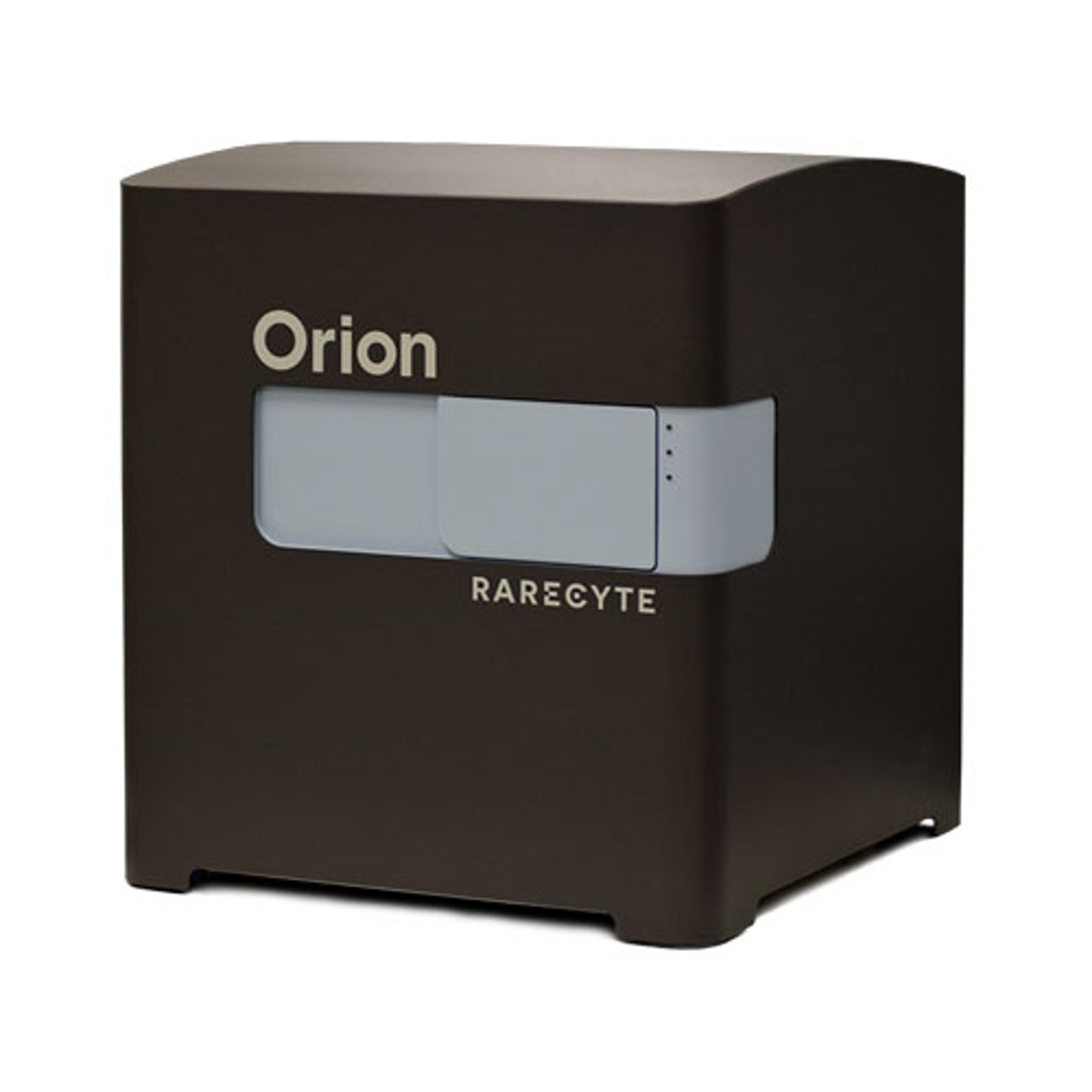
Orion Multiplex Imaging Instrument
RareCyte, IncThe Orion Platform delivers spatial biology and cellular phenotyping at a speed and resolution needed for translation from research to the clinic. It utilizes single round immunofluorescence staining and imaging of standard FFPE or fresh frozen tissue to deliver subcellular quantitation of biomarker targets across a whole slide in only a few hours. Combining speed and resolution with the additional benefits of industry-standard H&E and IHC modes, comprehensive phenotypic profiling of patient cohorts can be generated for characterization of tissue architecture, tumor heterogeneity, and the immune response. Use your own antibodies, run panels from an extensive biomarker list, or engage RareCyte services for custom panels and programs.