Label-free quantification of tumor spheroid drug responses in InSphero ARCTis™ Plates

5 Aug 2026

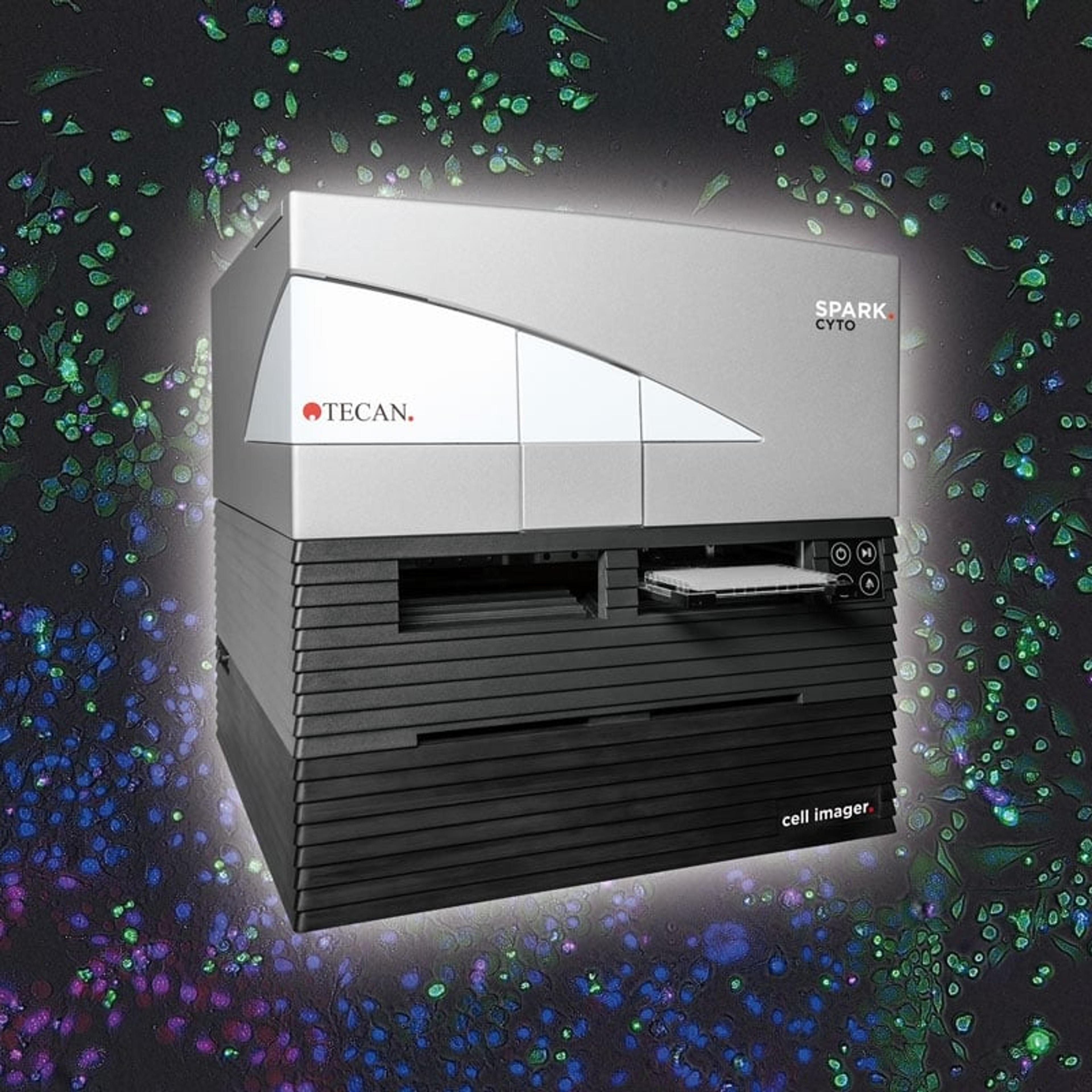
This Tecan application note demonstrates how label-free imaging can be used to monitor tumor spheroid drug responses in InSphero ARCTis™ Plates. Using the Spark® Cyto imaging microplate reader, HCT 116, T-47D, and A549 tumor spheroids were treated with varying concentrations of Staurosporine and monitored over time to assess dose-dependent changes in spheroid area.

Learn how automated brightfield imaging and 3D AI-based analysis can support kinetic monitoring of spheroid growth and treatment response without the need for labeling. The workflow is compared with endpoint CellTiter-Glo® 3D viability measurements and live/dead staining, showing how label-free imaging can provide complementary insight into spheroid viability, morphology, and drug response dynamics.

Discover how Spark Cyto can help streamline 3D cell culture analysis by enabling automated, label-free monitoring of physiologically relevant tumor models for high-throughput screening and translational research.

Resource details:
Resource type: Application note
Page count: 5
Read time: 8 mins

Spark® Cyto

Tecan

Spark Cyto is the first live cell plate reader to detect biological, chemical or physical events – capturing the maximum amount of data possible from each plate well, at the same time and under the same conditions.

(8)

Links

Tags

Cell-Based AssaysCell-based assays are used to monitor the presence, quantity and activities of a desired cellular analyte including drug molecules or biomarkers. This can reveal information on cell health (apoptosis, cytotoxicity, viability and proliferation assays), cell metabolism, cell migration and cell signaling mechanisms. Find the best cell-based assay products, kits and equipment with our peer reviewed product directory: compare products, check customer reviews and receiving pricing direct from manufacturers.High-Content ScreeningHigh-content screening (HCS), also known as high-content analysis (HCA), is a high-throughput technique used in drug discovery to identify substances that alter the phenotype of cells. HCS uses fluorescent microscopic imaging and automated image analysis to investigate cellular events such as apoptosis, cell viability, GPCR activation, oxide production, neurite outgrowth, and cell signaling. Find the best fluorescent labeling reagents, cellular assays, and high-content imaging systems in our peer-reviewed product directory: compare products, check customer reviews and receive pricing direct from manufacturers.Live Cell ImagingLive cell imaging is the study of living cells using microscopes and high-content imaging systems. This technique provides in-depth insight into fast and complex biological processes, by allowing dynamic imaging of living cells instead of acquiring an individual image at a single point in time.3D Cell CultureCulturing cells in three dimensions allows more physiologically relevant observations to be made regarding cellular interactions and gene expression. 3D models provide a more accurate representation of cells in vivo and are more stable than 2D cultures, making them very useful for long-term studies.SpheroidsSpheroids are clusters of cells that have been grown in 3D culture to be used as <i>in vitro</i> model systems. These 3D microtissues can be used for toxicology testing, DMPK studies and many other applications involving cell analysis.OrganoidsOncologyThe branch of medical science that deals with the diagnosis and treatment of cancer is known as oncology.