Multiplex data utilizing a single-step staining and imaging workflow to investigate multiple sample types
17 Jun 2024Understanding the tumor microenvironment has proven particularly important for oncology and immune oncology studies. Resolving tissue complexity across a statistically relevant number of patient biopsies at a cellular level has historically been challenged by image resolution, the number of targets that can be simultaneously assessed and sample throughput. These barriers have been broken with the generation of high plex, whole-slide immunofluorescence (IF) imaging data in a single cycle using the Orion™ platform to deliver multiplex biomarker quantitation in just hours. RareCyte gives some specific examples of how the Orion one-step stain-and-scan approach has been used to rapidly generate high-quality, subcellular quantitative IF data for multiple biomarkers across whole tissue sections. Plus, RareCyte describes how this, combined with traditional H&E on the same platform, can be used to provide accretive insights across the same cells and tissue microenvironments from the same biopsy sample.
Related products
Request Quote for All Products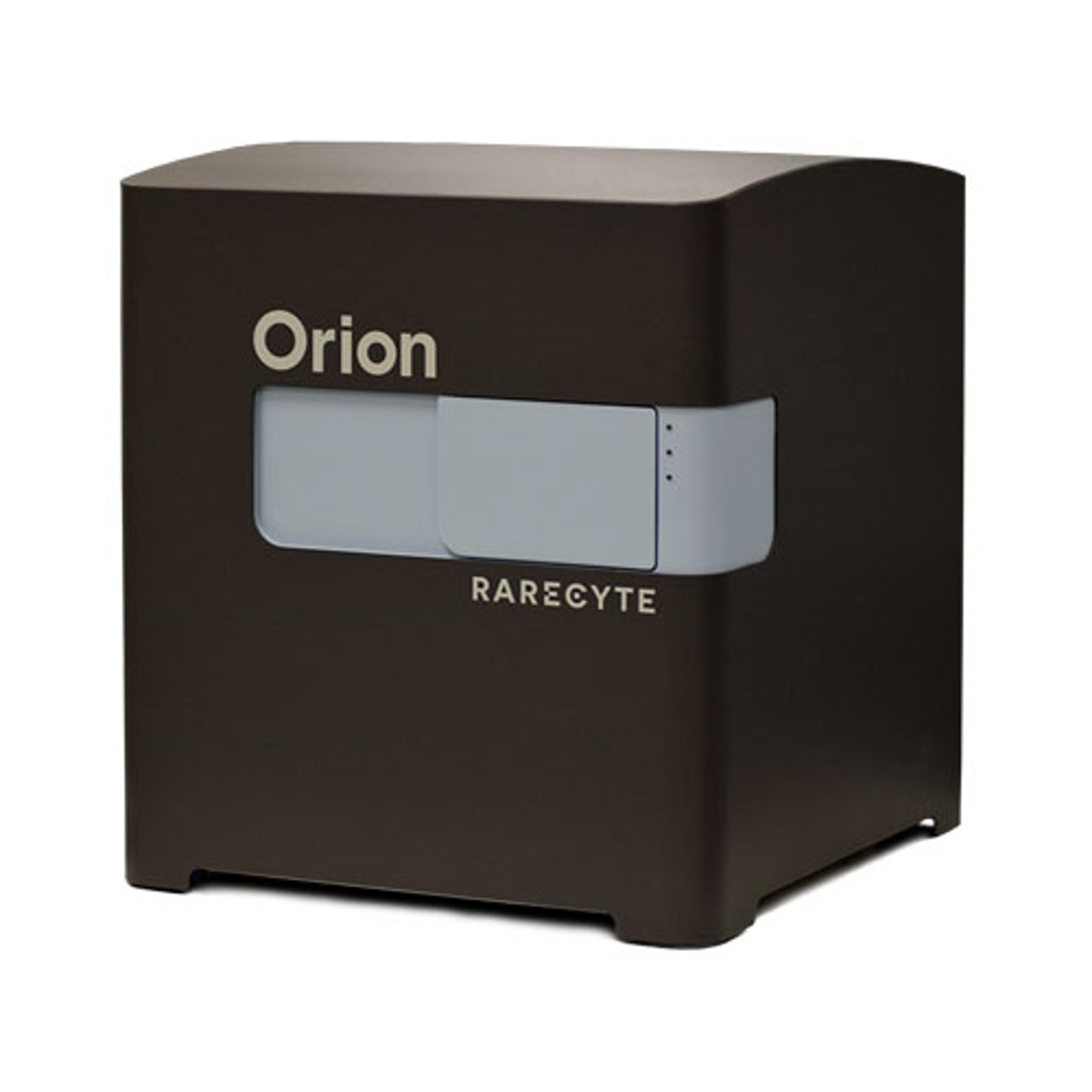
Orion Multiplex Imaging Instrument
RareCyte, IncThe Orion Platform delivers spatial biology and cellular phenotyping at a speed and resolution needed for translation from research to the clinic. It utilizes single round immunofluorescence staining and imaging of standard FFPE or fresh frozen tissue to deliver subcellular quantitation of biomarker targets across a whole slide in only a few hours. Combining speed and resolution with the additional benefits of industry-standard H&E and IHC modes, comprehensive phenotypic profiling of patient cohorts can be generated for characterization of tissue architecture, tumor heterogeneity, and the immune response. Use your own antibodies, run panels from an extensive biomarker list, or engage RareCyte services for custom panels and programs.