Cell DIVE Multiplexed Imaging Solution
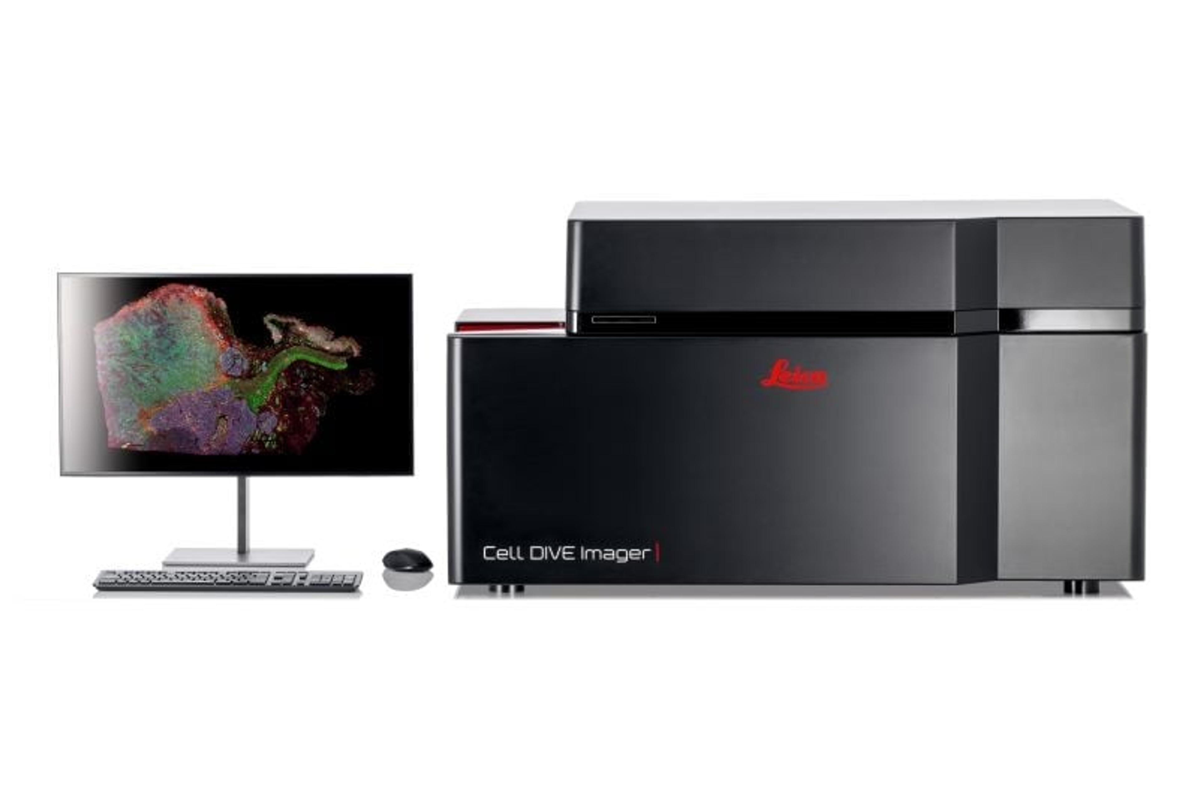
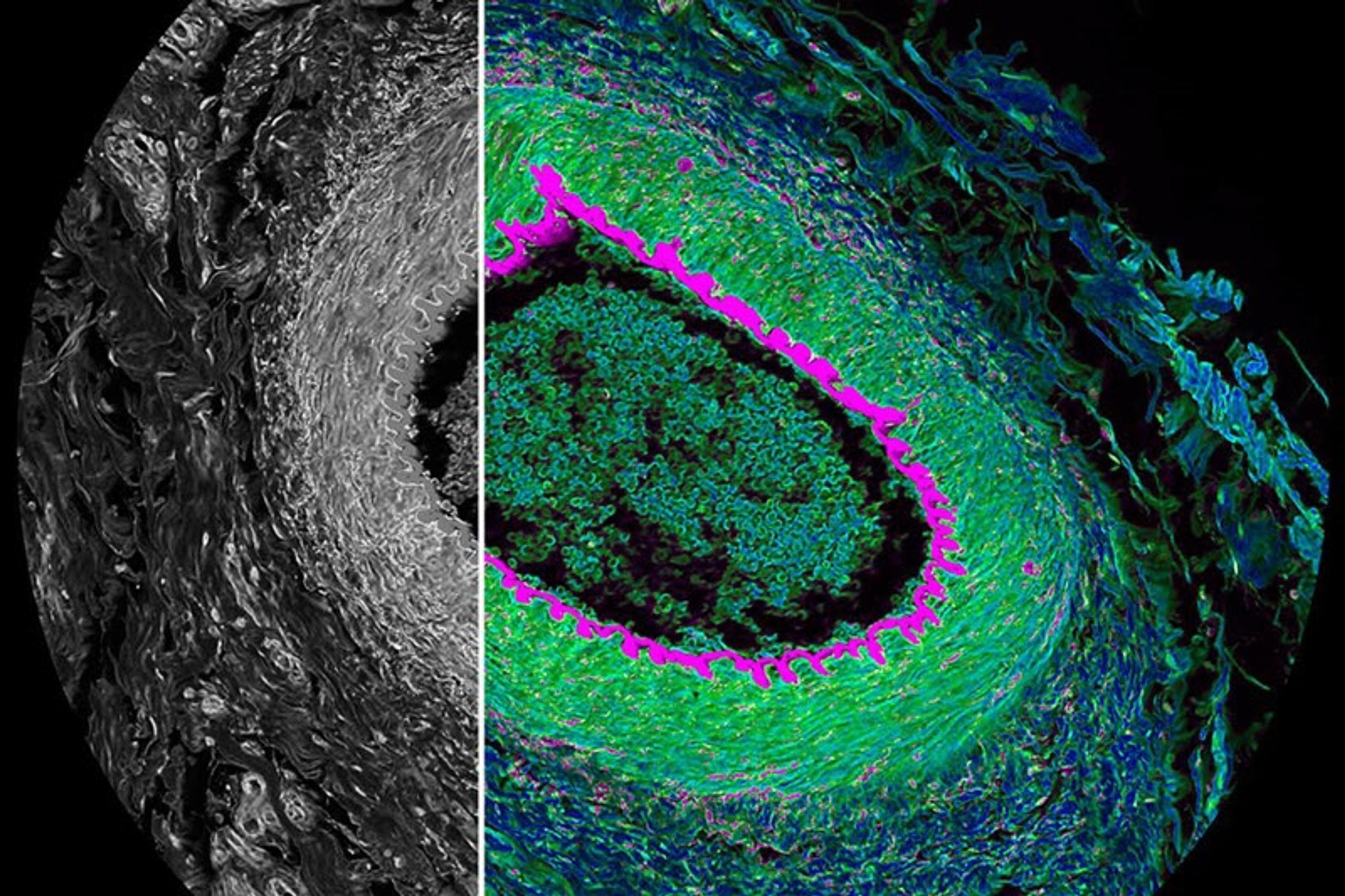
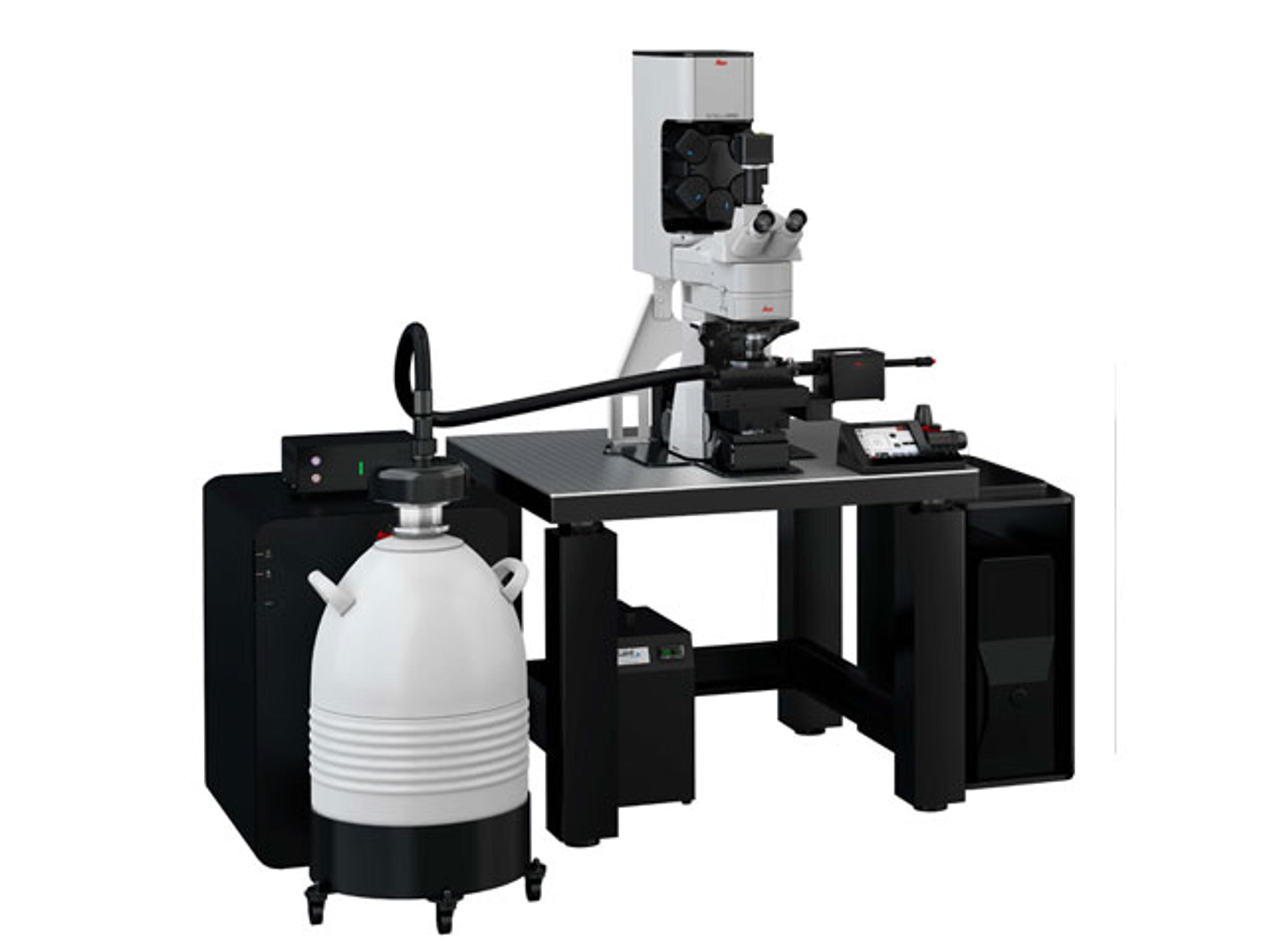
Leica Microsystems EuropeCell DIVE multiplexed imaging is an antibody-based hyperplexed technique addressing spatial cell biology and function within the tumor microenvironment.
Cell DIVE multiplexed imaging is an antibody-based hyperplexed technique addressing spatial cell biology and function within the tumor microenvironment.
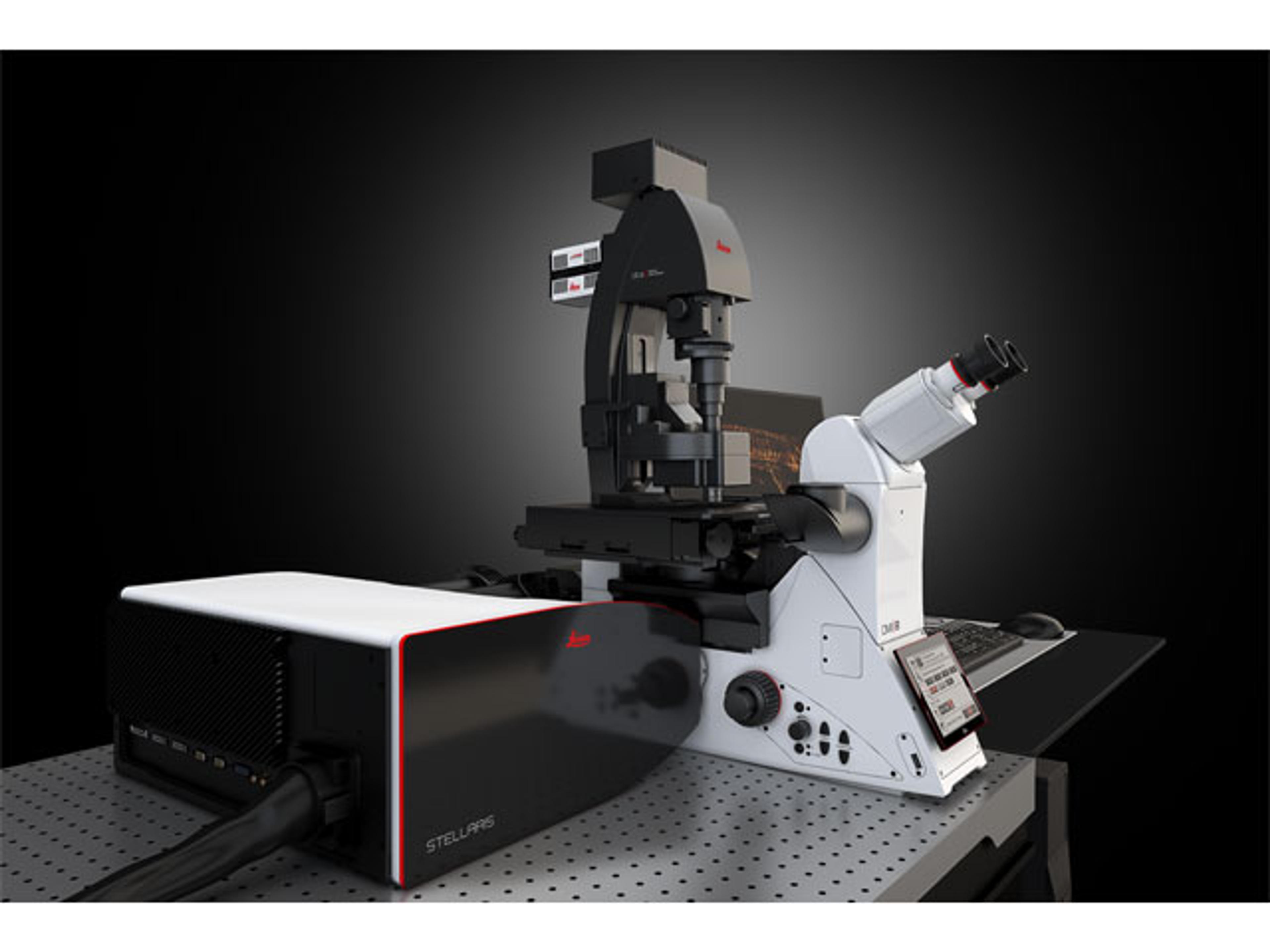
STELLARIS 8 FALCON (FAst Lifetime CONtrast) is the future of functional imaging. Harness the power of fluorescence lifetime to investigate cellular physiology and explore dynamics in living cells.
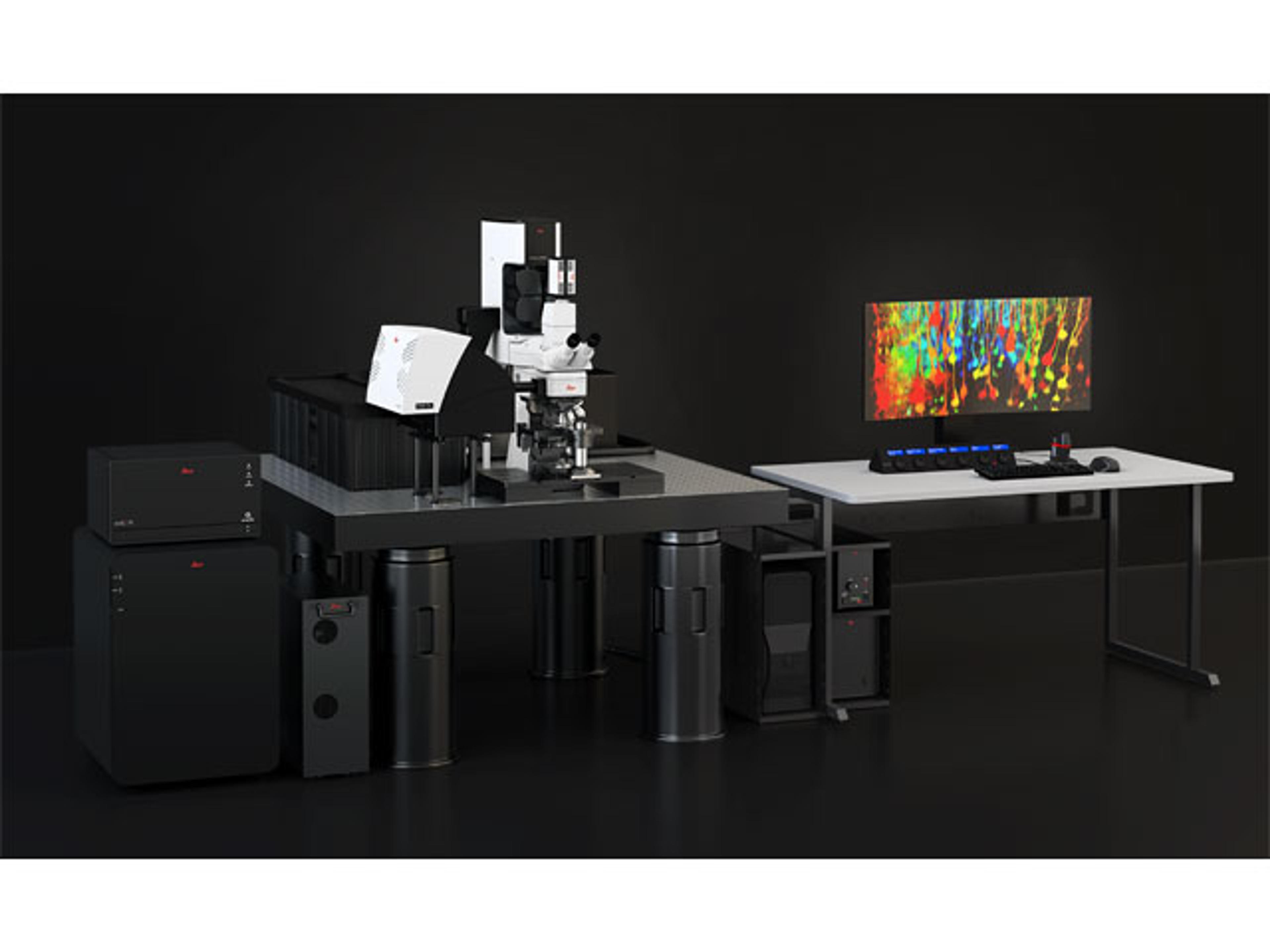
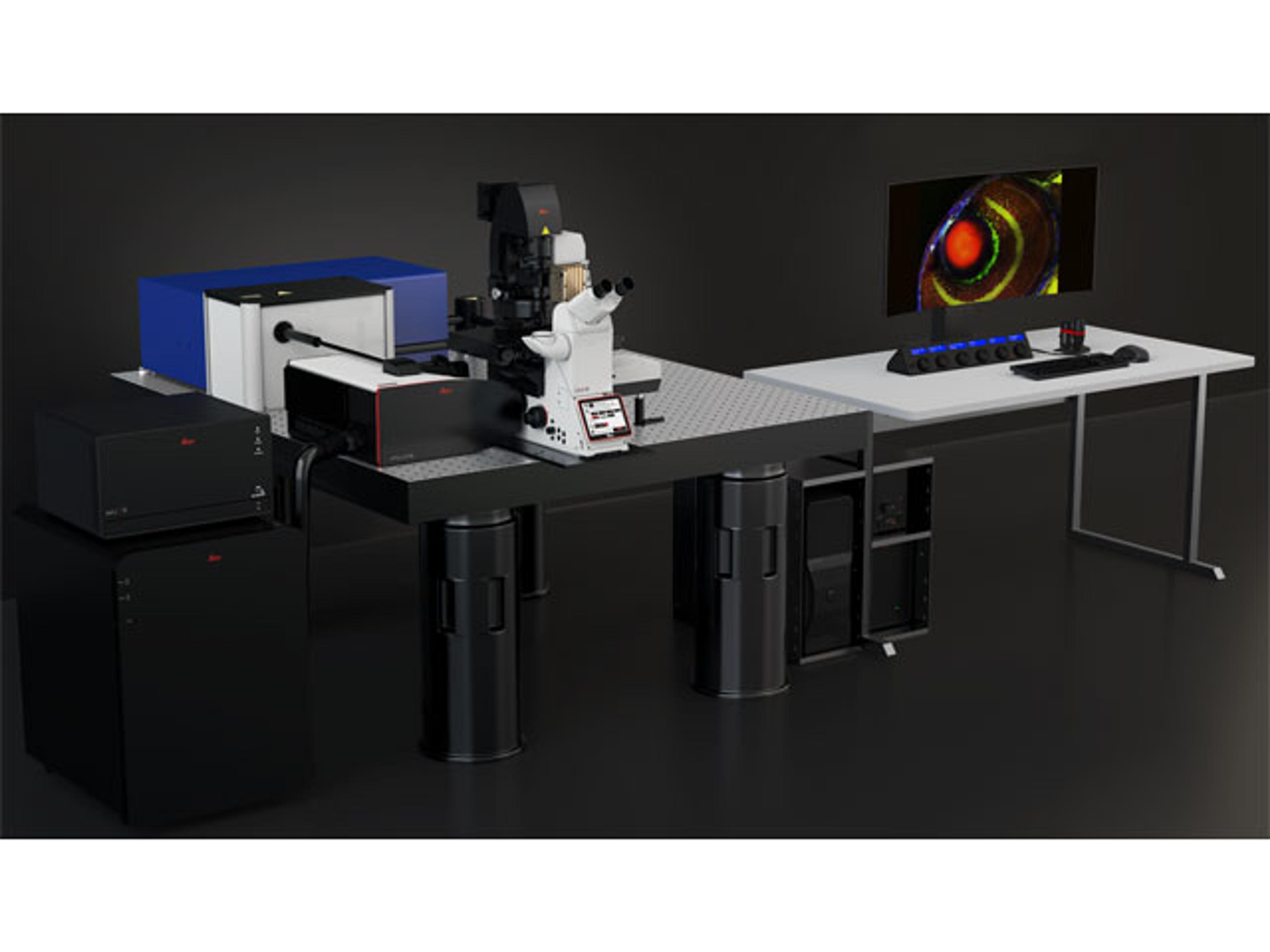
DIVE and STELLARIS have now merged to provide you with the power of flexible multicolor multiphoton imaging.
STELLARIS LightSheet (DLS) unites in one place a confocal system and a light sheet microscope – a unique combination aimed to make your research more versatile. DLS and STELLARIS enable even gentler imaging giving you the ability to perform fast and gentle volumetric light sheet imaging and to improve your live cell imaging applications.
When you need to study structures that cannot be visualized with traditional fluorescent microscopy methods, the STELLARIS 8 Coherent Raman Scattering (CRS) microscope enables you to implement label-free chemical imaging into your workflow to answer those challenging research questions.
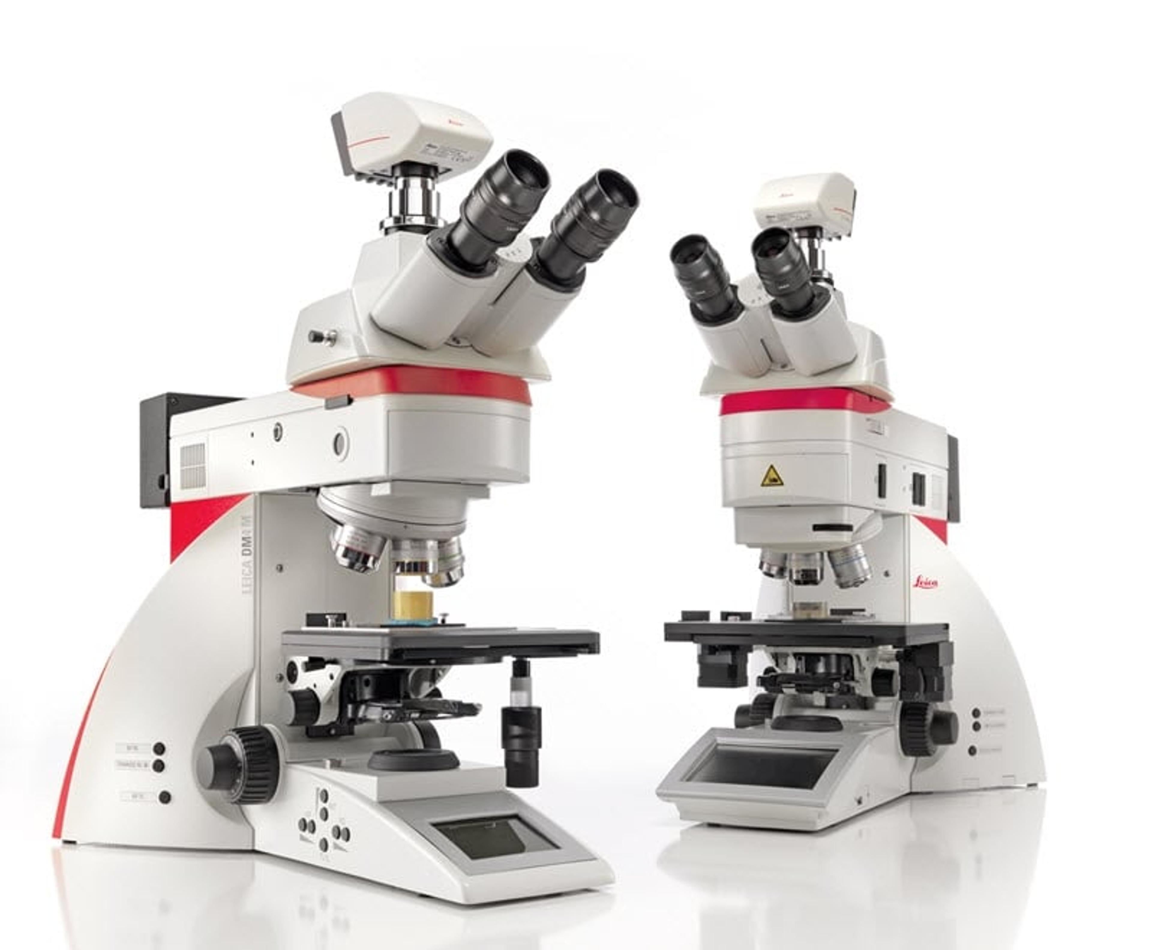
Do you need to image, measure, and analyze similar features across many samples and materials science and analysis? The Leica DM4 M and Leica DM6 M are the Industrial Microscopes for you, whether microscopy novice or professional.
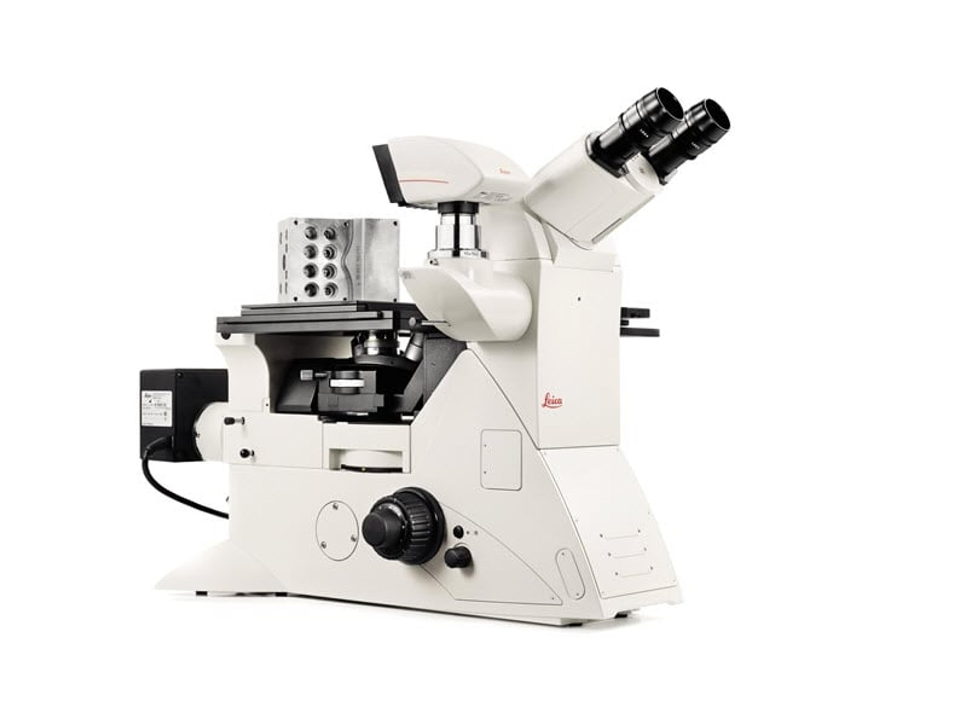
Inverted Microscopes for Industry
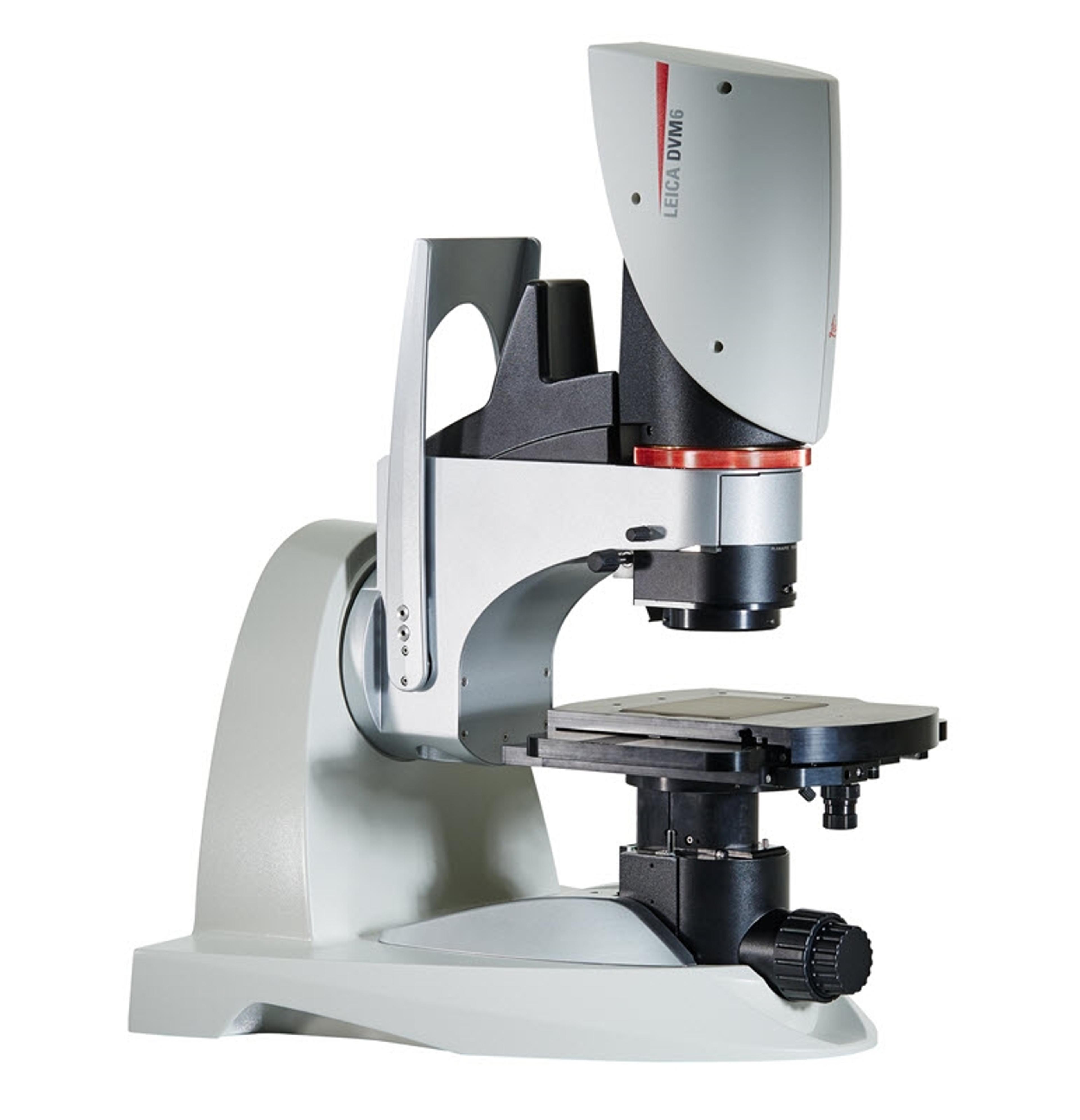
If you work in quality control/assurance, failure analysis, research and development, or in forensics, searching for the detail can take up a lot of your time in microscopy. The DVM6 digital microscope is a fast, reliable and easy to use solution that combines outstanding optics, intuitive operation, and smart software to save you time.
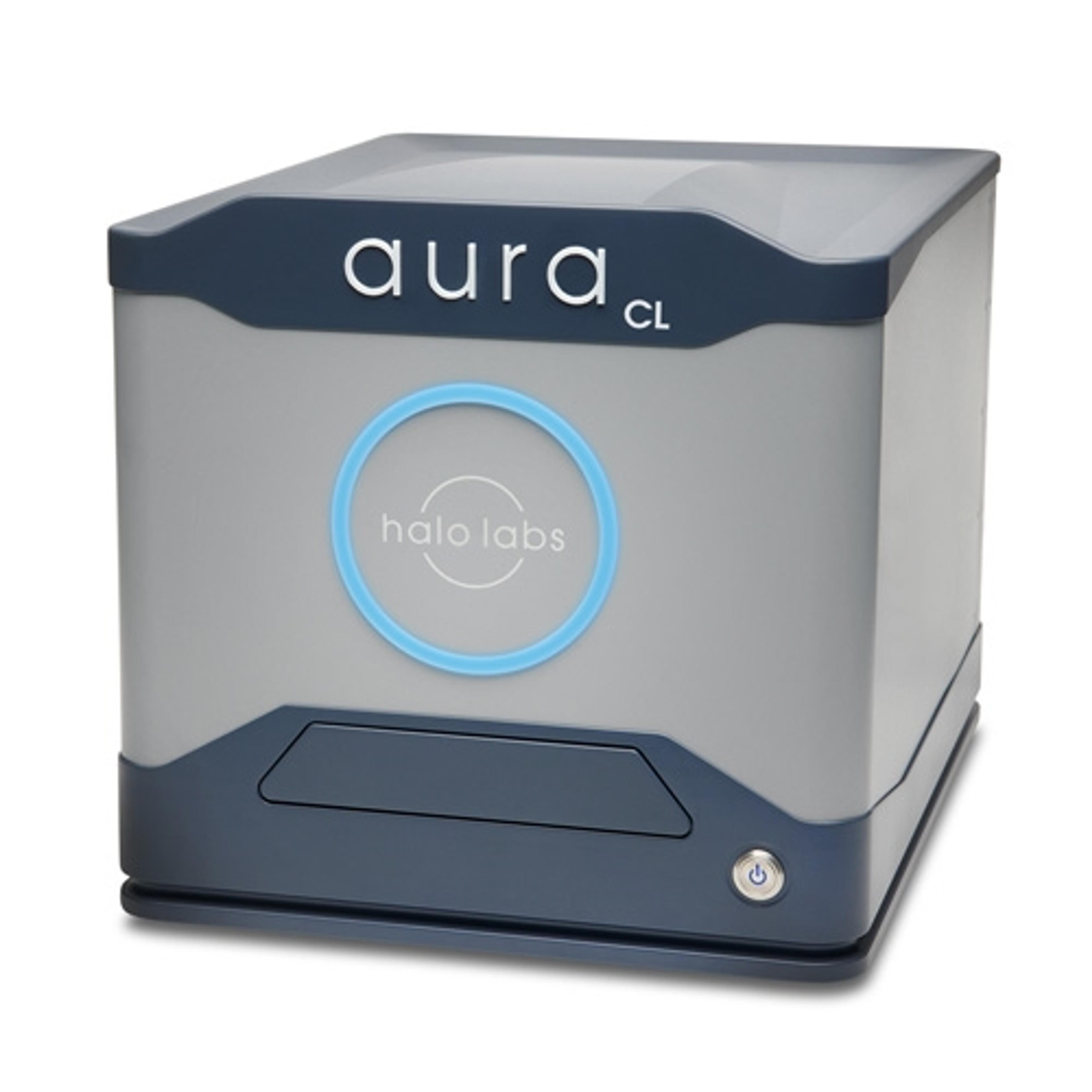
Aura® CL systems couples Fluorescent Membrane Microscopy and Backgrounded Membrane Imaging to help you understand what’s in your cell therapy. It detects, counts, sizes, and IDs cellular aggregates and subvisible particles and ID’s cellular from other particles without any machine learning so you can maximize the purity, safety, and efficacy of your therapeutic.
Aura® is the particle and aggregate detection system that combines Backgrounded Membrane Imaging which images 100% of your sample to give you count, size, and morphological information, with up to 2 channels of Fluorescence Membrane Microscopy. Identify if particles are protein or not with the first FMM channel and determine if your sample contains lipids, hydrophobic entities, or other aggregates of your choice with a second…
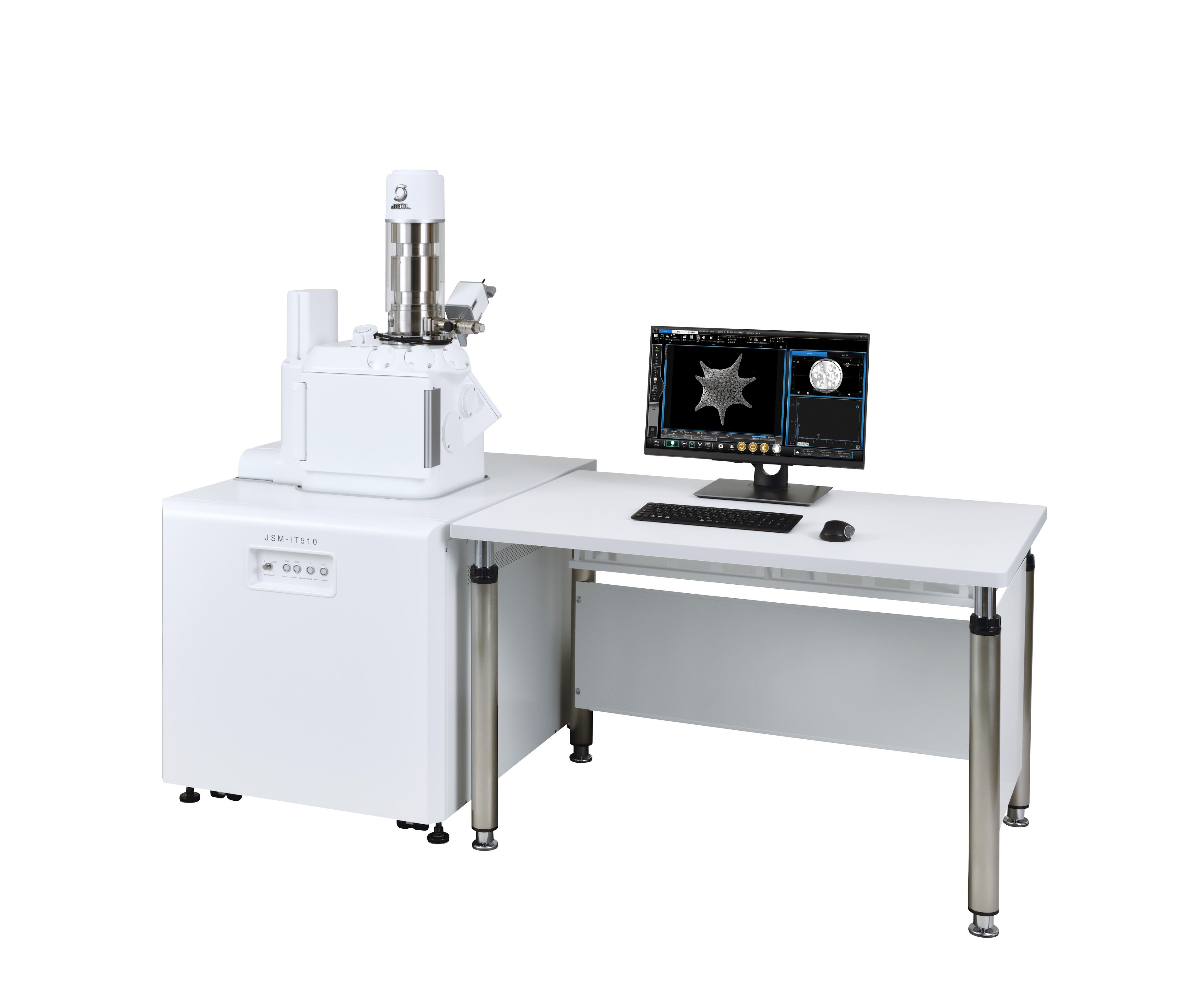
The newest generation InTouchScope™ SEM enhances productivity with Simple SEM automation. Automated image collection at multiple locations and conditions simplifies workflow for the most routine tasks. Embedded Signal Depth display enhances understanding of analytical spatial resolution. Seamless navigation from optical to SEM imaging, Live EDS (both spectrum and X-ray maps) and auto functions from alignment to focus produce…
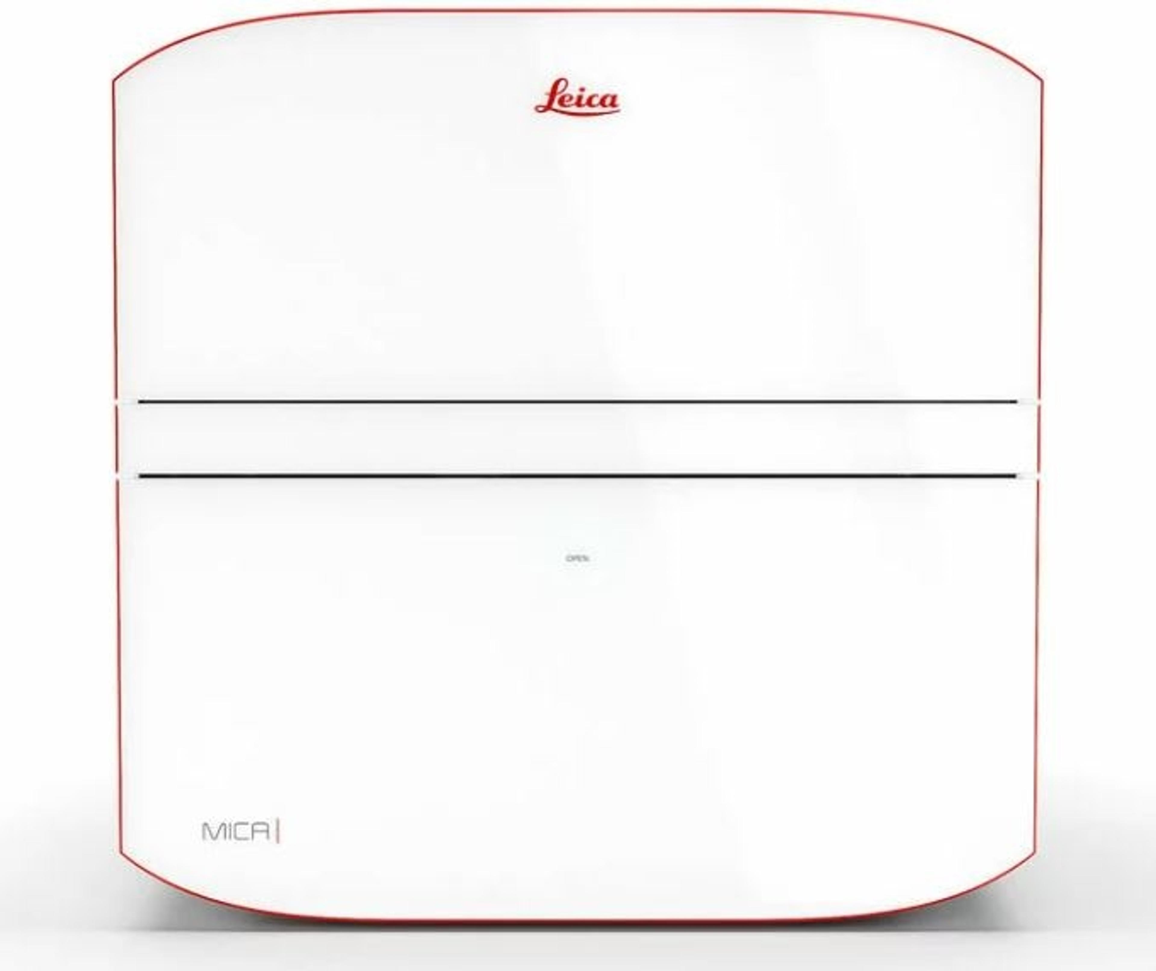
Mica is the world’s first imaging Microhub. More than a highly automated microscope, it unites widefield and confocal imaging in a sample protecting, incubating environment. With the simple push of a button, you have everything you need - all in one place - to supercharge fluorescence imaging workflows and get meaningful scientific results faster.
Leica's cryo-electron tomography workflow solution: boost your productivity with a seamless sample preparation workflow.
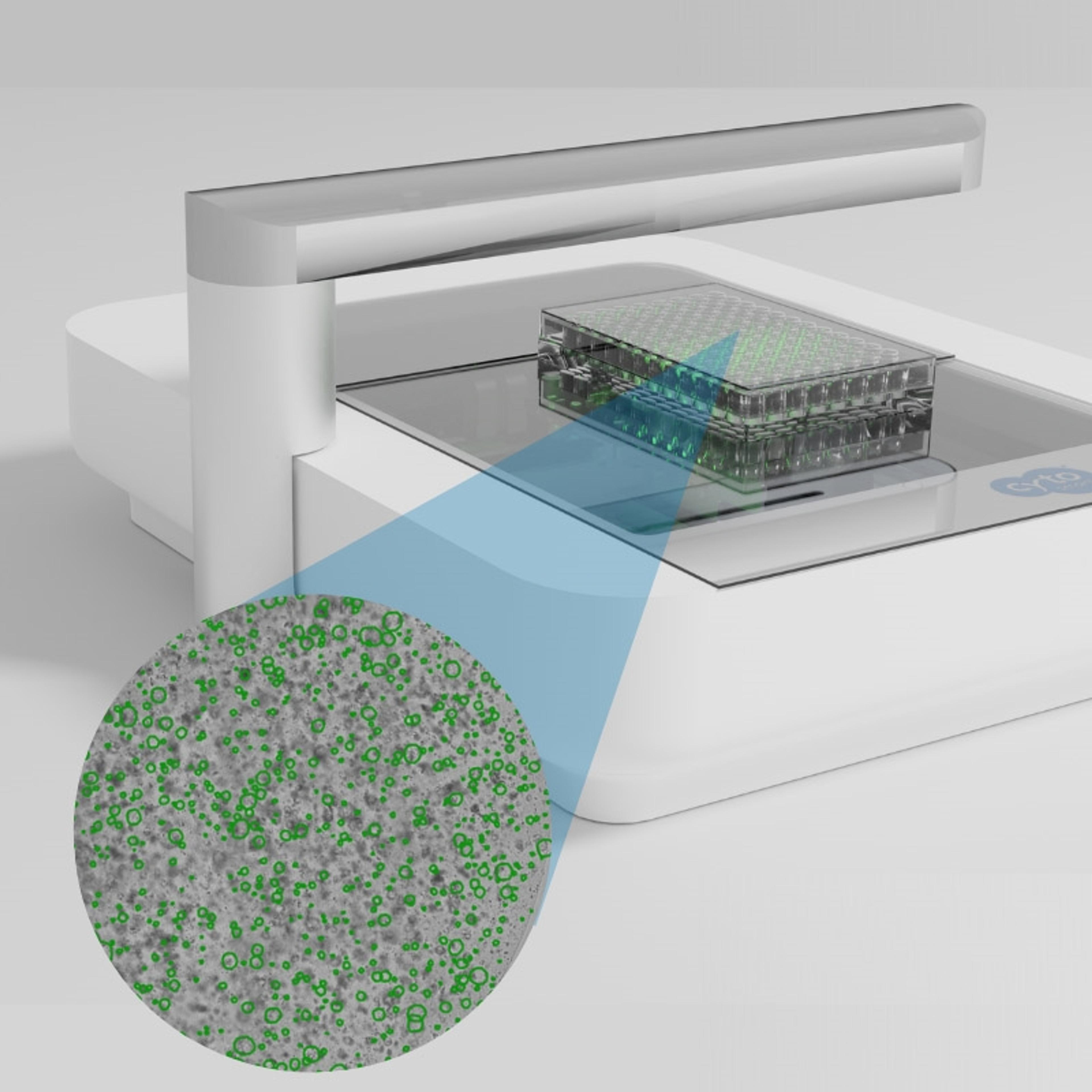
The Organoid Analysis Module for the CytoSMART Omni systems is an advanced machine-learning algorithm for robust organoid quantification, tracking, and analysis.
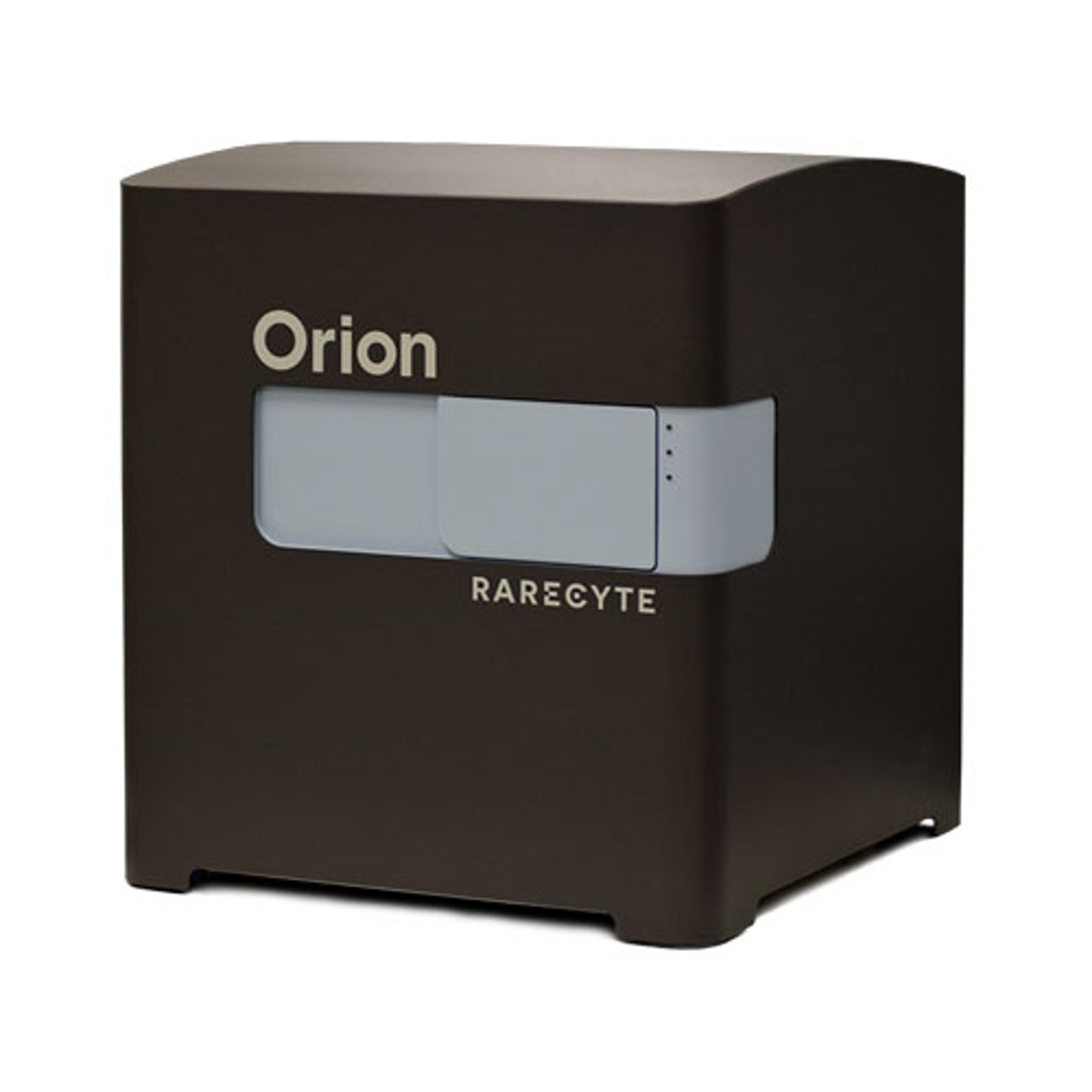
The Orion Platform delivers spatial biology and cellular phenotyping at a speed and resolution needed for translation from research to the clinic. It utilizes single round immunofluorescence staining and imaging of standard FFPE or fresh frozen tissue to deliver subcellular quantitation of biomarker targets across a whole slide in only a few hours. Combining speed and resolution with the additional benefits of industry-stan…
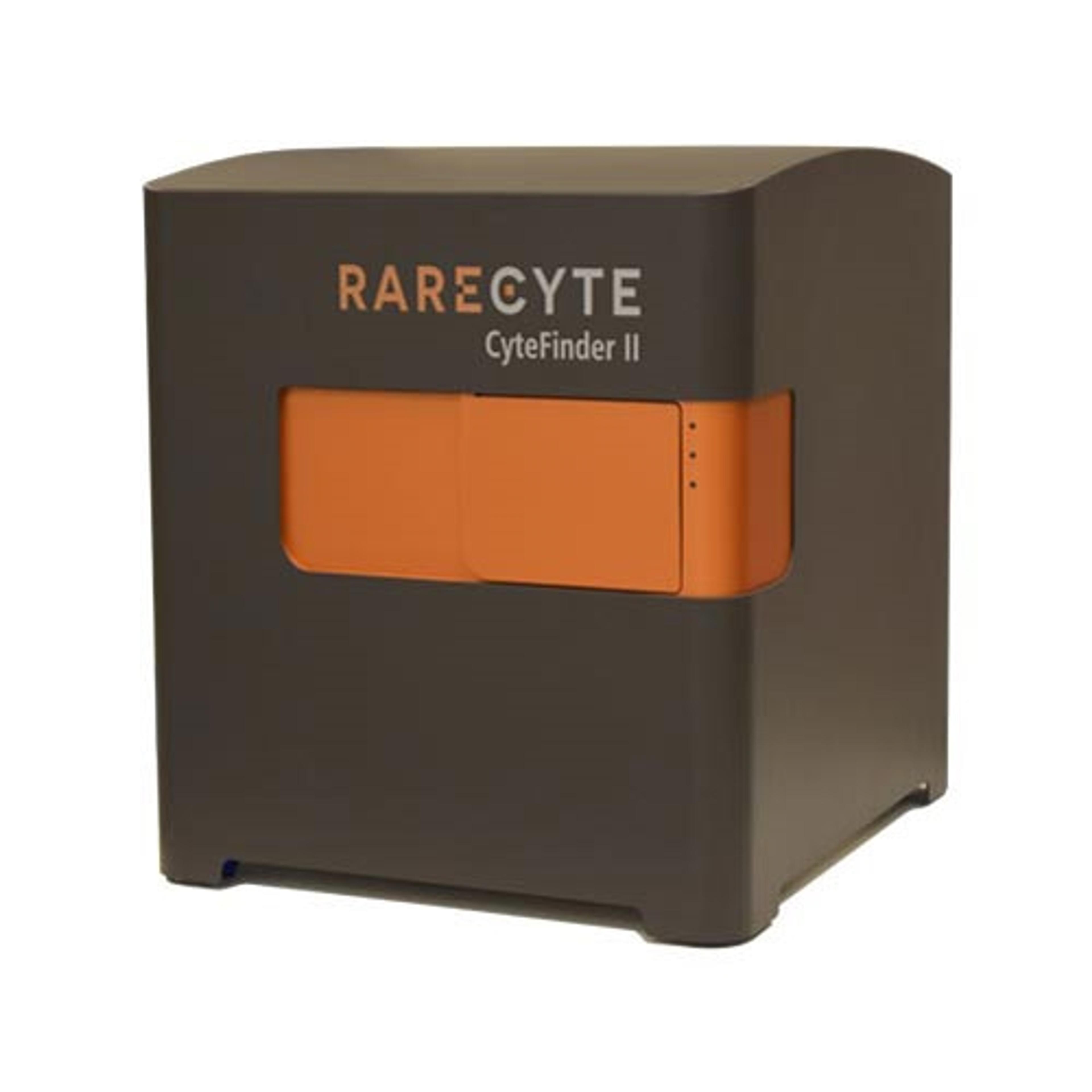
CyteFinder II Instruments are high speed, whole slide imaging systems with options for liquid biopsy analysis and multiplexed tissue imaging. Choose between the CyteFinder II with CytePicker® module for single cell or tissue micro-region retrieval, or the CyteFinder II HT for unattended scanning up to 80 slides at a time. High speed, multi-channel systems optimized for rare-cell detection and analysis Integrated m…
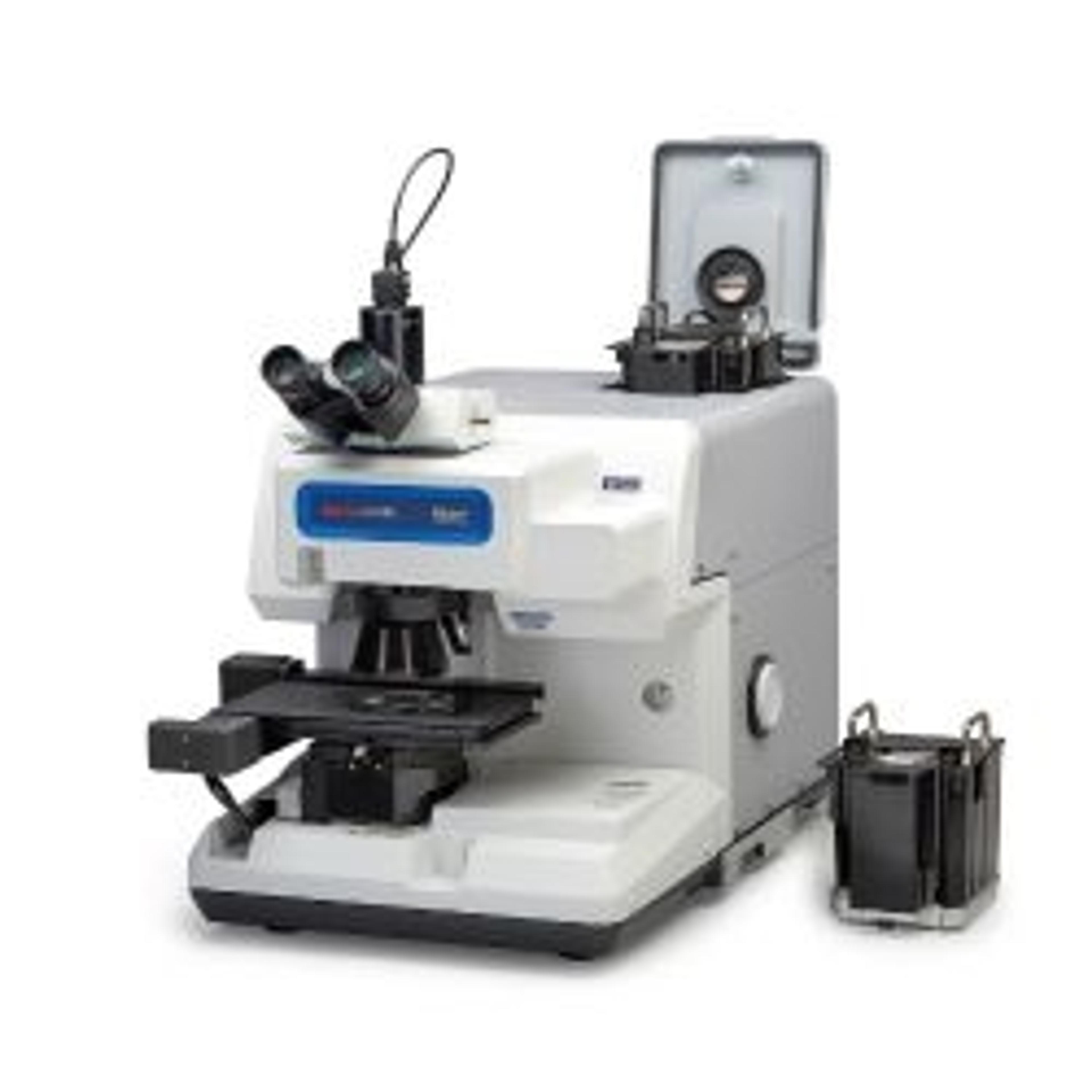
The Thermo Scientific™ Nicolet™ RaptIR+™ FTIR Microscope offers precision and agility to help streamline sample analysis by quickly generating actionable results. Homing in on the intricacies of a sample to find the answer you need is often a lengthy and difficult process. Any amount of time saved while searching for the solution makes a world of difference in delivering results.
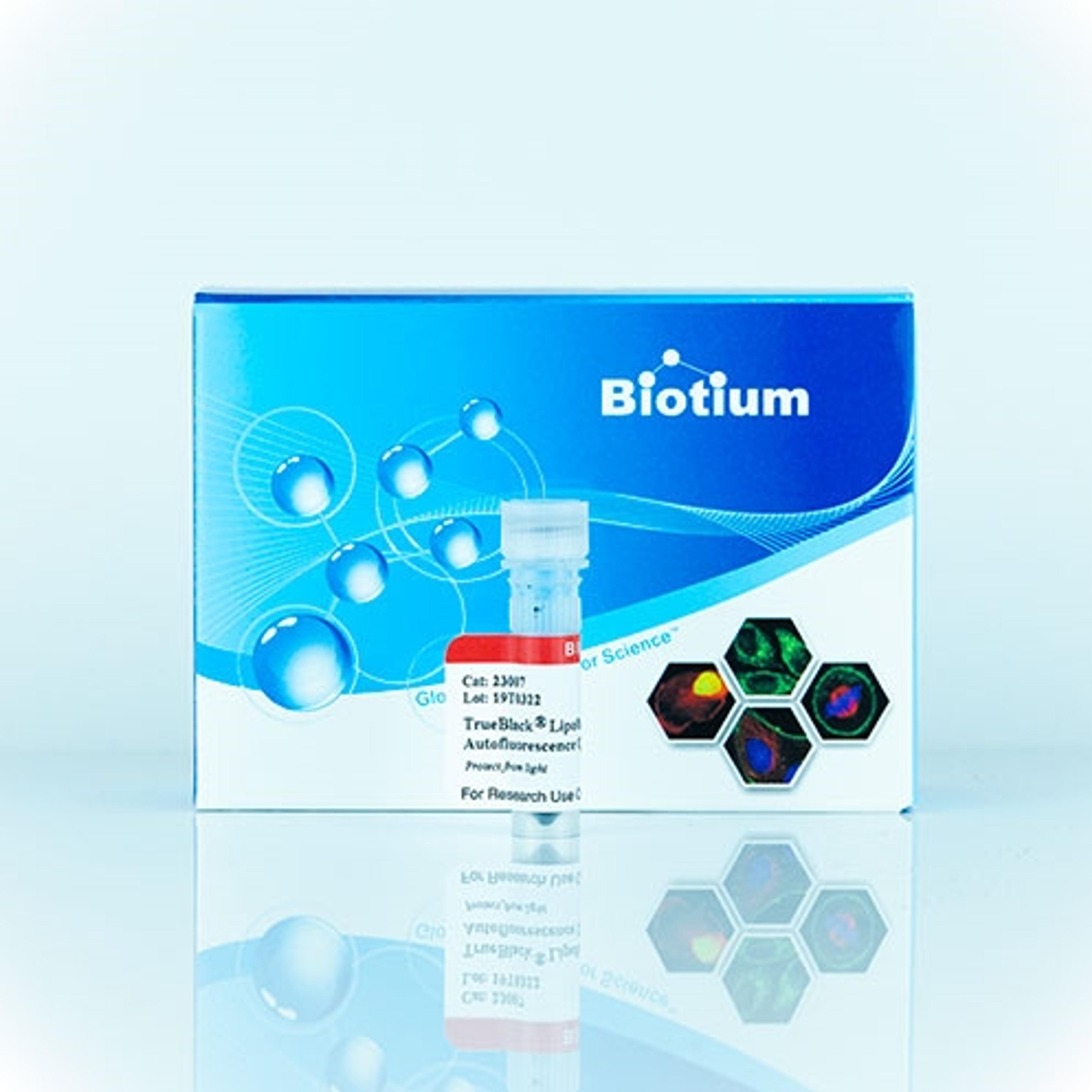
This unique reagent quenches lipofuscin autofluorescence in tissue sections for immunofluorescence staining and can also help to reduce autofluorescence from other sources like extracellular matrix.
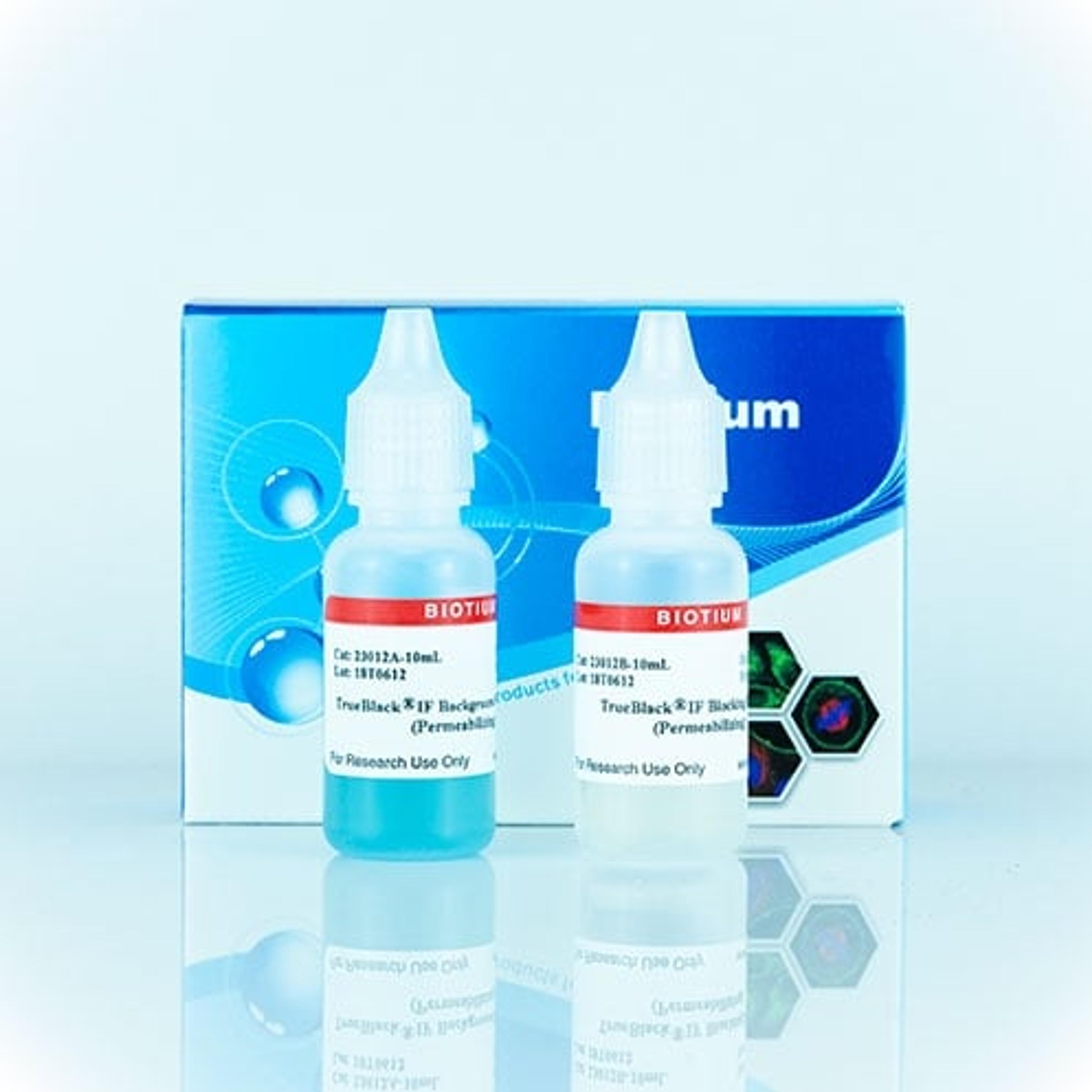
A buffer set that blocks non-specific antibody binding as well as direct interaction of fluorescent dyes on antibodies with cells or tissue sections.
We offer a wide selection of AFM tips of various shapes, tip geometries, materials and coatings, spring constants, frequencies, etc. to fit your specific application. Whether we are manufacturing probes for the budget-conscious shopper or high-end performance applications, meeting a high standard of quality is priority to us. Our dedication to manufacturing probes, coupled with our expertise in AFM, ensures that we are unique…
Aura® GT helps you realize the promise of gene therapies, without using a lot of your viral vector sample throughout the development process. Avoid conditions and formulations that facilitate viral vector aggregates, monitor DNA leakage, and ensure product quality with just 5 µL of sample per data point.
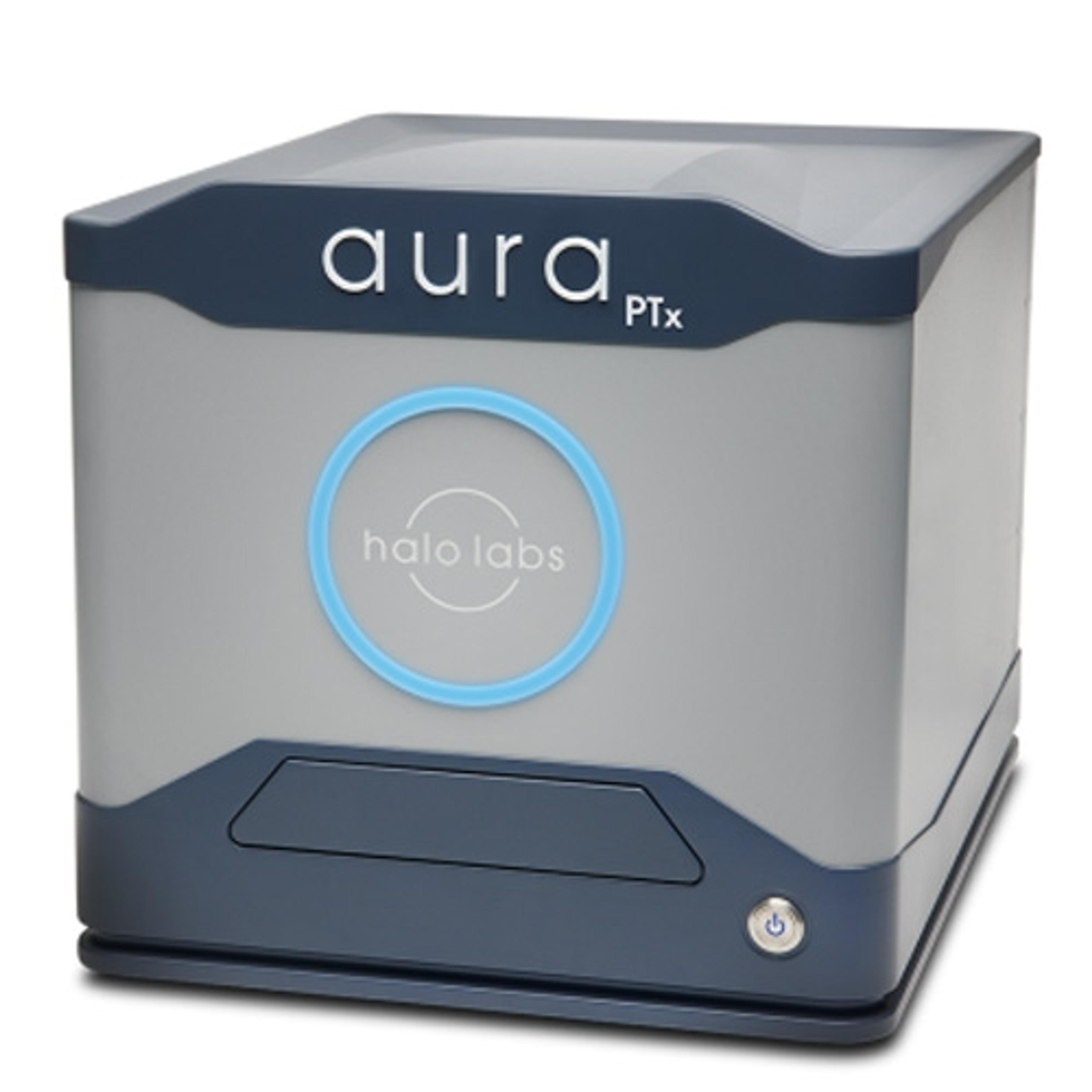
Aura® PTx gets you to your best formulation faster! Backgrounded Imaging (BMI) technology to give you accurate count, size, and morphology information about particles in your sample. Couple that with Fluorescence Membrane Microscopy (FMM), which gives you definitive ID of protein aggregates and an easy way to monitor polysorbate degradation.
With new advances in field of view, sensitivity, wavelength, and sample accommodation, the Ultima 2Pplus delivers an ideal combination of flexibility, resolution, imaging depth and speed, allowing simultaneous imaging, stimulation and electrophysiology protocols with greater efficiency and effectivity. The system is designed specifically for intravital imaging, with fully motorized control of the objective X-Y-Z position, as…