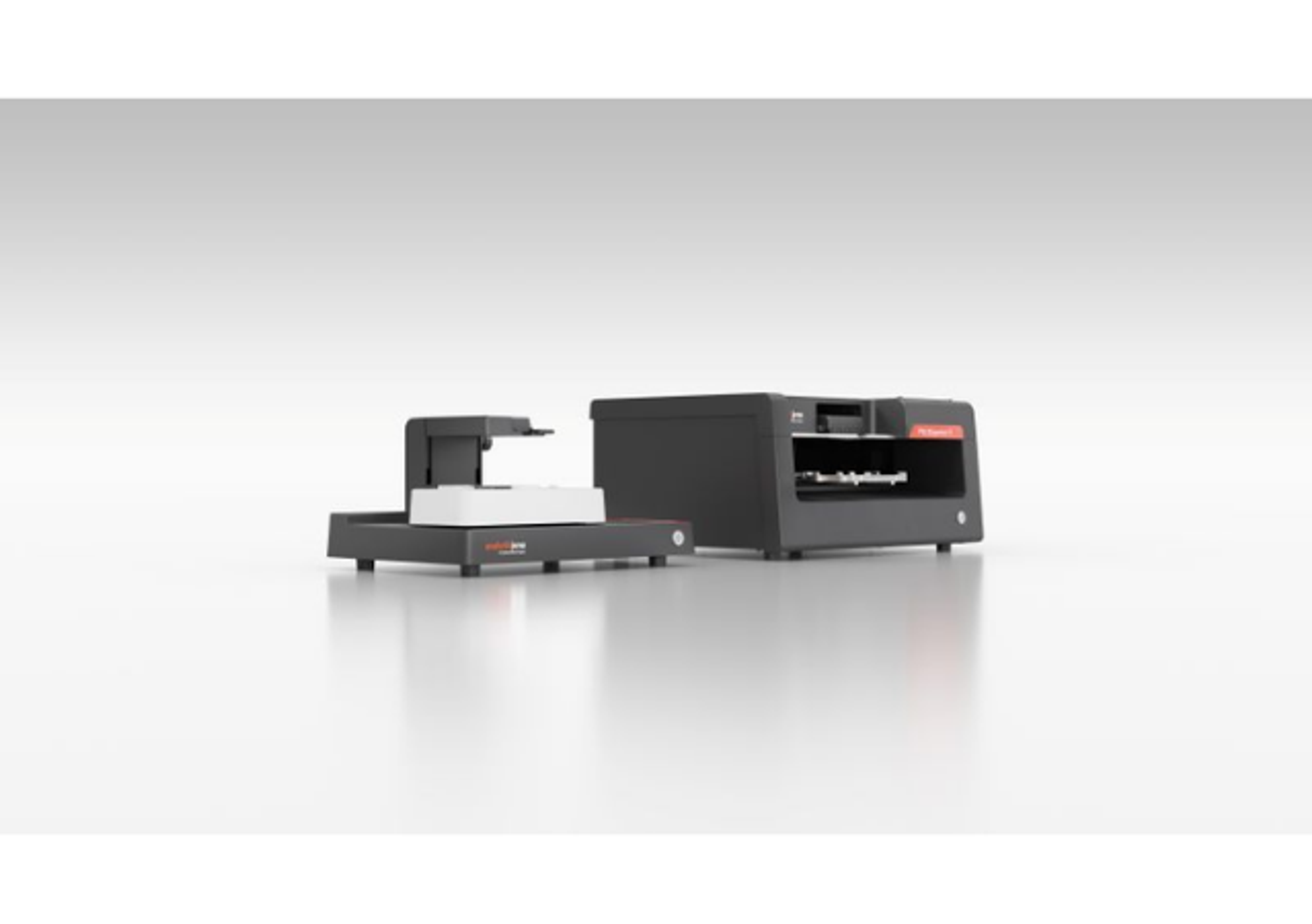
Cell DIVE Multiplexed Imaging Solution
Cell DIVE multiplexed imaging is an antibody-based hyperplexed technique addressing spatial cell biology and function within the tumor microenvironment.
Multiplexed Imaging Solution Cell DIVE

The supplier does not provide quotations for this product through SelectScience. You can search for similar products in our Product Directory.
Cell DIVE multiplexed imaging is an antibody-based hyperplexed technique addressing spatial cell biology and function within the tumor microenvironment.
Deepen your understanding of the tissue microenvironment with multiplexed imaging technology
Cancer is complicated. Immunotherapy, while promising, is still only 30% effective. Researchers need a greater understanding of the cellular architecture of normal and diseased tissues to develop better treatments and more accurately predict disease progression. Multiplexed, or hyperplexed, imaging is the latest technology that clearly visualizes, identifies, and quantifies significant biomarkers. Go from answering the question, “Is it cancer?” to gaining the ability to stratify tumors by cell type, biomarker profile, and specific features.
Cell DIVE is a precise, open multiplexing solution that allows you to follow where the science leads.
- Highly multiplexed imaging without expensive and restrictive commercial panels
- Tissue-preserving imaging without damaging, stripping or photobleaching
- Get more information out of a single experiment with an extra-large imaging area
- Rapid multiplexing at scale with customizable automation
- Accurate cell detection and automatic clustering with up to 30 markers with Aivia AI image analysis software

Spatial proteomic analysis of immune cells in Alzheimer’s Disease human brain using multiplexed imaging and AI-assisted phenotyping
Explore how Cell DIVE multiplexed imaging creates an intricate cellular map of Alzheimer's disease (AD) brain tissue. By visualizing diverse cellular phenotypes—including astrocytes, microglia, AD-specific markers, and various immune cells—we delve into the cellular complexity of AD pathology. AI-powered tools within Aivia are employed to characterize unique neuro-immune interactions, enhancing our understanding of the immune landscape in Alzheimer's.
A focal point of the study is the spatial analysis of β-amyloid plaque neighborhoods, where astrocytes, microglia, and immune cells localize and exhibit specialized expression profiles. By comparing immune cell activity in regions proximal and distal to plaques, we identify distinct spatial immune dynamics within AD brain tissue. The findings highlight how multiplexed imaging, combined with AI-driven analysis, provides critical insights into cellular interplay in AD, offering potential pathways for targeted therapeutic interventions.
Probing the human Alzheimer's cortical section using spatial multiplexing
A critical challenge of Alzheimer’s disease (AD) pertains to understanding the spatial microenvironment of AD brain tissue, specifically mapping the different neuronal cell types and expression markers associated with the disease. This type of spatial profiling is crucial for uncovering cellular relationships that could provide insights into disease mechanisms and potential therapeutic strategies. This application note from Leica Microsystems sets out to address these challenges and enhance understanding of AD by utilizing the Cell DIVE multiplexed imaging and AI-powered spatial analysis workflow. This workflow, combining cell-type specific markers, multiplexed tissue imaging, and AI-guided analysis will provide researchers with a deeper understanding of the spatial heterogeneity in the AD brain, paving the way for novel insights.
Empower spatial biology research with Cell DIVE
Explore how Cell DIVE is designed to transform spatial biology research with its advanced multiplexing platform. This innovative solution can enhance the analysis of complex tumor microenvironments by enabling high-resolution imaging of multiple biomarkers. Overcome traditional limitations with Cell DIVE's iterative staining techniques and customizable protocols, and unlock new insights into cancer progression and immuno-oncology.
Spatial proteomic analysis of Alzheimer's disease human brain using multiplexed imaging
Find out how iterative staining and imaging of human adult brain tissues with Cell Signaling Technology antibodies and the Cell DIVE multiplexed imaging system enable spatial characterization of an Alzheimer's Disease human brain.
A comprehensive workflow for large multiplexed 2D images
The Cell DIVE system, developed by Leica Microsystems, allows precise, highly multiplexed imaging of tissue sections without damaging them, preserving their integrity. Leica Microsystems presents a comprehensive solution for obtaining deeper spatial insights through large multiplexed 2D imaging. The workflow supports rapid and scalable multiplexing, capable of analyzing over 60 biomarkers on a single tissue section using Aivia AI image analysis software. This software enables accurate cell detection, automatic clustering, and AI-powered phenotyping, making it easier to interpret complex data from large imaging areas.
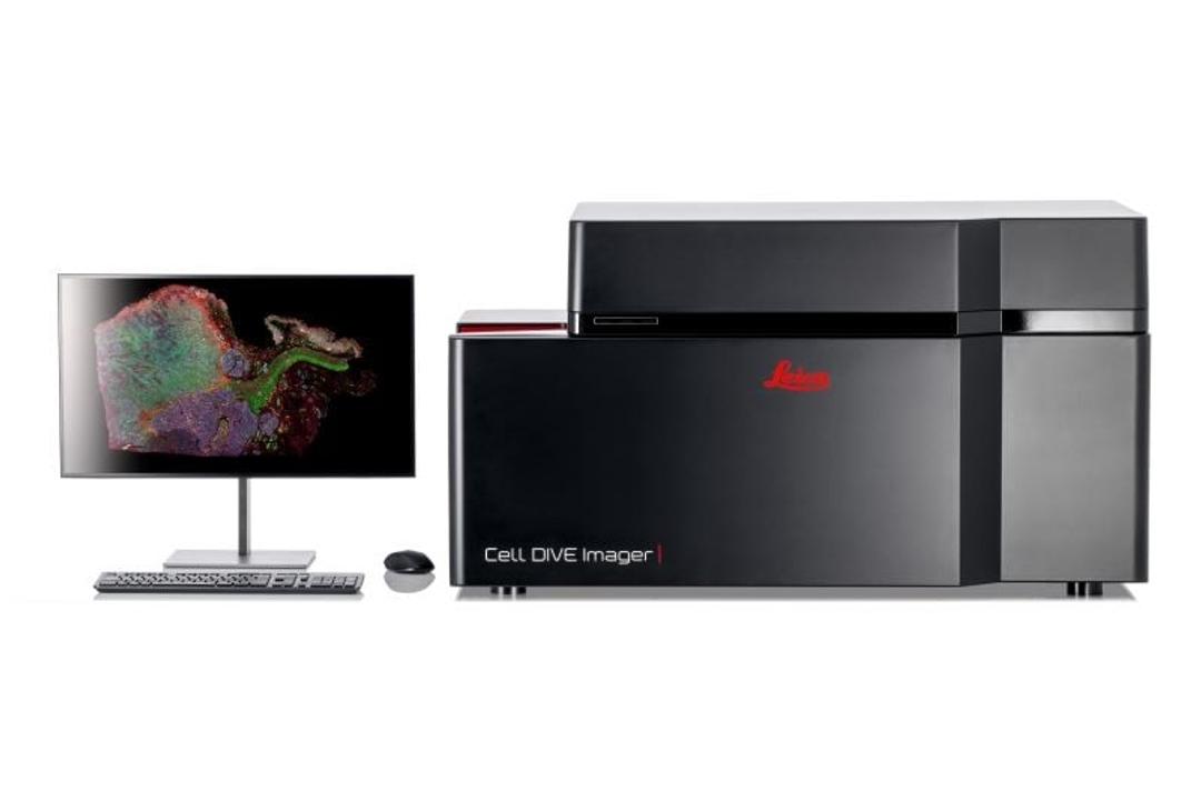
Cell DIVE open multiplexed imaging solution
The Cell DIVE system integrates software, hardware, and laboratory steps to iteratively stain and image tissues, allowing the detection of over 60 biomarkers from a single tissue section. Leica Microsystems introduces a comprehensive and adaptable multiplexed imaging solution designed to map cellular phenotypes, expression profiles, and morphology within tissue samples. The system features 350+ validated antibodies, scalable automation options, and a patented workflow that ensures reproducibility and precision.
Tumor-immune landscape exploration using AI
Cancer tissues are complex in organization. Beyond the tumor itself, the tumor microenvironment is often composed of many tissue and cell types, each with potential significance for cancer progression. Spatial biology approaches that allow the researcher to visualize and position many biomarkers in their organismal context offer an efficient way to dig deep into hard questions in cancer biology. Leica Microsystems presents a spatial biology workflow to dive into a colon adenocarcinoma tissue using Cell DIVE to visualize and Aivia software to study the cells within the tumor microenvironment.
Spatial landscape of the tumor microenvironment in pancreatic ductal adenocarcinoma using multiplexed imaging and AI based analysis
Pancreatic tumors are highly heterogeneous, often with more aggressive regions that are responsible for invasion and metastasis. These aggressive pancreatic ductal adenocarcinoma cells (PDAC) undergo epithelial to mesenchymal transition and create subregions in the tumor that evade treatments and provide a critical support niche for continued tumor growth and metastasis. In this scientific poster, Leica Microsystems presents a study in which biomarkers are utilized to probe tumor heterogeneity in pancreatic ductal adenocarcinoma. The spatial biology solution Cell DIVE™ and proprietary Spatial AI analytics software enabled the use of dozens of biomarkers to computationally reduce tumor heterogeneity and to spatially define cells in the microenvironment with hypoxic and normoxic regions of PDAC and in normal pancreas.

The power of spatial biology: A microscopy guide
Location is key to understanding biological mechanisms, from the inner workings of subcellular components to how cells form and interact across normal and diseased tissues.
Many application areas are seeing a growing trend toward spatial biology, which uses transcriptomics, imaging, and other approaches to put dissociated cellular information into spatial context.
In this free eBook, explore key microscopy techniques, across the spatial biology workflow, including:
- Multiplexing
- Laser microdissection
- Removing the blur to observe fine details
- Super-resolution microscopy
- Ultra-structural context
- AI-enabled spatial analysis
Plus, we look "under the microscope" at how these can be applied to study a wide range of spatial biology questions and gain expert insight into the imaging solutions designed to meet a variety of different research needs and priorities.