Aivia AI Microscopy Image Analysis Software
Access the future of AI microscopy. Using state-of-the-art, AI-first software architecture, Aivia is a uniquely innovative and complete 2-to-5D image visualization, analysis and interpretation platform designed for the reliable processing and reconstruction of highly complex images in just minutes. Aivia provides powerful 2D & 3D spatial relational analysis for objects across diverse types, morphological complexities, and hie…
Aivia AI microscopy software

The supplier does not provide quotations for this product through SelectScience. You can search for similar products in our Product Directory.
Take your image analysis further than ever before
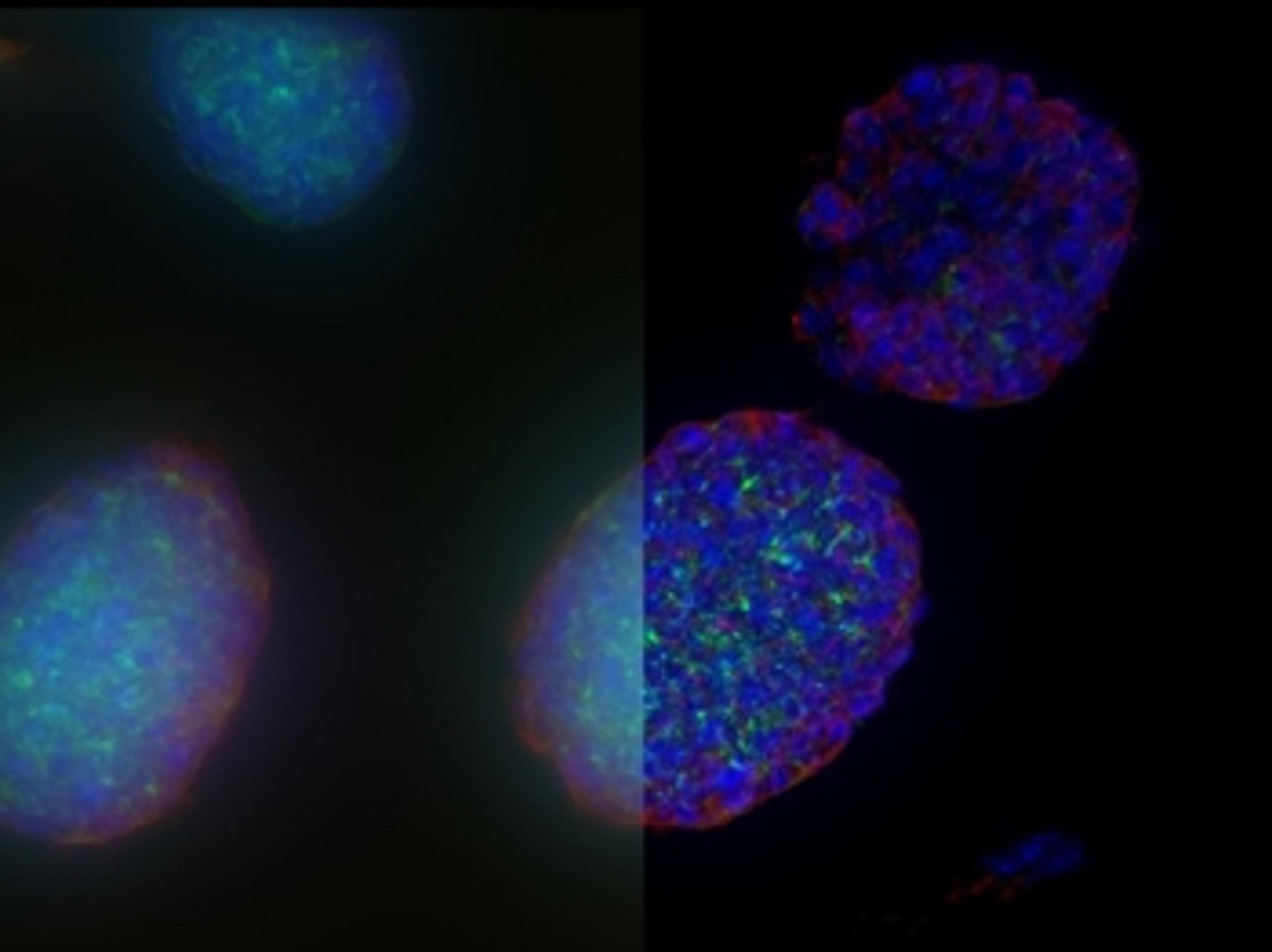
Subjectivity of analysis and poor reproducibility are key hurdles to be overcome for biological image analysis. Standard segmentation can lead to sub-standard results and require substantial manual curation which is subject to human error.
Aivia changes all of that.
Using state-of-the-art, AI-first software architecture, Aivia is a uniquely innovative and complete 2-to-5D image visualization, analysis and interpretation platform designed for the reliable processing and reconstruction of highly complex images in just minutes.
- Make AI-enhanced image analysis accessible for all - with no computer science expertise required
- Leverage machine learning capabilities to generate robust and reproducible segmentation results
- Realize powerful and fast 2-5D visualization and analysis to unleash the value of your data - all within a single platform

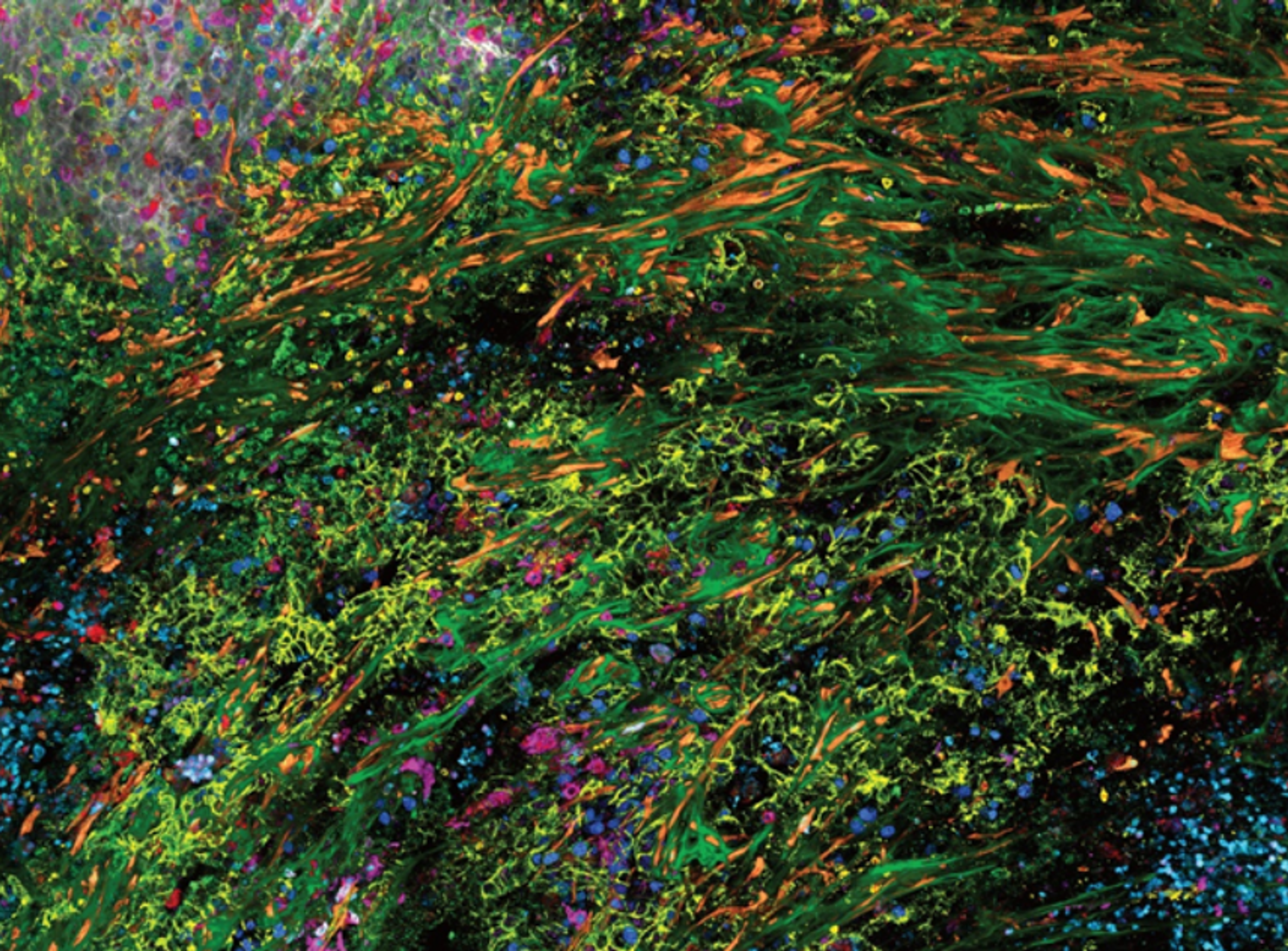
Probing the human Alzheimer's cortical section using spatial multiplexing
A critical challenge of Alzheimer’s disease (AD) pertains to understanding the spatial microenvironment of AD brain tissue, specifically mapping the different neuronal cell types and expression markers associated with the disease. This type of spatial profiling is crucial for uncovering cellular relationships that could provide insights into disease mechanisms and potential therapeutic strategies. This application note from Leica Microsystems sets out to address these challenges and enhance understanding of AD by utilizing the Cell DIVE multiplexed imaging and AI-powered spatial analysis workflow. This workflow, combining cell-type specific markers, multiplexed tissue imaging, and AI-guided analysis will provide researchers with a deeper understanding of the spatial heterogeneity in the AD brain, paving the way for novel insights.
Tumor-immune landscape exploration using AI
Cancer tissues are complex in organization. Beyond the tumor itself, the tumor microenvironment is often composed of many tissue and cell types, each with potential significance for cancer progression. Spatial biology approaches that allow the researcher to visualize and position many biomarkers in their organismal context offer an efficient way to dig deep into hard questions in cancer biology. Leica Microsystems presents a spatial biology workflow to dive into a colon adenocarcinoma tissue using Cell DIVE to visualize and Aivia software to study the cells within the tumor microenvironment.
Access 3D high-plex spatial information across scales
Leica Microsystems unveils next generation STELLARIS with SpectraPlex
AI-powered software allows deeper spatial insights from 3D images
Aivia's improved deep learning model from Leica Microsystems accelerates cell detection by up to 78%
Seeing in 3D: How cell culture imaging just got easier
Dr. Falco Krüger discusses the challenges of 3D cell culture imaging, enabling technologies and the future of AI microscopy