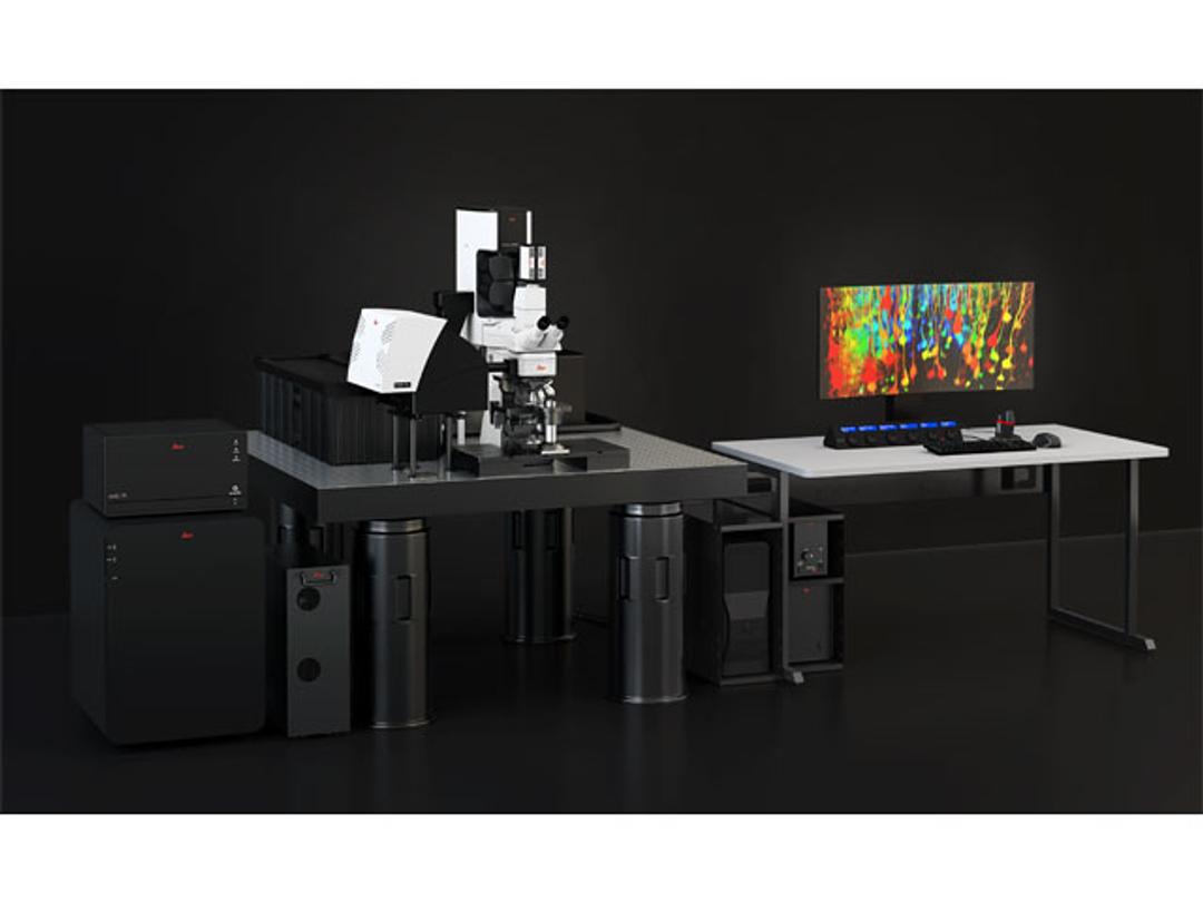
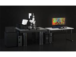
STELLARIS DIVE Multiphoton Microscope
DIVE and STELLARIS have now merged to provide you with the power of flexible multicolor multiphoton imaging.

The supplier does not provide quotations for this product through SelectScience. You can search for similar products in our Product Directory.
Multicolor Multiphoton gets you closer to the truth
The combination of multiple distinct fluorescent labels is increasingly used to study dynamic interaction and spatial relationships among cells and molecules. The goal is to understand a myriad of complex biological events, such as cellular connectivity, cell phenotyping, protein interaction or co-expression, and co-localization. Scaling up such studies to whole organs or tissues requires suitable large-volume multicolor microscopy methods. DIVE and STELLARIS have now merged to provide you with the power of flexible multicolor multiphoton imaging. Furthermore, you can expand the potential of your experiments with additional label-free imaging capabilities.
Seamlessly integrated into the confocal software interface, STELLARIS 8 DIVE offers the benefits of high speed and excellent navigation so that dynamic processes in complex samples can be studied with ease.
STELLARIS 8 DIVE – A rainbow of possibilities for your research
Brochures

STELLARIS Confocal Microscope
Leica Microsystems’ family of confocal microscopes – STELLARIS 5 and STELLARIS 8 – takes confocal imaging to a new level. Now you can see more, discover more, and do more than ever before, with STELLARIS.
Quantify meaningful changes in the metabolic status of single cells
Metabolic imaging based on fluorescence lifetime provides insights into the metabolic dynamics of cells, but its use has been limited as expertise in advanced microscopy techniques was needed. Now, STELLARIS 8 DIVE FALCON makes metabolic imaging accessible to every scientist, thanks to the integration of phasor analysis to easily use fluorescence lifetime information.
FLIM phasor analysis is a robust analytical method to generate a visual fingerprint of the lifetime of a fluorophore that can be mapped back onto an image to highlight function in cells. Download this application note for a summary of studies that use FLIM phasor analysis to reveal connections between cellular metabolism and pivotal cellular functions in health and disease.
In vivo multi color imaging of a confetti breast cancer mouse model
In this application note, from Leica Microsystems, find out how to gain deep insights into the development of cancer using spectrally tunable 2-photon microscopy.