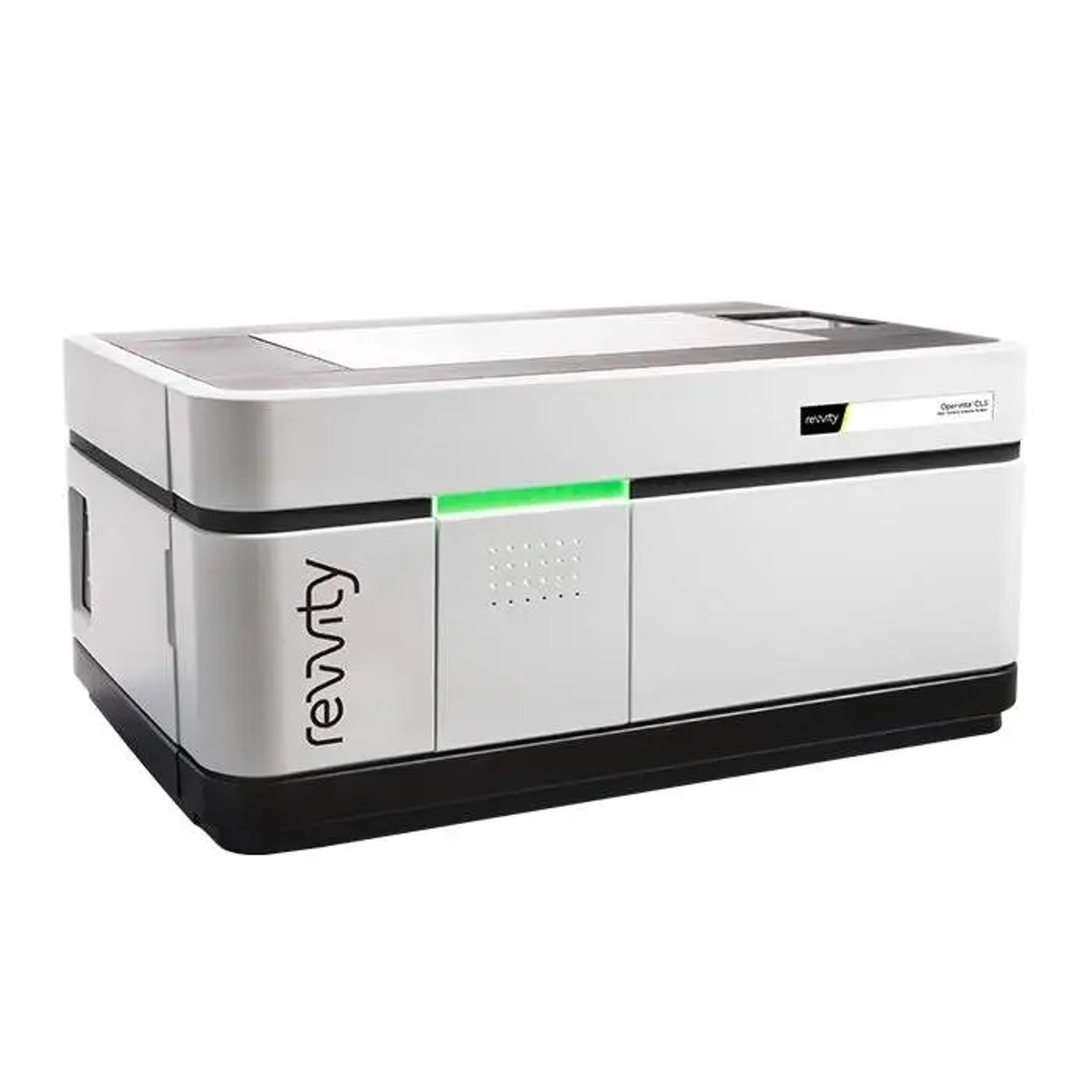
Operetta CLS™ High-Content Analysis System
Uncover deep biological understanding in your everyday assays and innovative applications using the Operetta CLS™ high-content analysis system. Featuring a unique combination of technologies, the system delivers all the speed, sensitivity and resolution you need to reveal fine sub-cellular details. And with our simple, powerful Harmony software, Operetta CLS™ lets you find even subtle phenotypic changes.


The supplier does not provide quotations for this product through SelectScience. You can search for similar products in our Product Directory.
Great quality instrument.
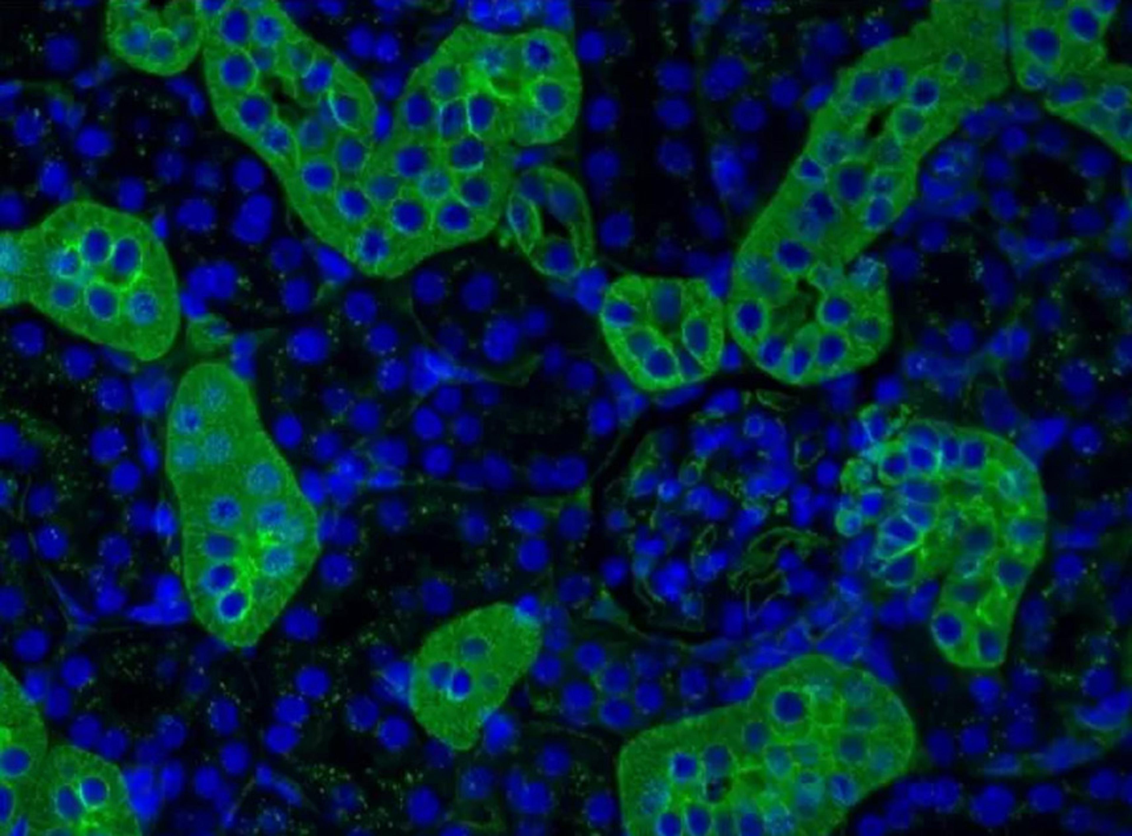
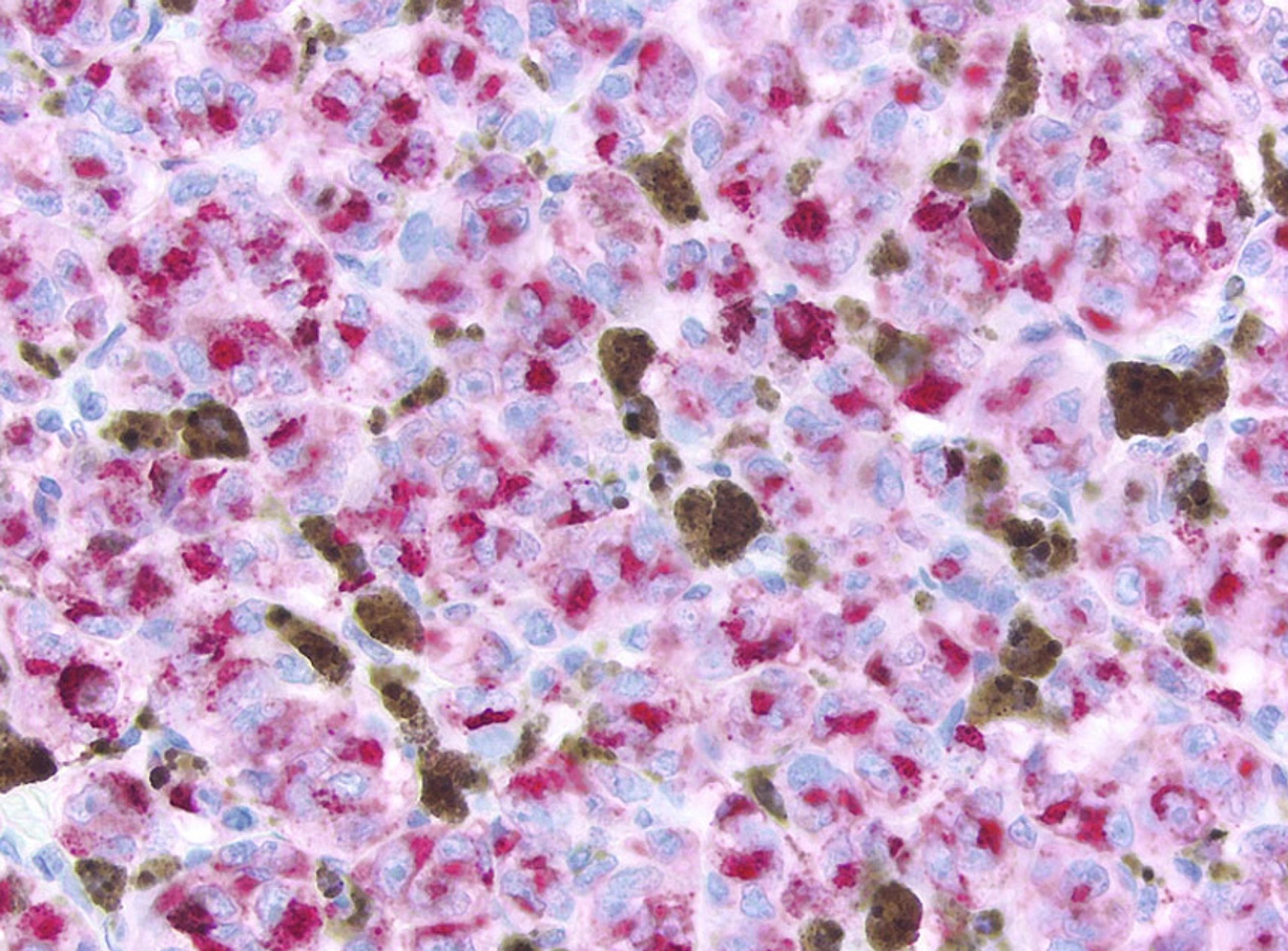
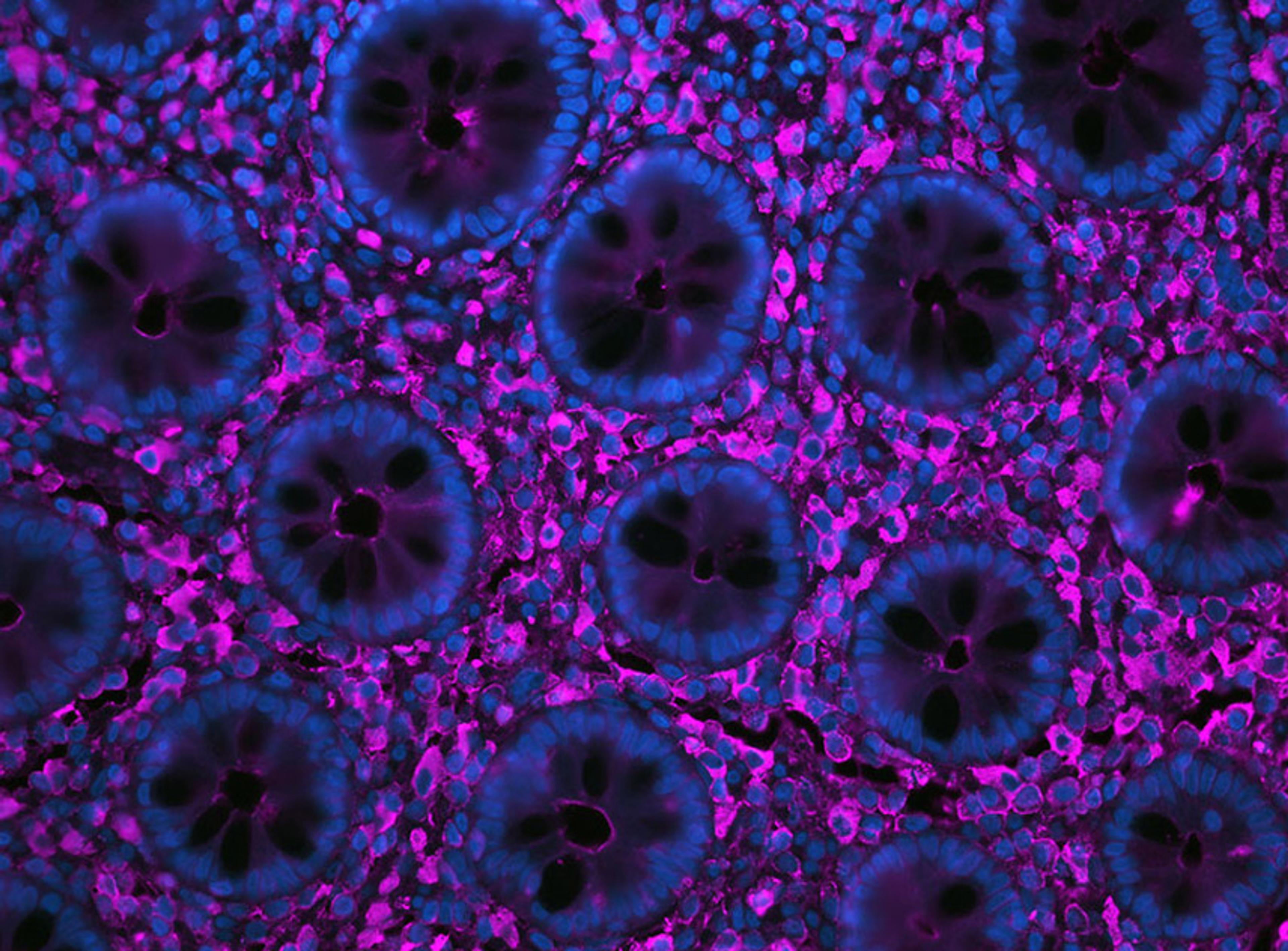
High content screening
Achieved high quality confocal images with water objective and powerful data analysis software.
Review Date: 20 Jun 2020 | Revvity
The Operetta CLS system combines speed and sensitivity with the powerful and intuitive data analysis you’ve come to trust from the Operetta platform. The all-new Operetta CLS delivers everything you need from the high-content analysis. What’s more, the Operetta CLS system is part of our comprehensive HCS workflow – everything from HCS systems and microplates to automation and informatics for every application. All from one knowledgeable, trusted vendor. Put that together with our Harmony® high-content imaging and analysis software – the easy-to-learn, easy-to-use software that empowers biologists to do their own analysis – and you have everything you need to run your every day (and complex) analyses right away.
Applications
From everyday assays to more demanding applications, the Operetta CLS high-content analysis system delivers just the right combination of flexible excitation, sensitive optics, and advanced software features to enable you to gain deeper biological insight from all your critical applications.
Fixed-cell assays
- Up to eight high-power excitation sources and user-accessible emission filters enable maximum flexibility of fluorescent stains and labels, plus the system features widefield and spinning-disk fluorescent imaging.
Live-cell assays
- The spinning disk confocal optics and synchronized LED illumination provides stable excitation and minimize phototoxicity and bleaching for meaningful live-cell assays. You can also choose brightfield or digital-phase contrast imaging modes.
Complex cellular models
- The large-format sCMOS camera with water-immersion objectives provides sensitivity and high resolution, while advanced software helps you address the imaging and analysis challenges presented by complex cellular models.
Advanced assays
- FRET is a powerful tool for investigating conformational changes and protein-protein interactions. The Operetta CLS system’s sensitive imaging and dedicated analysis tools for ratiometric imaging, facilitate robust results.
Phenotypic fingerprinting
- The Operetta CLS combines high-resolution imaging with advanced software tools to help create robust phenotypic fingerprints of the subtle differences at the core of successful phenotypic assays.
Your Complete HCS Workflow
- Get superior results with our PhenoPlate™ microplates for high content screening
- Use together with our PhenoVue™ cellular imaging reagents including cell painting kits
- Improve throughput and productivity by automating your Operetta CLS system, or benefit from our automated cellular workflows and drug discovery workflows
- Export results automatically into Signals Image Artist™ Image Analysis and Management platform, so you can access, re-analyze, store, and share all your cell image data from Operetta CLS and other HCS systems
- Combine with Signals VitroVivo for powerful multivariate statistical methods and unsupervised machine learning techniques so you can identify parameters that best define distinct cellular fingerprints

Quantitative Analysis of 3D Microtissue Growth and Biomarker Intensity
In this application note, a high content screening application to analyze different characteristics of 3D microtissues with the Operetta® High Content Imaging System is presented. The sensitivity of tumor microtissues to treatment with cytotoxic drugs was visualized and quantified several cancer-associated biomarkers by staining the microtissues with different in vivo NIR agents.
Cytotoxicity Studies on 3D Primary Liver Microtissues
This application note presents a proof of concept study to demonstrate the feasibility of a 4-color toxicity assay using living Troglitazone and Staurosporine treated primary human liver microtissues. In this assay, the combination of High Content Imaging with Multimode Detection provides complementary readouts for assessing phenotypic changes and potential toxicological pathways beyond general cytotoxicity markers.
The Benefits of Automated Water Immersion Lenses for High-Content Screening
High-content screening (HCS) combines automated microscopy and multiparametric analysis of cellular events. Most HCS assays are based on fluorescence detection, whereby air is the typical objective medium. However, water has a higher refractive index and this application note demonstrates that water immersion objectives can provide significantly brighter images from typical HCS samples compared to air objectives.
Automation of 3D Spheroid Production, Cell Culture and Analysis
Reproducible cell seeding and reliable formation of similar-sized 3D microtissues are essential to enable collection of robust data when adapting more biologically relevant, complex 3D models to high-throughput workflows. This application note demonstrates a standardized, cost effective and automated means of producing and characterizing 3D spheroid microtissues, using the InSphero GravityPLUS™ Hanging Drop System and the Zephyr® G3 automated workstation to automate the liquid handling steps involved in microtissue seeding and transfer to the GravityTRAP™ Plate for long-term culture and imaging
PerkinElmer Drug Discovery Screening Solutions
Watch this video to find out about high throughput drug discovery screening solutions from PerkinElmer, including; state-of-the art imaging and detection instruments, assay technologies and reagents, and versatile automation systems.
How PerkinElmer is helping to combat COVID-19
It's comprehensive SARS-COV-2 offerings span RT-PCR, high throughput RNA extraction, automation, ELISA and lateral flow based serology testing.