Shamrock 500 Spectrographs
The Andor Shamrock SR-500i imaging platform is the latest addition of the Andor family of spectrographs based on Czerny-Turner optical design. The optimized optical design provides exceptional performance for multi-track Spectroscopy.The Shamrock 500i is available as a pre-aligned, pre-calibrated camera/spectrometer detection solution allowing for seamless integration in user set-up. The Spectroscopy-dedicated software interfa…


The supplier does not provide quotations for this product through SelectScience. You can search for similar products in our Product Directory.
The Andor Shamrock SR-500i imaging platform is the latest addition of the Andor family of spectrographs based on Czerny-Turner optical design. The optimized optical design provides exceptional performance for multi-track Spectroscopy.
The Shamrock 500i is available as a pre-aligned, pre-calibrated camera/spectrometer detection solution allowing for seamless integration in user set-up. The Spectroscopy-dedicated software interface provides an interactive and intuitive platform for simultaneous detector(s) and spectrograph real-time control.

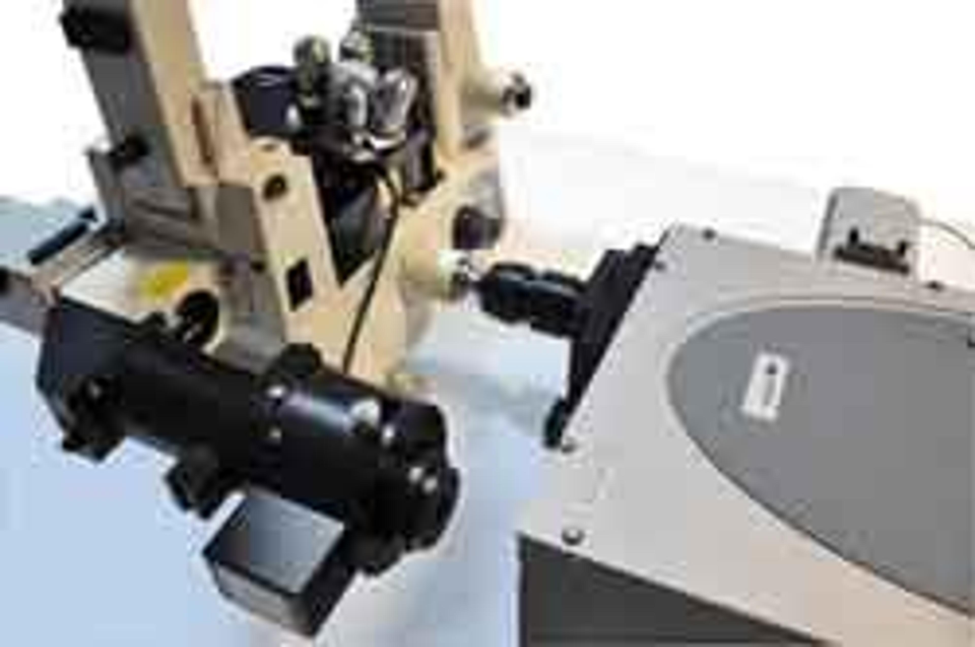
Using a Surface-Forces-Apparatus to Measure Force-Distance Profiles Across Confined Ionic Liquids
In this application note, the interfacial forces between two atomically smooth mica surfaces across the ionic liquid 1-ethyl-3 methylimidazolium-bis trifluorosulfonylimide (EMIM BTI) are measured using an SFA2000 from SurForce LLC. For about 40 years now, the Surface-Forces-Apparatus is a well-established technique to measure distances, normal or friction forces between interacting surfaces across fluid media. It was the first method to directly measure and quantify nowadays well-known interfacial phenomena like Van-derWaals forces, hydrophobic forces, hydration repulsion and electric-double-layer interactions.
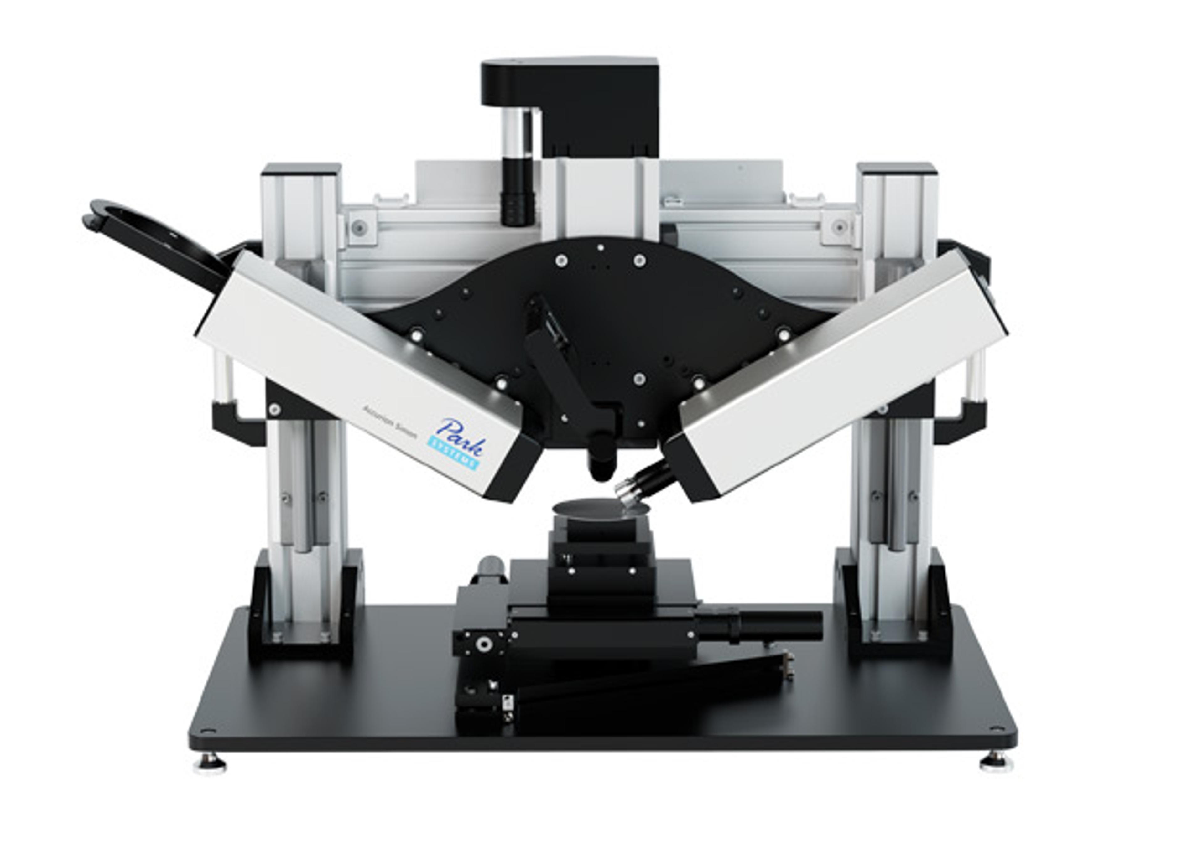
UV/VIS Spectroscopy of Membrane Proteins Encapsulated into Artificial Bilayer Lipid Membranes
An artificial bilayer lipid membrane system was employed, featuring the oriented encapsulation of membrane proteins in a functionally active form. Nickel-nitrilo-tri-acetic acid-functionalized silica nanoparticles of a diameter of around 25 nm were used to attach the proteins via a genetically engineered histidine-tag in a uniform orientation. Subsequently the proteins were reconstituted within a phospholipid bilayer, formed around the particles by in-situ dialysis to form so-called proteo-lipobeads (PLBs). With a final size of about 50 nm, the PLBs could be employed for UV/VIS spectroscopy studies, particularly of multi-redox center proteins, since effects of light scattering are negligible. Andor Technology imaging instruments were employed including the Shamrock SR-303i and Newton CCD and EMCCD Cameras.
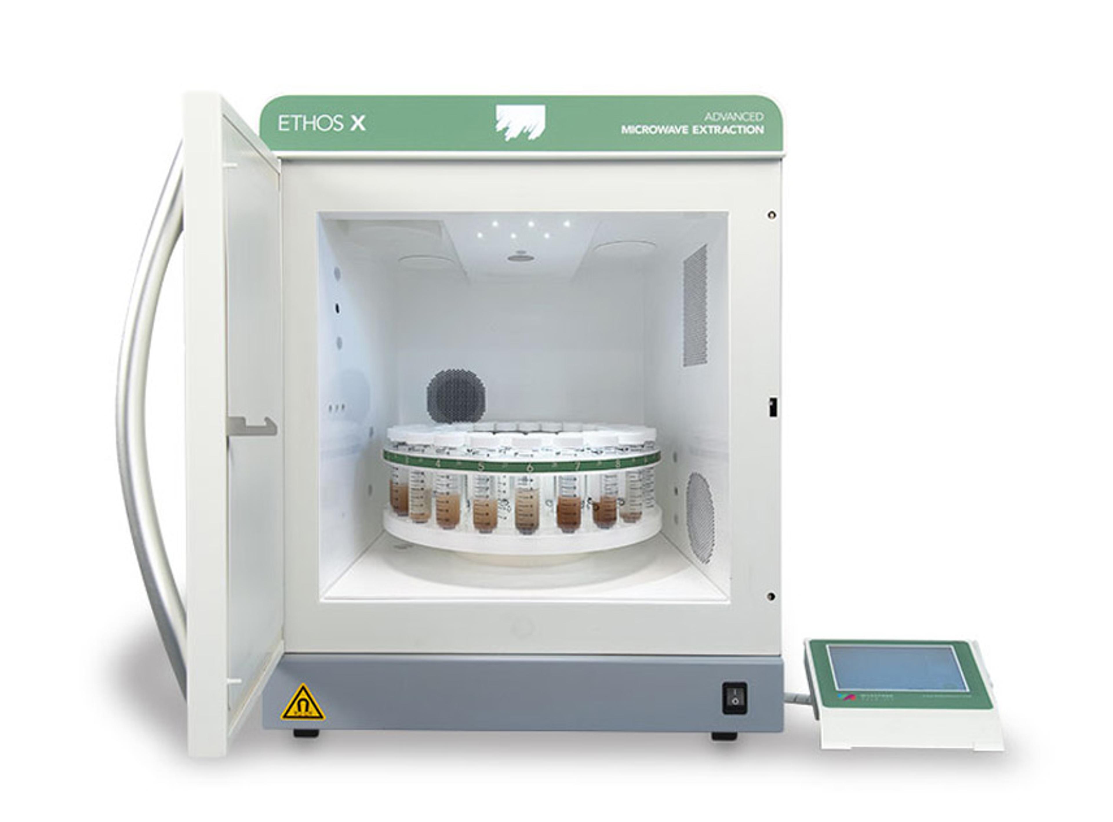
Probing Molecular Structure with Low Frequency Raman Spectroscopy
This application note describes method for the analysis of L-Cystine using low-frequency raman spectroscopy. The ability of this technique to distinguish between different polymorphic structures is also demonstrated through the analysis of three different forms of carbamazepine.