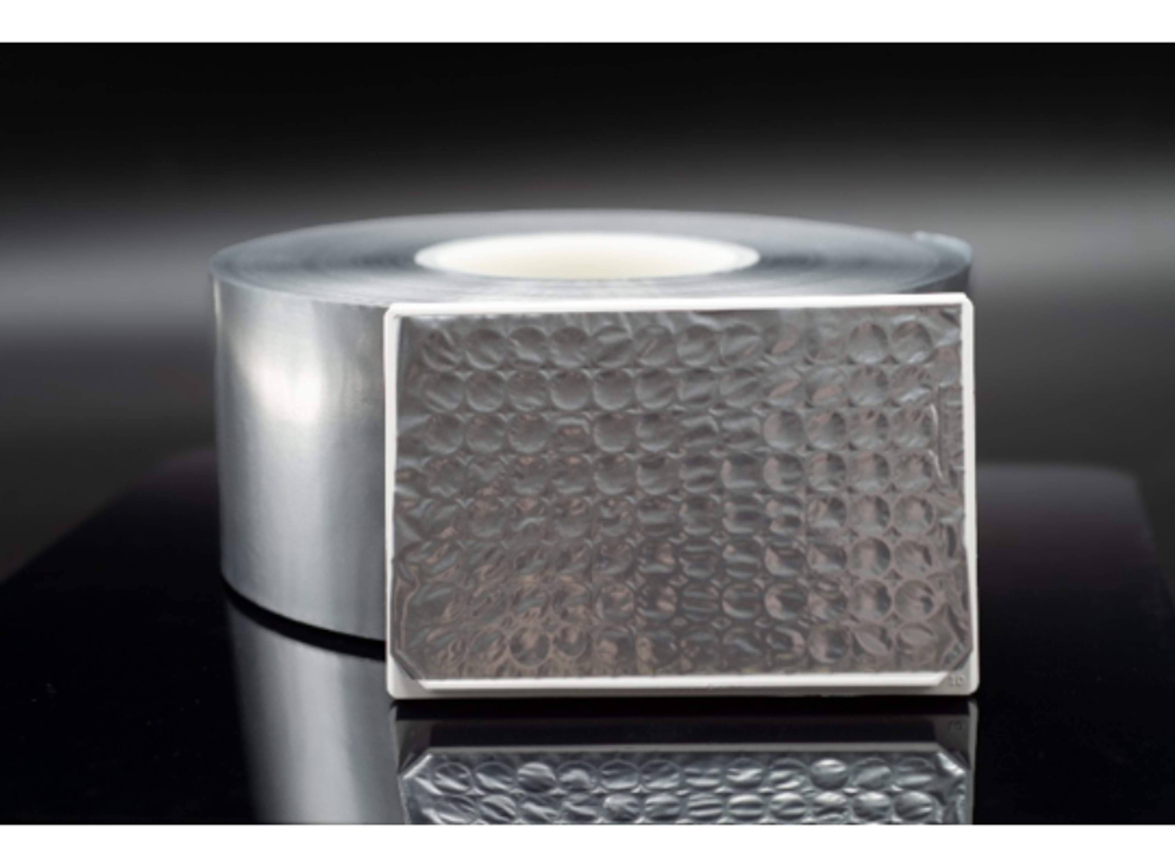
RNAscope™ Technology
Independent of your genes of interest, thes easy RNA in situ hybridization assays use common reagents and protocols; providing universal assay conditions. All RNA-ISH kits contain reagents in a convenient ready-to-use (RTU) format, and chromogenic dyes are available to interrogate a single or two genes simultaneously. Most versatile are the RNAscope Multiplex Fluorescent Reagent Kits, enabling you to interrogate 1, 2 or 3 gen…


The supplier does not provide quotations for this product through SelectScience. You can search for similar products in our Product Directory.
To maintain continuous and sustainable development
Health Service
Amazing and great review webinar.
Review Date: 23 May 2022 | Advanced Cell Diagnostics
Very useful product.
In situ fluorescence hybridization
Very useful application for temporal gene expression analysis in cells or tissue.
Review Date: 8 Feb 2021 | Advanced Cell Diagnostics
Independent of your genes of interest, thes easy RNA in situ hybridization assays use common reagents and protocols; providing universal assay conditions. All RNA-ISH kits contain reagents in a convenient ready-to-use (RTU) format, and chromogenic dyes are available to interrogate a single or two genes simultaneously. Most versatile are the RNAscope Multiplex Fluorescent Reagent Kits, enabling you to interrogate 1, 2, or 3 genes simultaneously.
Four flexible solutions for all your research needs.
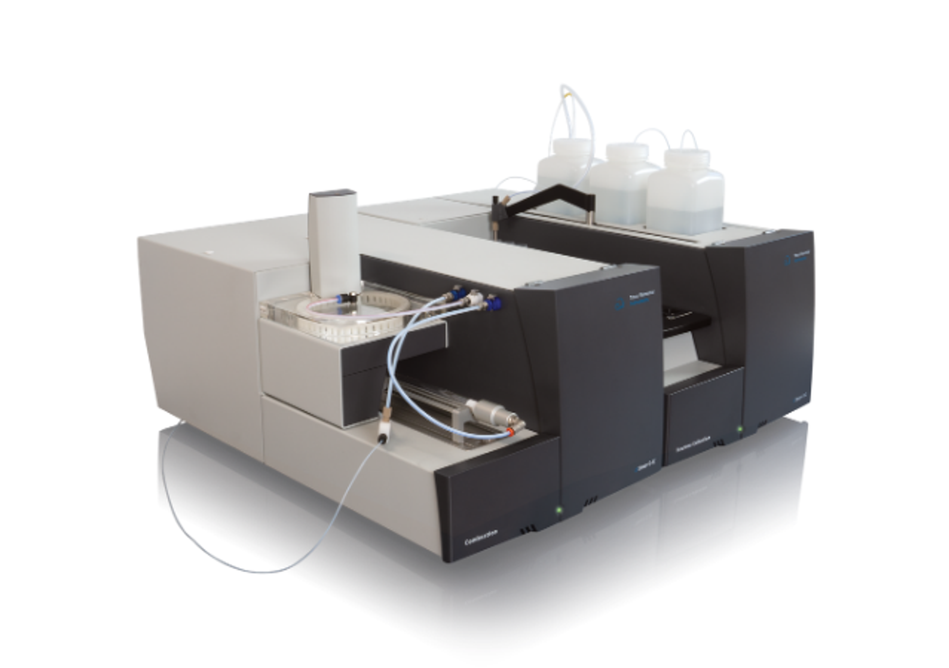
Automated RNAscope ISH assays are also available for Leica Systems (LS) and Ventana Systems (VS).
Brochures

Simultaneous visualization of RNA and protein targets using the new Co-detection Kit
In this product brochure, discover how ACD’s new Co-detection Kits are designed to allow researchers to simultaneously examine cell-type specific gene expression and identify cellular sources of secreted proteins. The new workflow is designed to allow the inclusion of a wider range of antibodies to be combined with RNA ISH enabling researchers to acquire more data and conserve precious samples.
Combined workflow of GeoMx Cancer Transcriptome Atlas and RNAscope assays
This application note demonstrates the concordance between the RNAscopeTM and GeoMx® assays and highlights the detailed molecular analysis that can be performed with this type of high-plex spatial data. Also described are qualified RNAscopeTM probe combinations selected for cell type markers shown to be compatible with the GeoMx® CTA assay.
Quantitative RNAscope image analysis guide
In this application note, Advanced Cell Diagnostics (ACD) provides examples of exemplary assays, discusses experimental considerations, addresses common challenges, introduces HALO® image analysis solutions, and answers common quantitative image analysis questions. Plus, learn how RNAscopeTM technology is designed to enable RNA target expression analysis within intact cells and tissues with high sensitivity and specificity.
RNAscope technology enables the incorporation of spatial mapping and confirmation of gene signatures into single cell RNA sequencing workflows
In this application note, the diverse cell types in the mouse striatum that have been previously identified by scRNA-seq were confirmed and spatially mapped using the RNAscopeTM Multiplex Fluorescent assay and the RNAscopeTM HiPlex assay. The major and minor gene signatures identified by scRNA-seq, including discrete D1 and D2 medium spiny neuron (MSN) subtypes, were confirmed.
Further cellular heterogeneity within the MSN subpopulations was marked by a transcriptional gradient, which was spatially resolved with the RNAscopeTM technology.
Upgrade your single-cell data with higher-plexing
RNA plays an important role in the regulation of cellular processes, making it an ideal biomarker for dynamic gene expression changes in both healthy cells and disease phenotypes. In situ hybridization (ISH) technology provides a powerful method to detect this gene expression, at single-cell resolution, within the spatial and morphological context of an intact tissue. Advanced Cell Diagnostics offer a comprehensive portfolio of ISH assays, with various levels of plexing, that can be applied to a wide range of applications, from neuroscience to immuno-oncology to cell therapy.
In this application eBook, we present:
- A series of case studies illustrating the research capabilities of high-plexing assays
- An outline of the benefits of the RNAscope™ technology
- Top tips on how to achieve accurate, reliable results for virtually any target species, tissue, or gene
Incorporation of spatial mapping and confirmation of gene signatures into single cell RNA sequencing workflows
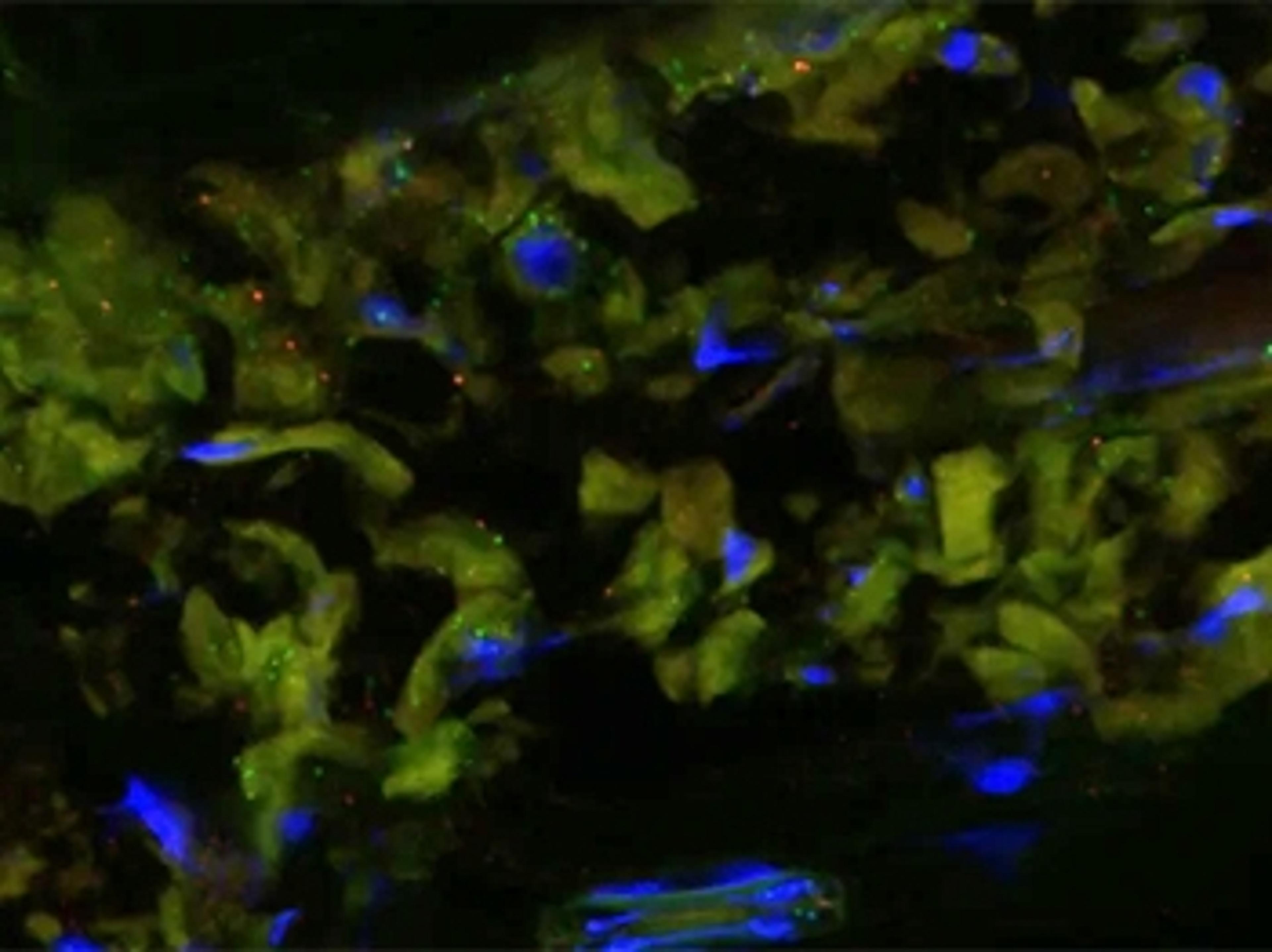
The RNAscope technology is an advanced in situ hybridization assay that allows for the visualization of single-cell gene expression targeting RNA sequences directly in tissues. The proprietary double Z probe design in combination with the advanced signal amplification enables highly specific and sensitive detection of target RNAs in fresh frozen, fixed frozen, and formalin-fixed paraffin-embedded (FFPE) cells and tissues, with each dot representing a single RNA transcript.
Preclinical Safety, Biodistribution, and Tumor Infiltration Analysis of CAR-T Cell Targets Using in situ Hybridization Technology
Chimeric antigen receptor (CAR) T cell therapy has proven to be highly effective in treating hematologic malignancies, and major efforts are being made to achieve similar efficacy in solid tumors. This poster evaluates a method for tracking and monitoring CAR+ T cells within the context of intact tissue and tumor to understand the mechanisms underlying off-tumor toxicity and efficacy in tumor killing.
A Novel RNA <em>In Situ</em> Hybridization Method for the Detection of Mycobacteria in Clinical FFPE Samples
In this poster, Advanced Cell Diagnostics outlines a highly sensitive and specific RNA in situ hybridization (RISH) assay using the RNAscope technology for the detection of mycobacteria rRNA in FFPE tissues and distinction of mycobacteria tuberculosis (MTB) and non-tuberculosis mycobacteria (NTM).
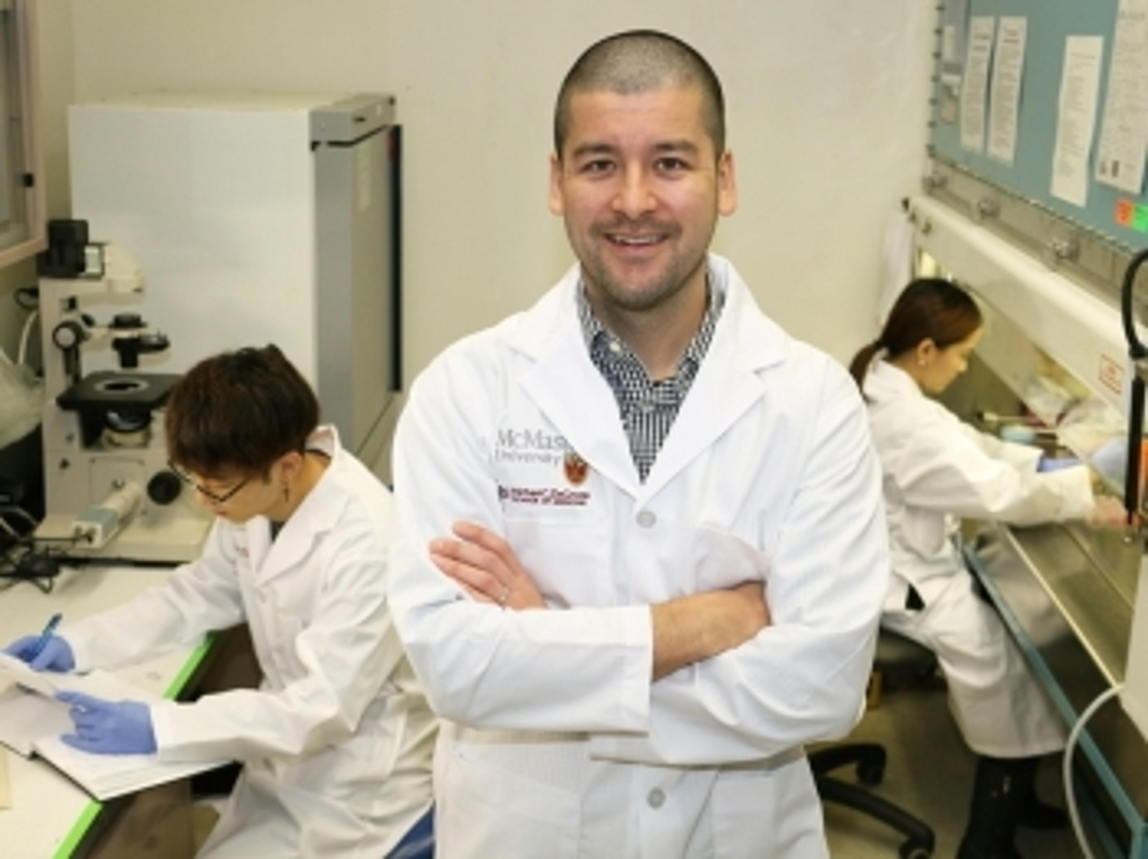
Single-Cell Biology Research with RNAscope Technology
In this application note, Advanced Cell Diagnostics highlights the publications that utilize RNAscope assays to validate and spatially map single-cell RNA seq results in tissue samples.
Validation and Spatial Mapping of Diverse Cells Identified by scRNA-seq
In this poster, Advanced Cell Diagnostics highlights the results of an in situ study that utilized single-cell RNA sequencing (scRNA-seq) to validate and map striatal cells identified in mouse brain tissue at single-cell resolution.
Complement Your scRNAseq Results with RNAscope Technology
In this application note, Advanced Cell Diagnostics highlights recent publications to illustrate how RNAscope assays can be used to complement single-cell RNA sequencing (scRNAseq) results, as well as spatially map and validate scRNAseq results in intact tissue.
Advancing neuroscience research with single cell spatial transcriptomics using RNAscope technology
Watch this presentation by Dr. Sayantani Basak, application scientist, Advanced Cell Diagnostics, a Bio-Techne brand, titled: Advancing neuroscience research with single cell spatial transcriptomics using RNAscope technology. This talk was presented at the SelectScience® Virtual Neuroscience Science Summit 2021.
The study of memory mechanisms to identify new therapeutic targets for PTSD
In this video, Dr. Mark Cembrowski, Assistant Professor at University of British Columbia, explores the importance of understanding the physical underpinnings of fear memory in order to find new therapies for fear-related memory disorders like PTSD. Kaitlin Sullivan, an MSc Student in the Cembrowski Lab, discusses how the lab uses single-cell RNA sequencing to look for neurons that are active during the creation and recollection of fear memory, in the hopes of being able to disrupt these processes.

New insights into reproductive dysfunction with advanced spatial genomics
Dr. Lique Coolen and Dr. Aleisha Moore, Kent State University, present their latest research into the mechanisms underlying spinal cord injury and neuroendocrine dysfunction
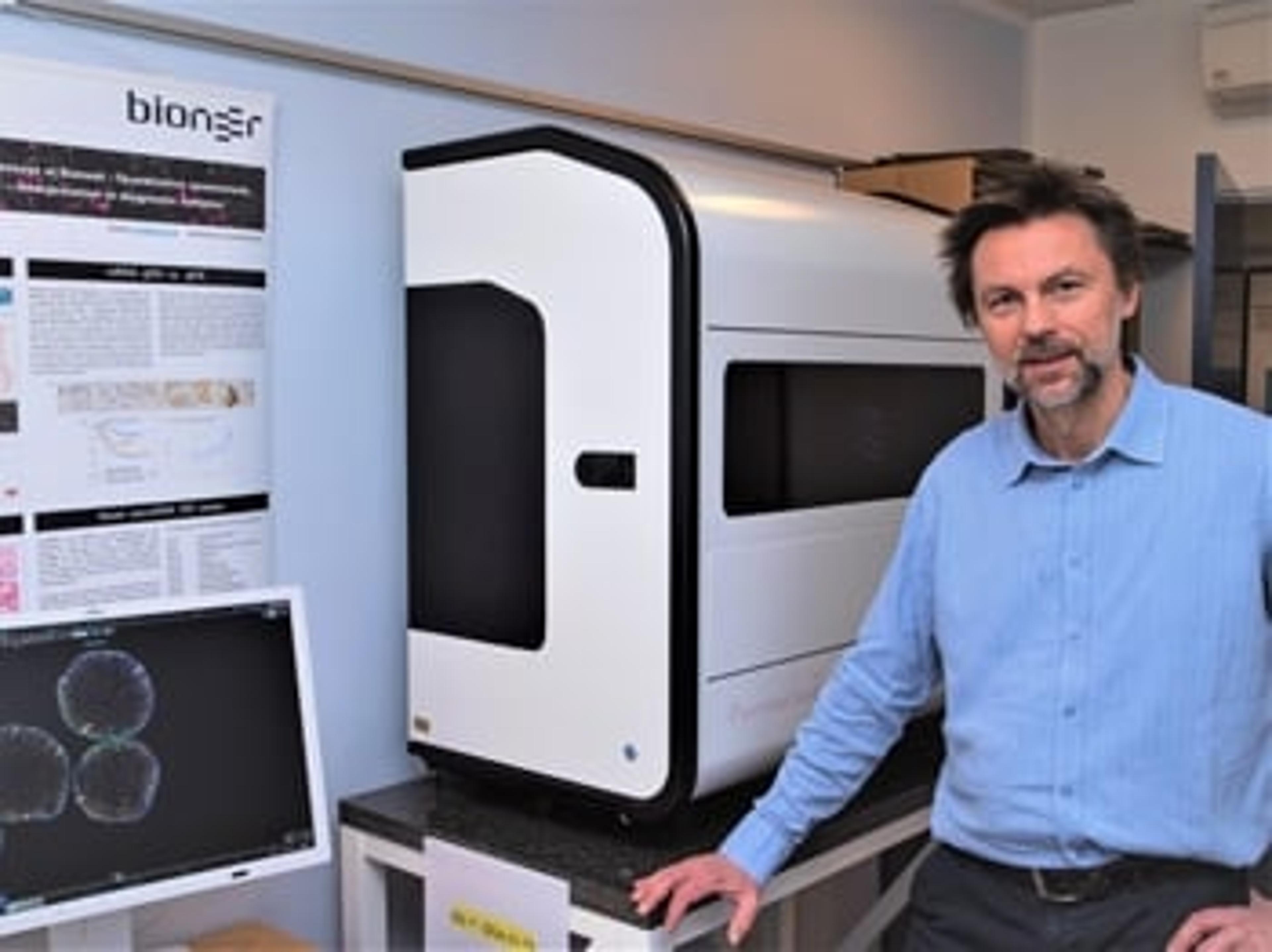
The breakthrough technology transforming RNA detection
Dr. Boye Schnack Nielsen shares tips for running successful in situ hybridization assays as he provides insights on how RNA detection methods have evolved over time

Understanding pain mechanisms: RNAscope technology for RNA expression analysis
Watch this on-demand webinar to discover the latest research on pain mechanisms using peripheral nervous system tissue from organ donors
From cannabis to COVID: Discover the power of RNA in situ hybridization
Dr. Jeremy Hirota takes us through the technology behind his translational research program for respiratory health

Early detection of prostate cancer through game-changing molecular biomarkers
Dr. Nallasivam Palanisamy, Henry Ford Cancer Institute, discusses his discovery of the first pseudogene-associated recurrent gene fusion in cancer and the potential for this robust biomarker for application beyond prostate cancer
Using in situ RNAscope analysis to visualize spatial gene expression in plants
Learn how RNAscope technology was adapted to study plant tissues and gain insights into plant immunity

Establishing the etiology and pathogenesis of viral disease with RNAscope
Don’t miss this on-demand webinar exploring how RNAscope technology can be used to detect viral infection and pathogenesis

Understanding pain mechanisms: RNA expression analysis of peripheral nervous system tissue
Learn about the technical aspects and challenges to using RNAscope technology for RNA expression analysis on rodent and human dorsal root ganglion (DRG)

Detecting pre-cancerous lesions: Duke researcher uses new RNAscope HiPlex technique to visualize the spread of cancer
Fluorescent RNA markers molecularly phenotype lesions, providing insight into development of pre-cancerous events

Musculoskeletal injury: New cellular targets to assist the tendon repair process
Discover how RNA in situ hybridization assays are being used to advance research into treatments for Achilles tendon injuries