MuVI SPIM
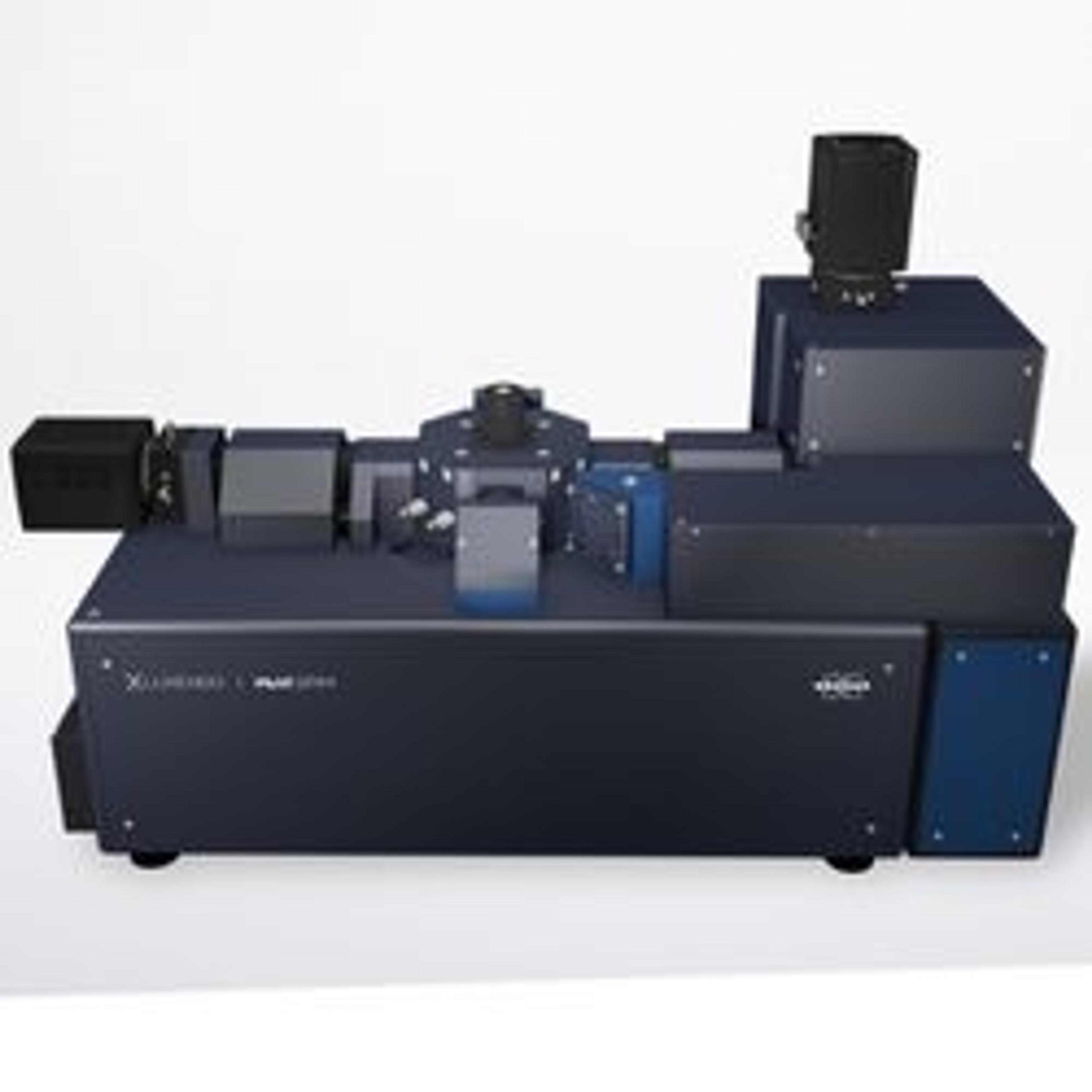
Bruker's MuVi Selective-Plane Illumination Microscope (SPIM) is the fastest multi-angle view system on the market. MuVi incorporates years of Luxendo light-sheet technology innovations to provide high-speed volumetric acquisition for analyzing dynamic processes in delicate live specimens, as well as offering compatibility with any clearing method. A variety of other advanced hardware and software integrations can be easily co…
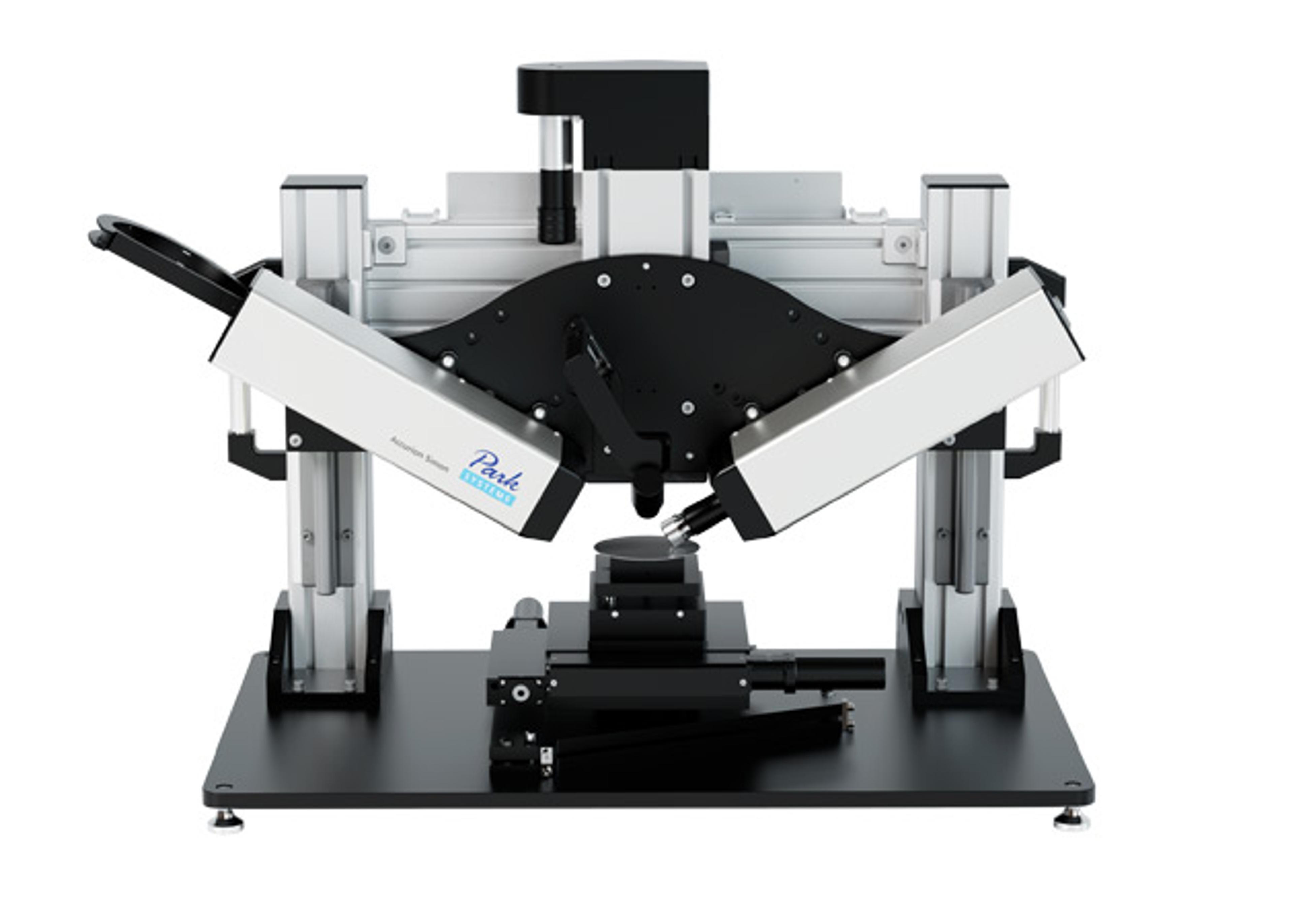
MuVI SPIM Light-Sheet Microscope


The supplier does not provide quotations for this product through SelectScience. You can search for similar products in our Product Directory.
Superior multiview imaging for live and cleared samples. --- Bruker's MuVi Selective-Plane Illumination Microscope (SPIM) is the fastest multi-angle view system on the market. MuVi incorporates years of Luxendo light-sheet technology innovations to provide high-speed volumetric acquisition for analyzing dynamic processes in delicate live specimens, as well as offering compatibility with any clearing method. Only the versatile and customizable MuVi SPIM provides 360° high-resolution imaging with simultaneous dual-color detection and up to six laser lines (405 to 785 nm). Users also have the flexibility for a variety of research in disciplines such as neurobiology and developmental biology with an add-on photomanipulation module for advanced photoablation, FRAP, or optogenetic experiments. To make the most of your research, after image acquisition Bruker provides full software support for image fusion, registration, tile stitching, and 3D visualization. The MuVi SPIM is also advancing with research trends by offering a large field-of-view, superior axial resolution, improved signal-to-noise ratios, low phototoxic effects, and impressive elimination of striping artifacts to enable scientists around the world to excel in their research.
Brochures

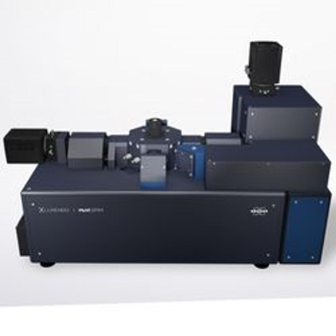
Luxendo MuVi SPIM
In this product brochure, Bruker Fluorescence Microscopy presents the MuVi Selective-Plane Illumination Microscope (SPIM), a fast multi-angle view system. MuVi incorporates years of Luxendo light-sheet technology innovations to provide high-speed volumetric acquisition for analyzing dynamic processes in delicate live specimens, as well as offering compatibility with any clearing method. A variety of other advanced hardware and software integrations can be easily configured to support evolving multidimensional experimental needs.
Considerations for cleared tissue imaging with light-sheet fluorescence microscopy in neuroscience research
A major goal in the field of neuroscience is to understand the connectivity of the brain across scales, from individual synaptic connections to whole organism connectomics. Visualizing the whole brain often requires imaging at depths that are not supported by conventional imaging methods, such as laser scanning confocal microscopy. In recent years, light-sheet fluorescence microscopy (LSFM) has emerged as an imaging technique capable of addressing a wide variety of applications in neuroscience due to its faster acquisition speed and larger imaging depth compared to confocal microscopy. Although early implementations of LSFM were optimized around imaging developmental processes in typical model organisms, technological advances in optics have allowed researchers to extend this technique to much larger samples, such as an entire mouse brain. Combined with emerging tissue clearing techniques, researchers are now able to investigate intact tissues, organs, and even entire organisms with subcellular resolution using light microscopy methods. The versatility of LSFM for neuroscience research has contributed to its rapid adoption and increasing popularity in the scientific community. However, there are multiple important factors one must consider when moving from standard imaging techniques to LSFM. In this application note, Bruker discusses the reasons why LSFM and clearing techniques are particularly useful for neuroscience research, important considerations for neuroscientists adopting this technology, and application examples of LSFM for cleared sample imaging.
Imaging Complex StructuImaging complex structure with Luxendo light-sheet microscopy and CLARITY tissue clearinge with Luxendo Light-Sheet Microscopy and CLARITY Tissue Clearing
Using conventional light microscopy techniques to image large or dense biological samples can be challenging due to light scattering properties of tissues and mismatched refractive indices. The combination of tissue clearing and light-sheet microscopy enables the fast, gentle, and high-resolution imaging deep within thick samples. Here, Bruker Fluorescence Microscopy presents a recap of the webinar presented by Sharla White, Ph.D., Vice President of Research and Development at ClearLight Bio, and the practical applications and best-known methods of combined light-sheet fluorescence microscopy (LSFM) and CLARITY tissue clearing are presented. An example of CLARITY tissue clearing on mouse intestines is also presented, showcasing how imaging with LSFM provides added insight to the unique structure, shape, and layers of the intestine.
Imaging breast cancer evolution in 3D organoid cultures with Luxendo light-sheet microscopy
Bruker's Luxendo light-sheet technology can image a wide range of live, fixed, and cleared biological samples from organoids and embryos to large whole organisms. Overcoming many of the barriers that conventional light microscopy faces, lightsheet fluorescence microscopy, also called selective plane illumination microscopy (SPIM), supports advanced research in the life sciences. SPIM works by de-coupling the fluorescence excitation and detection beams and together with a sheet of light for excitation, selectively illuminates a focal plane for high-resolution images without the danger of phototoxicity. Here, Dr. Martin Jechlinger, the Senior Scientist and Head of VISION Laboratory at the MOLIT Institute, talks about how his lab utilizes light-sheet microscopy to investigate 3D organoid cultures. Specifically, the molecular mechanisms underlying mammary tumor development during breast cancer, as well as what is happening when treatments are failing.
Light-sheet fluorescence microscopy for living plants
Imaging living plants involves challenges on several levels, including sample properties (e.g., aerial and roots, chlorophyll, cell walls), tissue visualization (e.g., dyes vs reporter lines), sample-friendly imaging (e.g., embedding, low phototoxicity, timelapses), and efficient data handling (e.g., processing and storage). Working together with end users and application specialists, Bruker Luxendo has the experience needed to deliver expert commercial LSFM solutions that can significantly aid the imaging of living and naturally growing plants. In this application note, Bruker highlights challenges with using microscopy for image acquisition of plants, challenges specific to LSFM, data processing challenges, and how Bruker Luxendo can support you in overcoming these obstacles to create meaningful data.