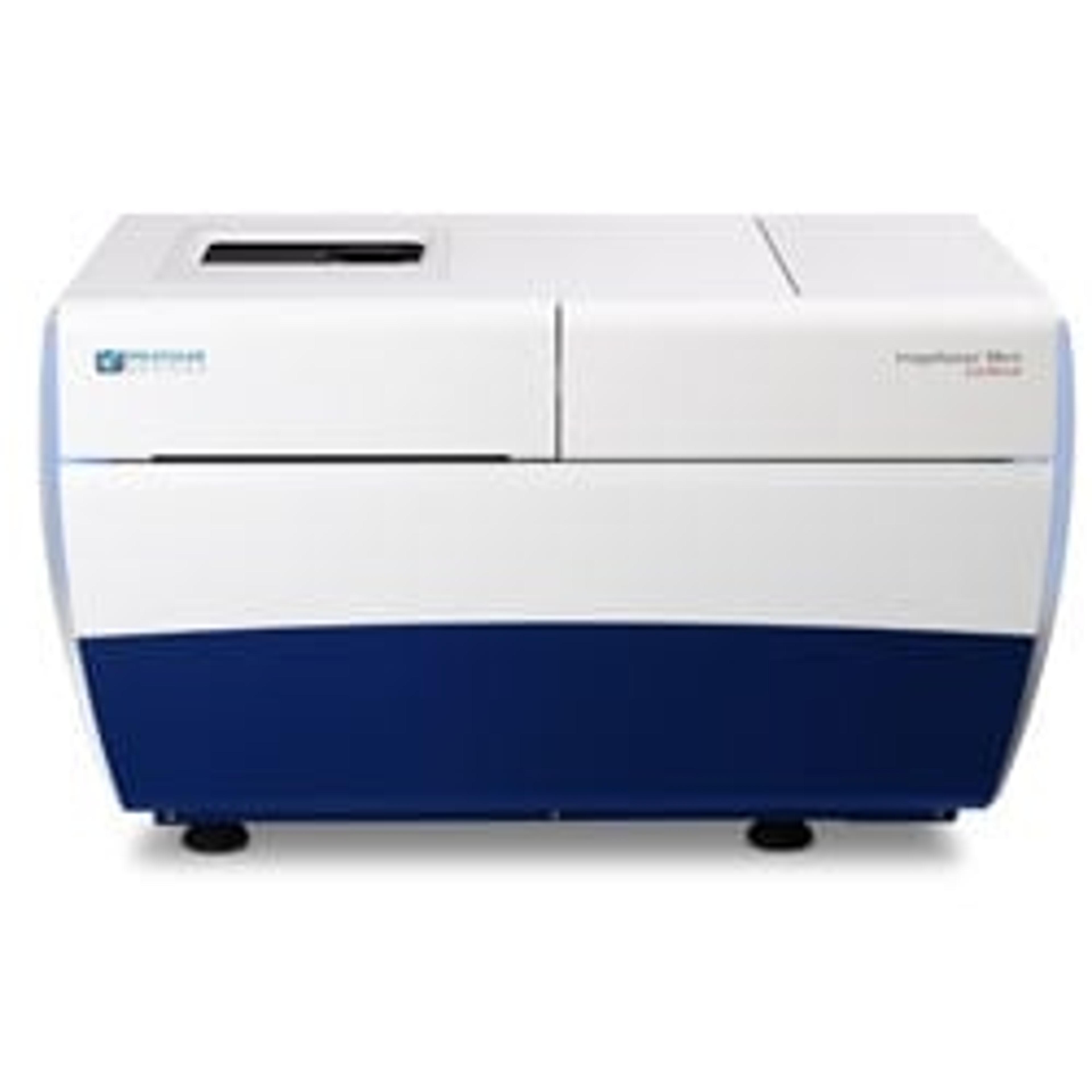
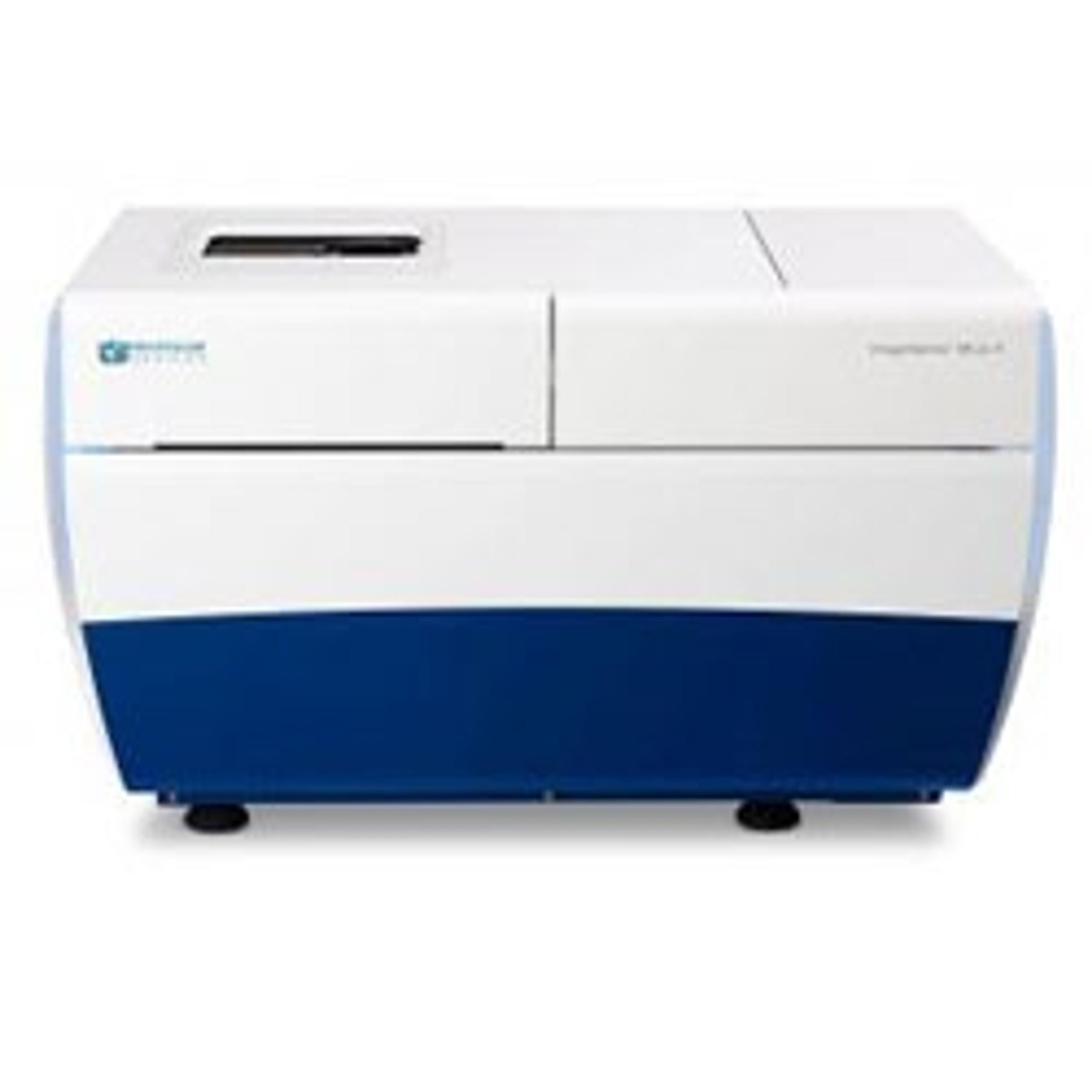
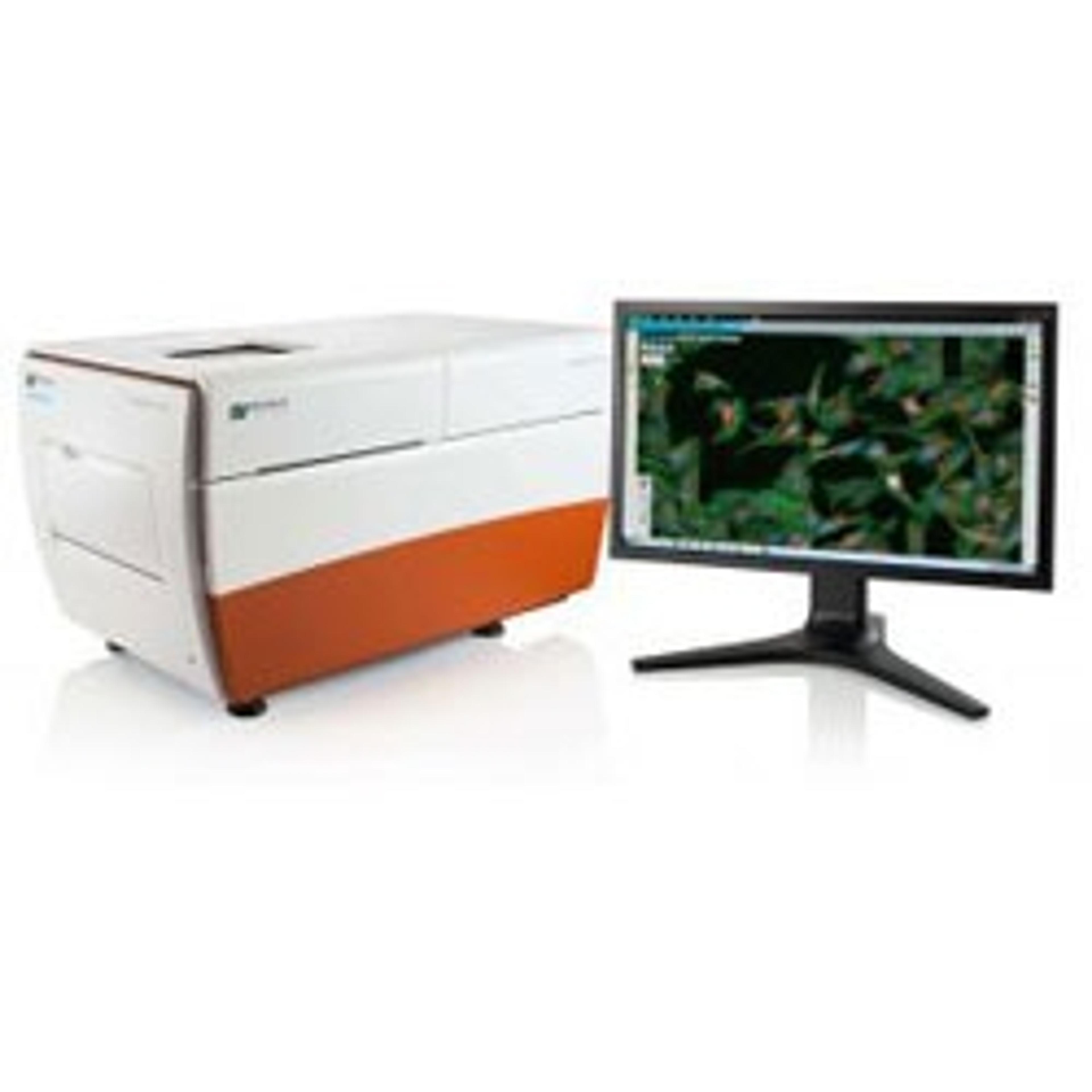
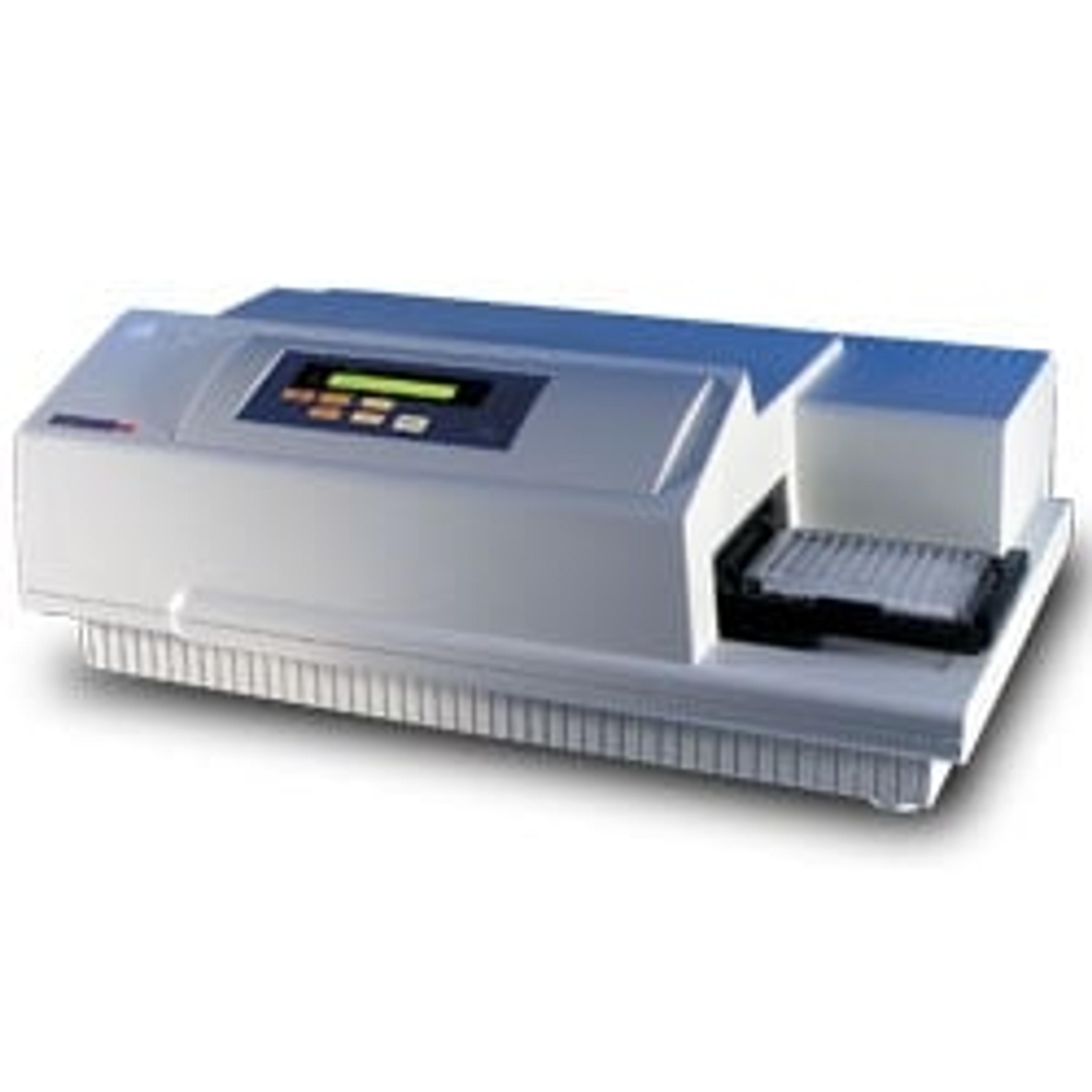
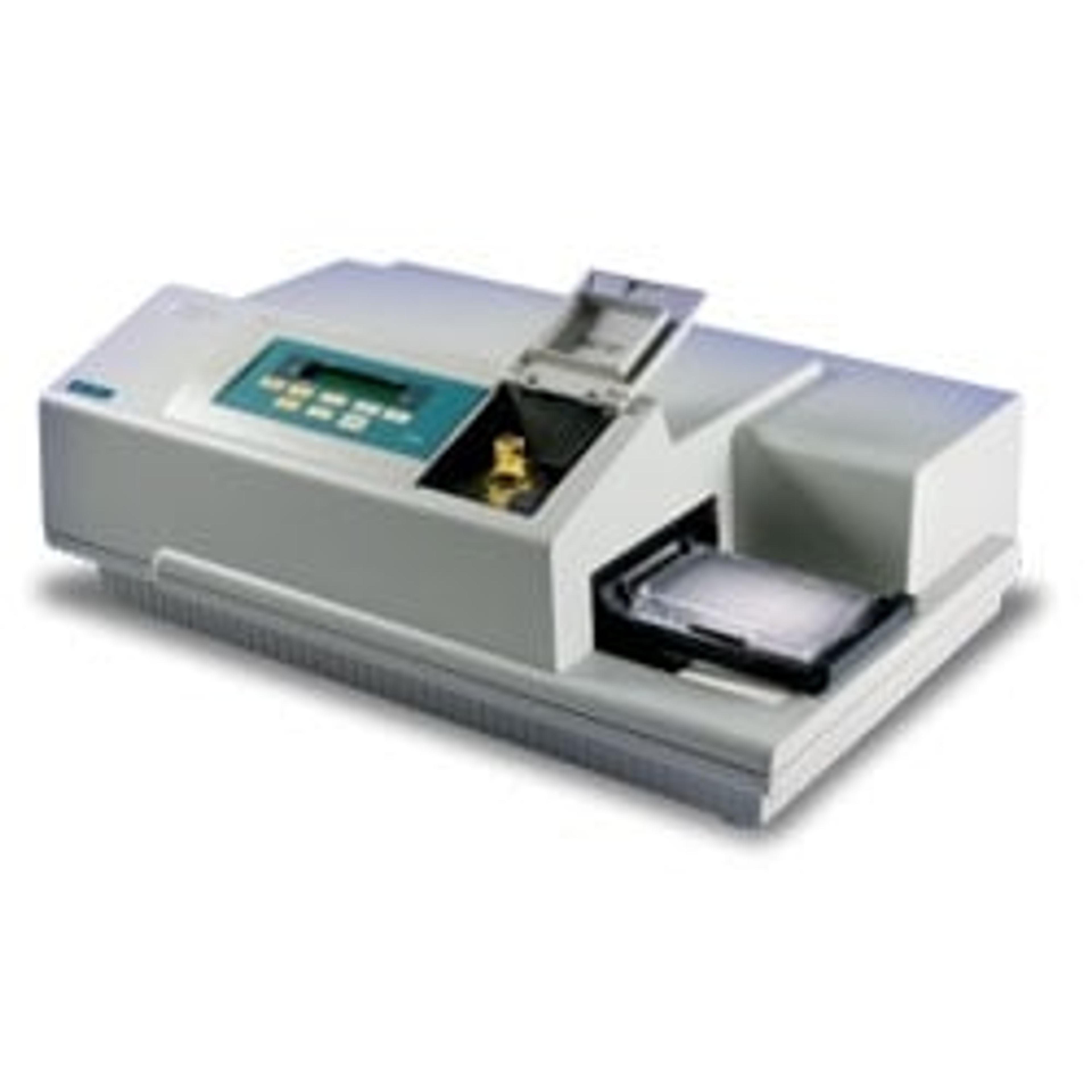
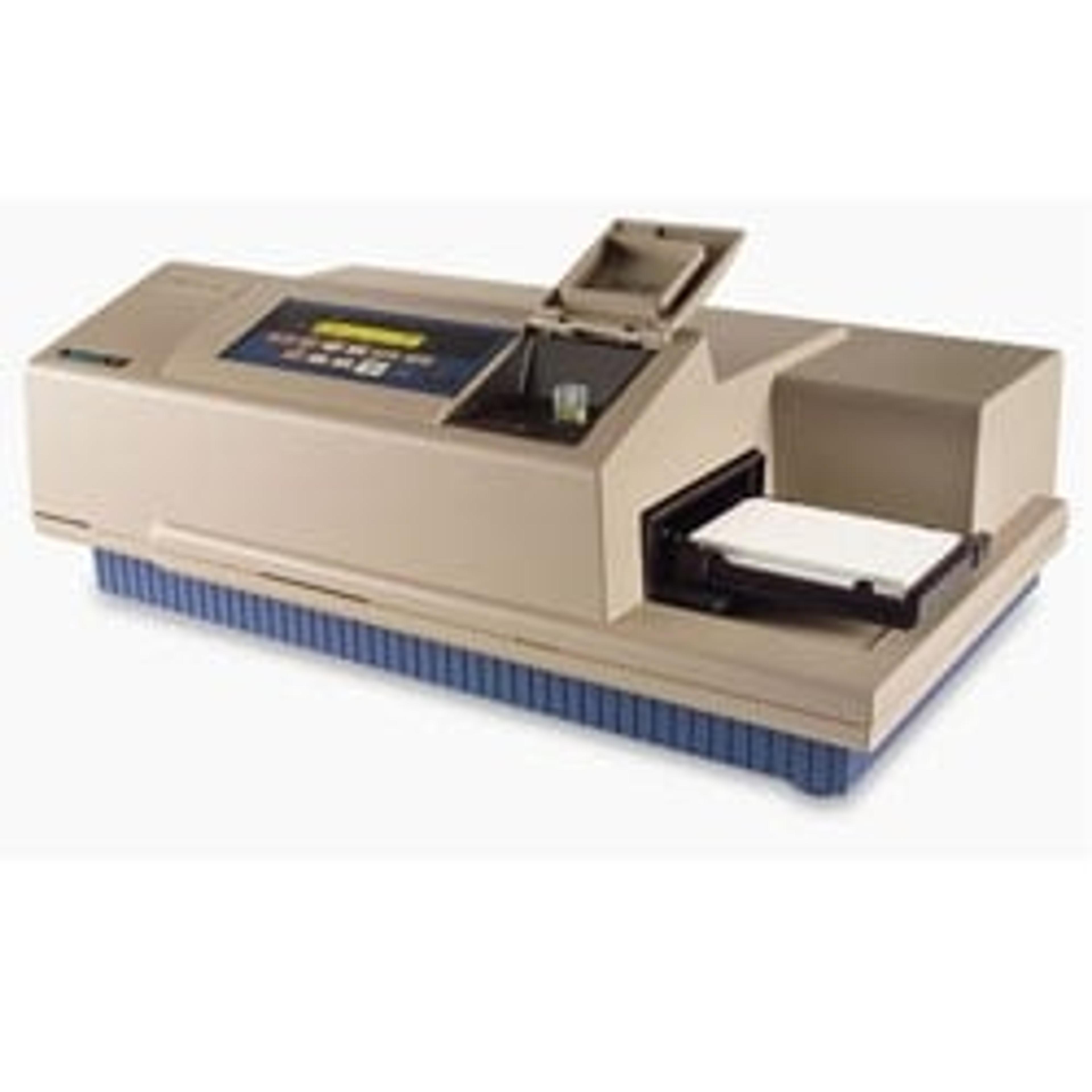
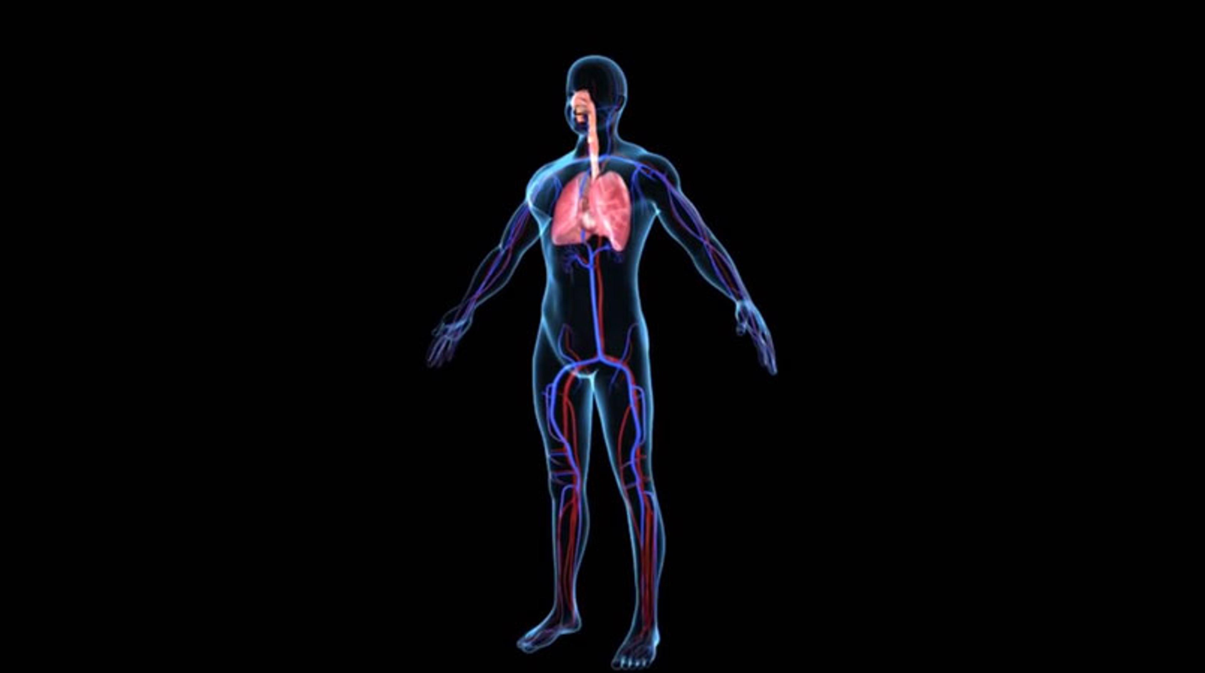
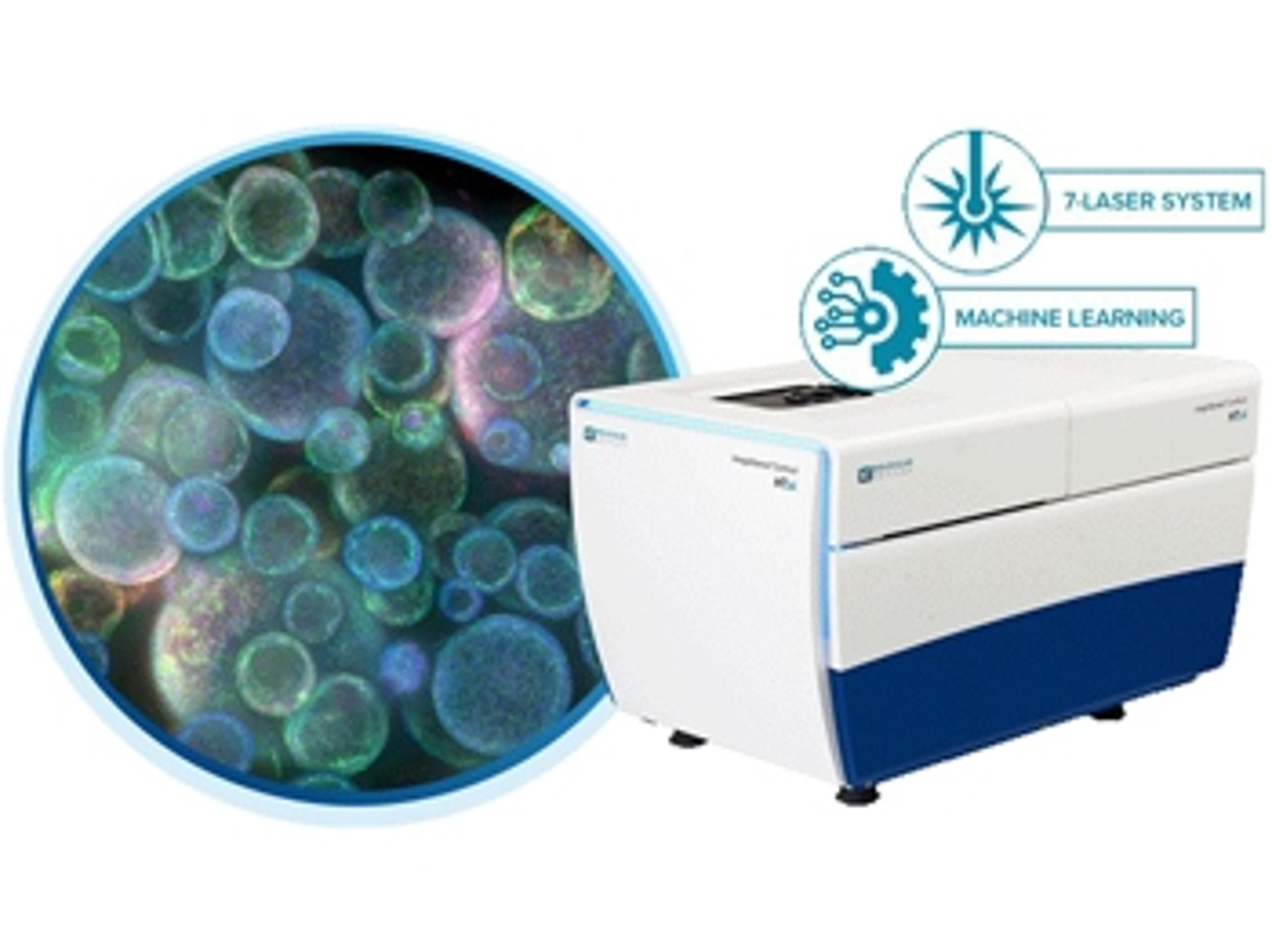
ImageXpress® Micro Confocal High-Content Imaging System
Explore the new dimension with the confocal system for your complex biology.


Receive your quote directly from the manufacturer.
Indispensable for our work!
Neurotoxicity quantification
The ImageXPress Micro is indispensable for our work in measuring the toxic response in neurons to chemotherapy agents. We are looking to find compounds that alleviate toxicities and could'nt achieve this end without the high quality images and analysis software of the MetaXpress software 2.0. The machine is fast in imaging even 9 sites per well in our 96 well plates and finishes a whole run in less than 30 minutes. The analysis can be done off site and runs in the background without tying up the real time imaging capacity. We love this machine!!
Review Date: 21 Nov 2019 | Molecular Devices®
The ImageXpress Micro from molecular devices has become essential for our research!
Drug discovery, cell phenotyping, foundational research
Extremely high-quality images and data analysis. The only limitation/drawback of the HCI system is that it is exquisitely sensitive to the quality of the imaged sample. The power of the HCI is simultaneously a challenge, because it requires one to invest considerable time in optimizing and perfecting staining conditions, acquisition parameters, and post-acquisition analysis. However, once done, the ROI for these steps is excellent. The ImageXpress Micro provides extremely high quality images.
Review Date: 11 Jul 2019 | Molecular Devices®
The software is quite user friendly and has nice analysis capabilities.
Confocal microscopy
It's a pretty cool machine with great capabilities for getting nice images. The software is quite user friendly as well and has nice analysis capabilities.
Review Date: 30 Apr 2019 | Molecular Devices®
Amazing results which are impossible to obtain with a regular microscope
Cell biology academic research
This is my second IXM, it is now more powerful than our previous IXM-XLS. The image quality dramatically increased, the system is extremely stable and easy to use. We are now performing more 3D and live images thanks to the spinning disc capabilities.
Review Date: 20 Jul 2018 | Molecular Devices®
The ImageXpress Micro Confocal system combines the ability to capture high-quality images without sacrificing throughput, reliability, or flexibility. Quickly and easily switch between confocal and widefield imaging modes to satisfy the specific throughput and sensitivity needs for your assay. Combined with MetaXpress® High-Content Image Acquisition and Analysis Software, the ImageXpress Micro Confocal system provides you with a complete multi-dimensional, high-throughput screening solution to help you discover your next landmark scientific breakthrough.For researchers looking to expand their laboratory’s capabilities, the ImageXpress Micro Confocal system leverages large field-of-view optics to map macro structures with minimal tiling. In addition, querying of large cell populations is accelerated, speeding up the characterization of highly heterogeneous samples or identification of rare sub-populations.
Key Benefits:
- Match acquisition mode to your assay needs with multiple confocal geometries and widefield mode
- Capture publication-quailty images without sacrificing throughput using our proprietary AgileOptix(TM) technology
- Acquire statistically relevant data faster with large field of view and > 3log dynamic range
- Expand your research capabilites with available options
The ImageXpress® Micro Confocal system represents the culmination of four generations of imaging expertise. Built on over 30 years of cell-based imaging experience, the ImageXpress Micro Confocal system is engineered for performance to reliably accelerate the pace of your research
Brochures

ImageXpress Micro Confocal: The confocal solution for your complex biology
In this brochure, Molecular Devices provides key features, applications and specifications of the ImageXpress® Micro Confocal system, a high-content solution that can switch between widefield and confocal imaging for improved quantification of live or fixed cell assays.
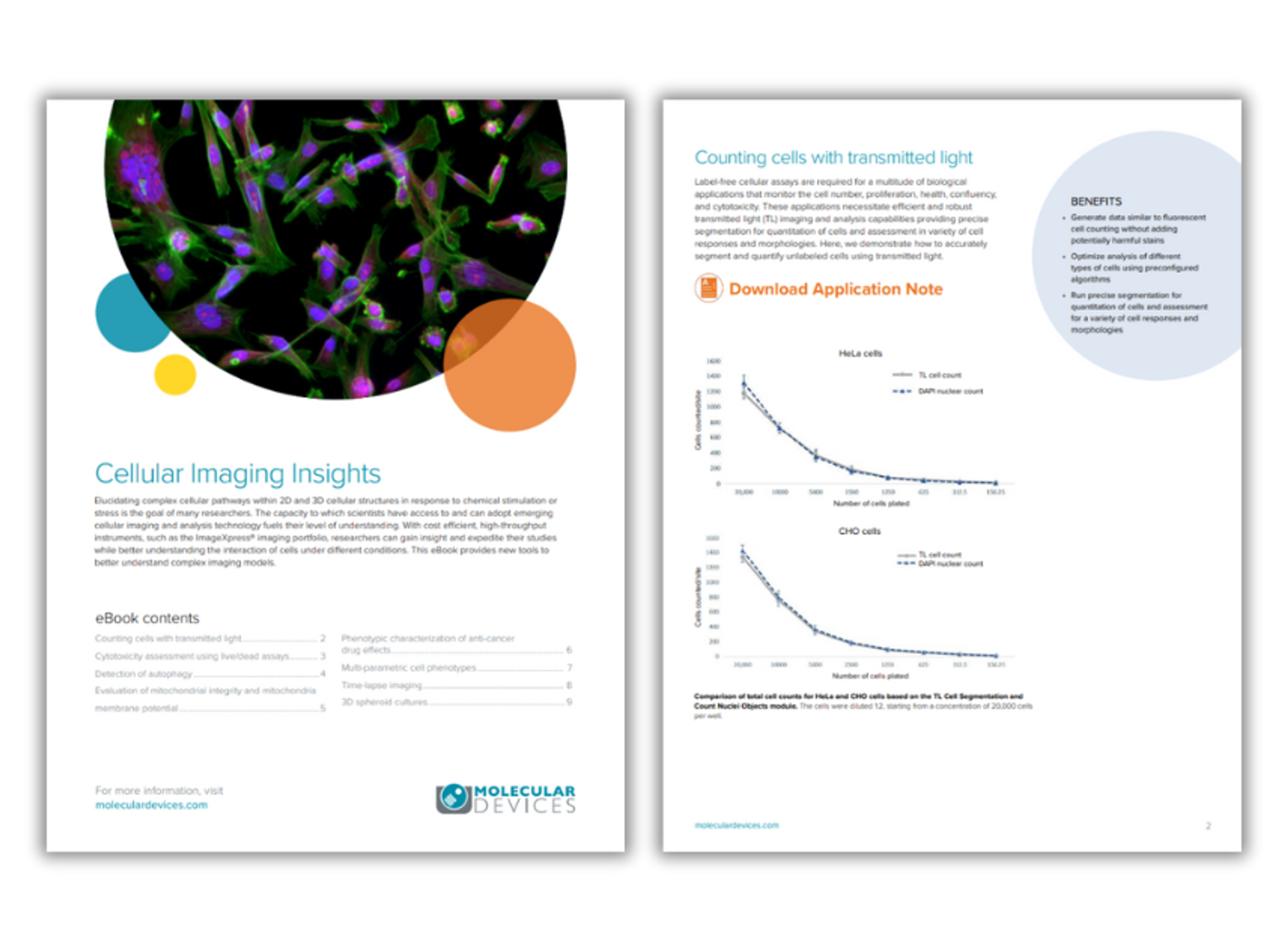
Cellular imaging insights
The goal of many researchers is to elucidate the complex cellular pathways within 2D and 3D cellular structures in response to chemical stimulation or stress. The capacity to which scientists have access to and can adopt emerging cellular imaging and analysis technology fuels their level of understanding. In this eBook, Molecular Devices provides an overview of the new tools that can be used to better understand complex imaging models.
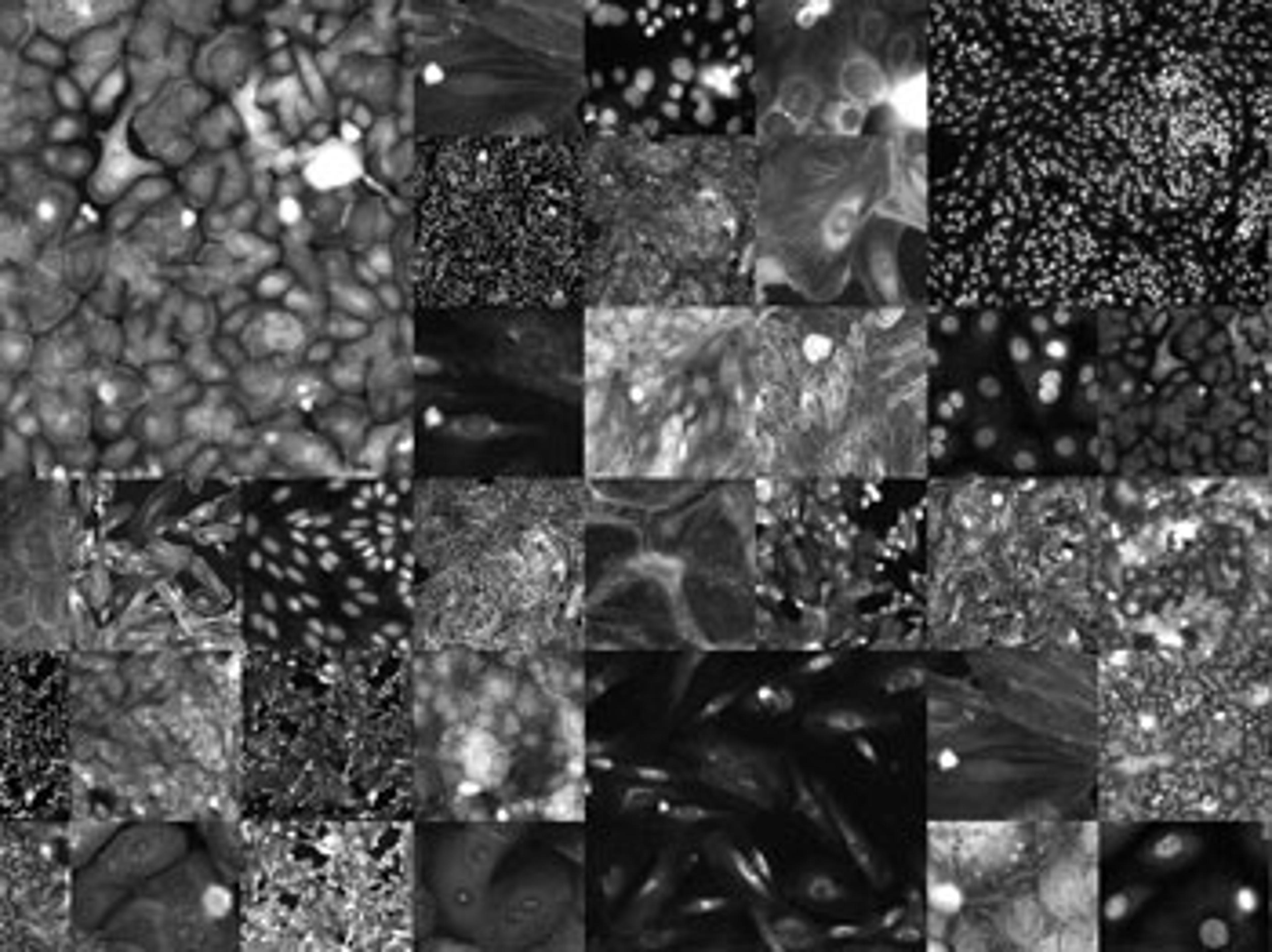
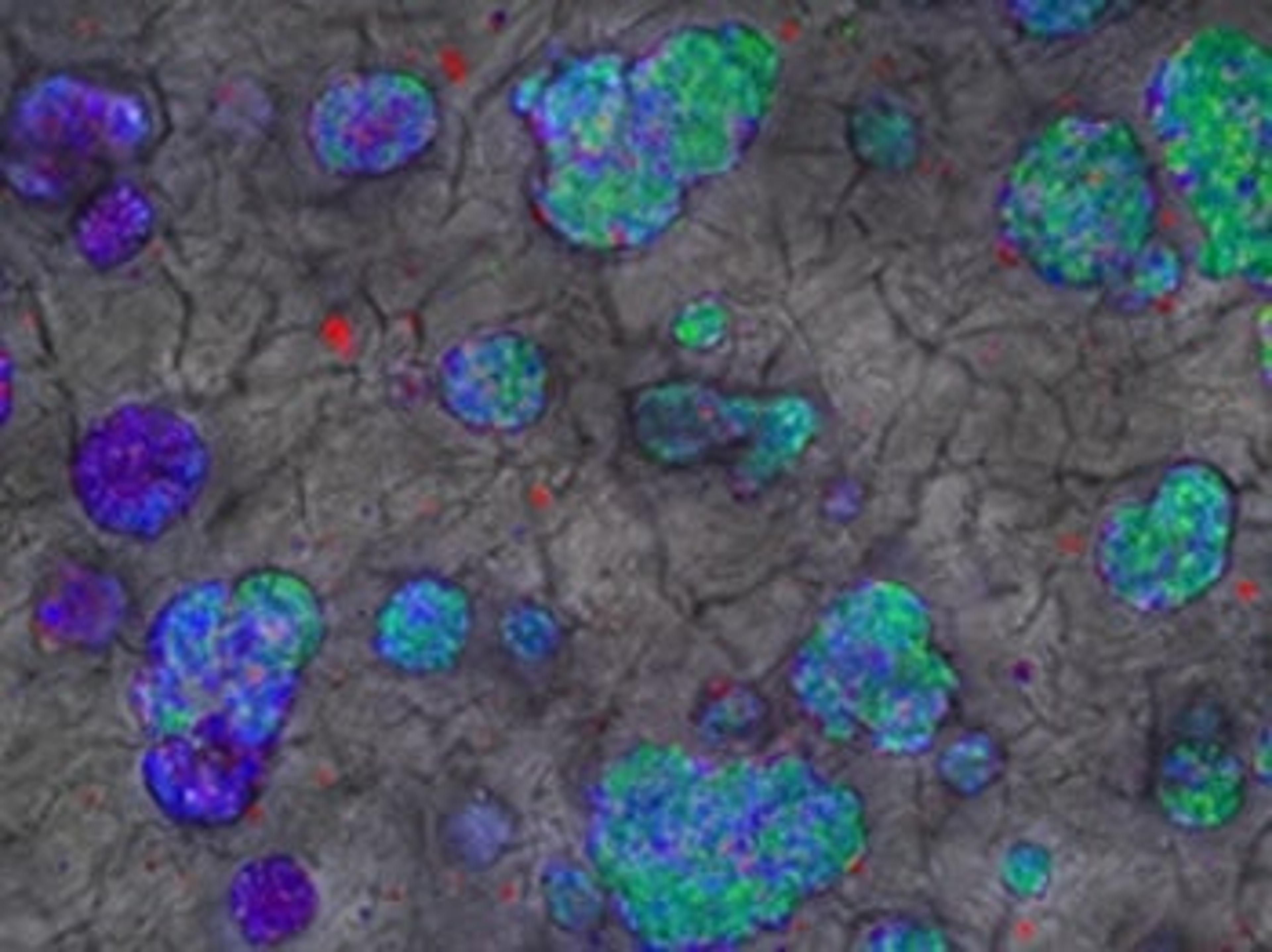
High-content phenotypic profiling using the Cell Painting assay
High-content phenotypic profiling is increasingly popular in research areas that span from gene function studies, drug discovery, and toxicology. The Cell Painting assay, often used in phenotypic profiling, uses up to six fluorescent dyes to label and visualize a variety of subcellular structures at the single cell level. The aim of this assay is to visualize as much of the cell as possible in order to construct a representative image of cell state. Molecular Devices shows how to simplify your Cell Painting assay workflow using the ImageXpress Micro Confocal system.
Automated monitoring of development and activity analysis of iPSC-derived 3D cerebral organoids
In this application note, Molecular Devices describes a method for semi-automated culturing and monitoring of cerebral organoids, as well as the testing of functional neuronal activity by means of recording Ca2+ oscillations.
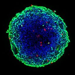
High-throughput confocal imaging of spheroids for screening cancer therapeutics
In this application note, Molecular Devices demonstrates how the ImageXpress® Micro Confocal system and MetaXpress® software allows rapid imaging and analysis of 3D spheroids in microplates for monitoring induced apoptosis and mitochondrial toxicity of anti-cancer drugs.
High-throughput screening of 3D cell cultures with multiple high density scaffold-free spheroids for cancer toxicity studies
In this application note, Molecular Devices demonstrates the use of the Elplasia 96-well plates with a 3D culture workflow that includes spheroid generation, compound treatment, cytotoxicity assay, high-content imaging on the ImageXpress® Micro Confocal High-Content Imaging System, and 3D image analysis using MetaXpress® High-Content Image Acquisition and Analysis Software. The ability to easily generate multiple spheroids and to seamlessly integrate the workflow with high-content imaging promises to have significant applications in drug discovery and compound toxicology.
Lung organoids as an assay model for in vitro assessment of toxicity effects by 3D high-content imaging and analysis
In this application note, Molecular Devices describes a high-content imaging method that monitors the growth and differentiation of lung organoids. 3D reconstruction allowed for further complex analysis of the organoid structure.
Monitor mitochondria dynamics and phenotype with high-content imaging
Mitochondria are the main energy source for cells and play a key role in regulating cellular metabolism. In this application note, Molecular Devices describes phenotypic assays for mitochondria phenotypes and structural re-arrangements that can be used for studies of mitochondria dynamics in cell-based assays.
High-content imaging for diverse 3D cell culture models
3D cell models are morphologically diverse with varying characteristics based on the cell type and the underlying research questions. In this application compendium, we bring you helpful case studies to perform high-content imaging on a range of 3D models and resolve common challenges experienced in 3D cell culture assays.
Learn how to capture in-depth and high-quality images of spheroids, stem cells, organs-on-chips and whole organisms and read case studies from the scientists developing 3D cell assays to delve into the complex biology of neurodegenerative disease, angiogenesis and tumor microenvironment.
The eBook covers 3D high-content imaging protocols for:
- Characterizing compound effects on 3D cells in extracellular matrix
- Screening cancer therapeutics in spheroids
- Morphological characterization of 3D neuronal networks in an organ-on-a-chip model
- Characterization of angiogenesis in an organ-on-a-chip model
- High-throughput imaging assays using zebrafish
- High-content 3D toxicity assay using iPSC-derived hepatocyte spheroids
Water immersion objectives for automated high-content imaging
Download this poster by Molecular Devices to find out if water immersion objectives, used to improve image quality in complex biological assays, could be used in a high-throughput environment.
High-Throughput Confocal Imaging of Spheroids for Screening Cancer Therapeutics
This application note demonstrates a method for cancer cell spheroid production, followed by protocols for staining and imaging of these spheroids for use in multi-parameter cytotoxicity studies.
Key partnership increases access to organoid models
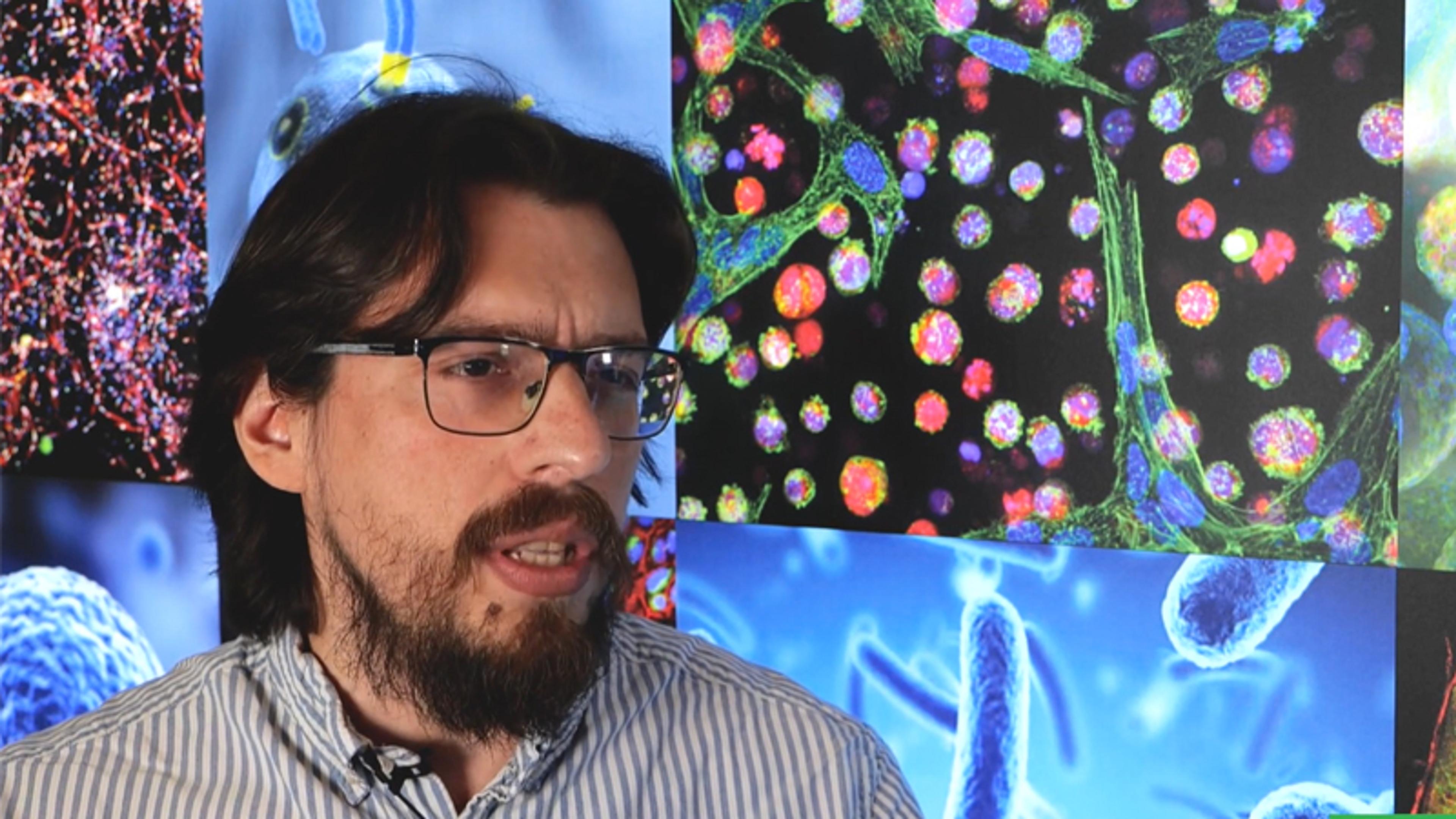
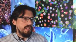
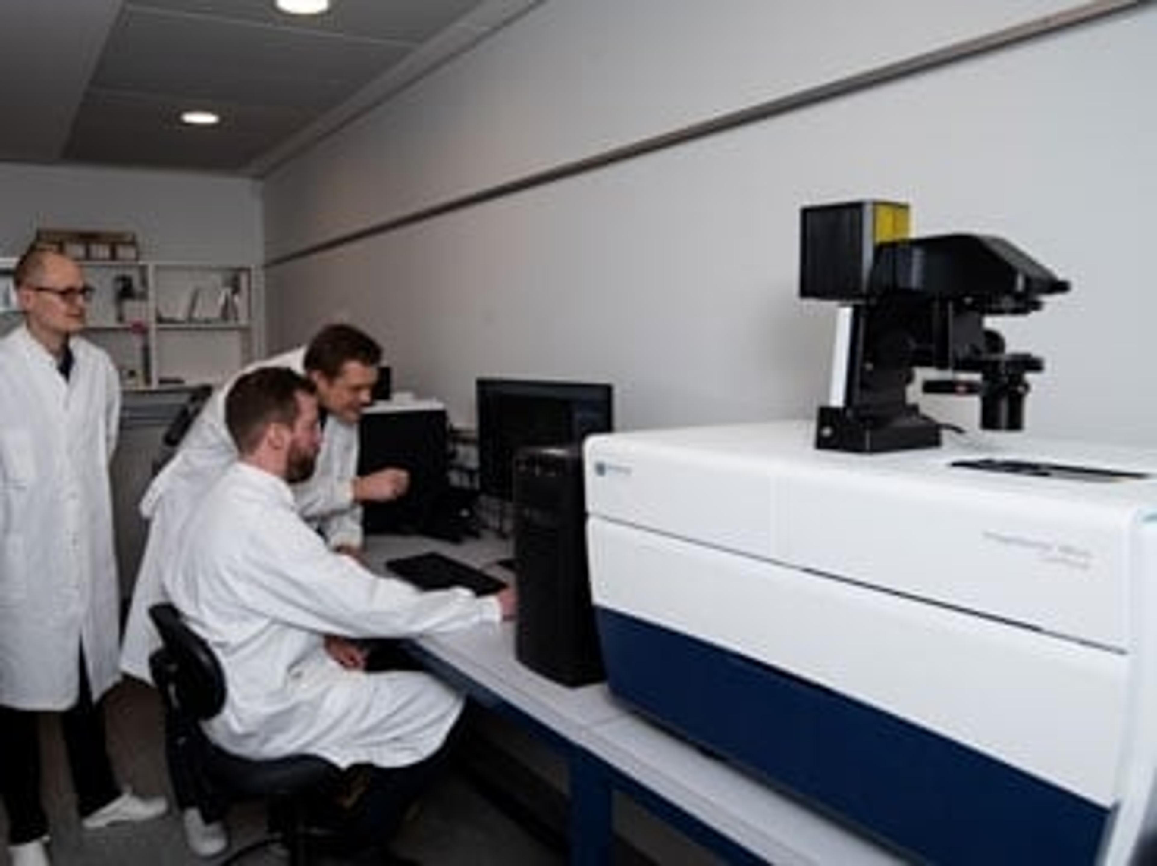
Patient-derived organoids address the urgent need for more predictive in vitro models for biopharmaceutical applications. However, challenges around standardization and scale up remain. Dr. Francisco Morales-Rodriguez shares insight into the partnership between HUB Organoids and Molecular Devices, which aims to increase the access of organoids for both academia and industry. Here, he highlights the integral role of the ImageXpress® Micro Confocal High-Content Imaging System and its suite of software solutions in providing high image quality, high throughput, and flexibility that HUB Organoids requires.
This video was filmed at SLAS Europe 2024.
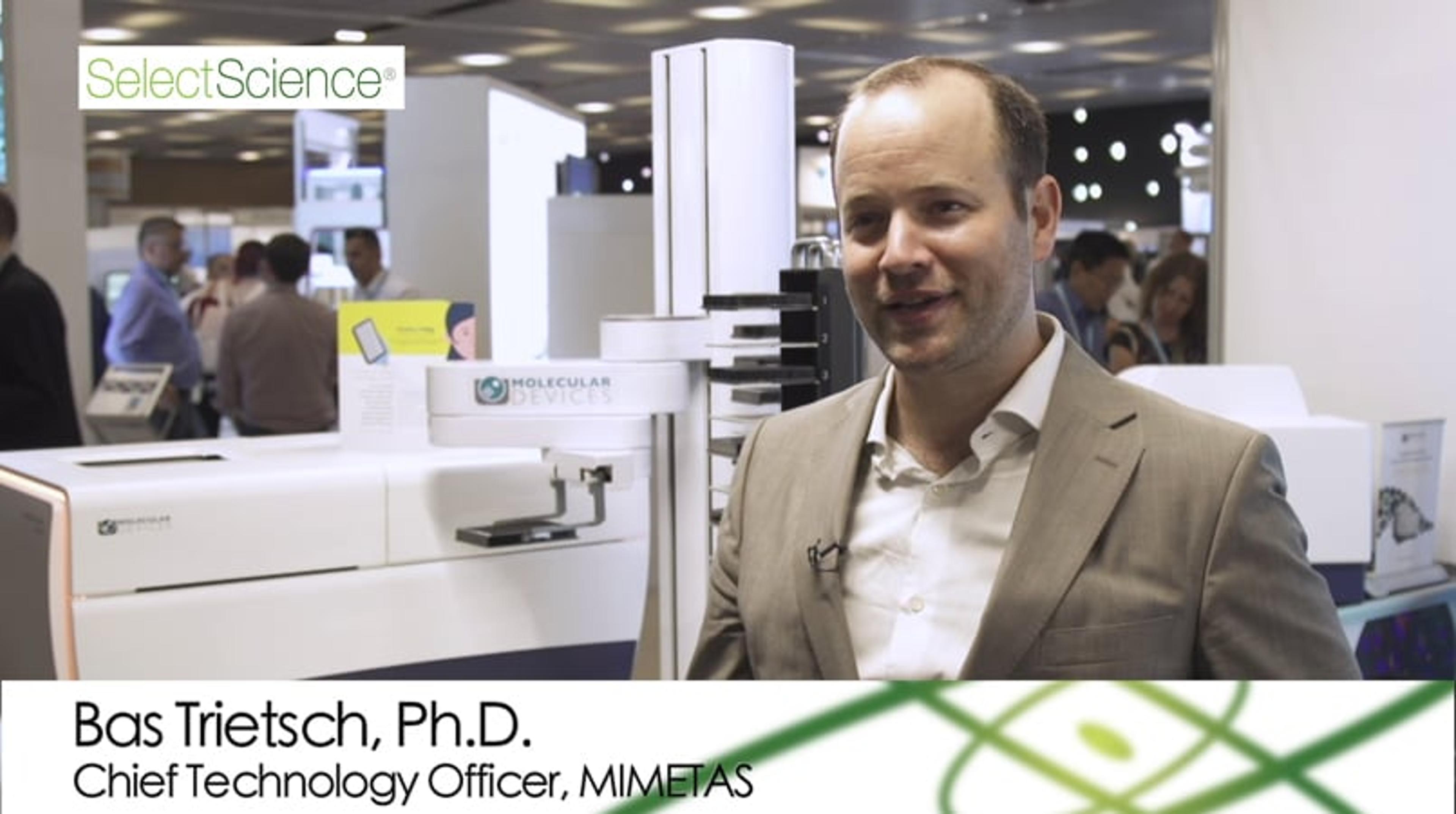
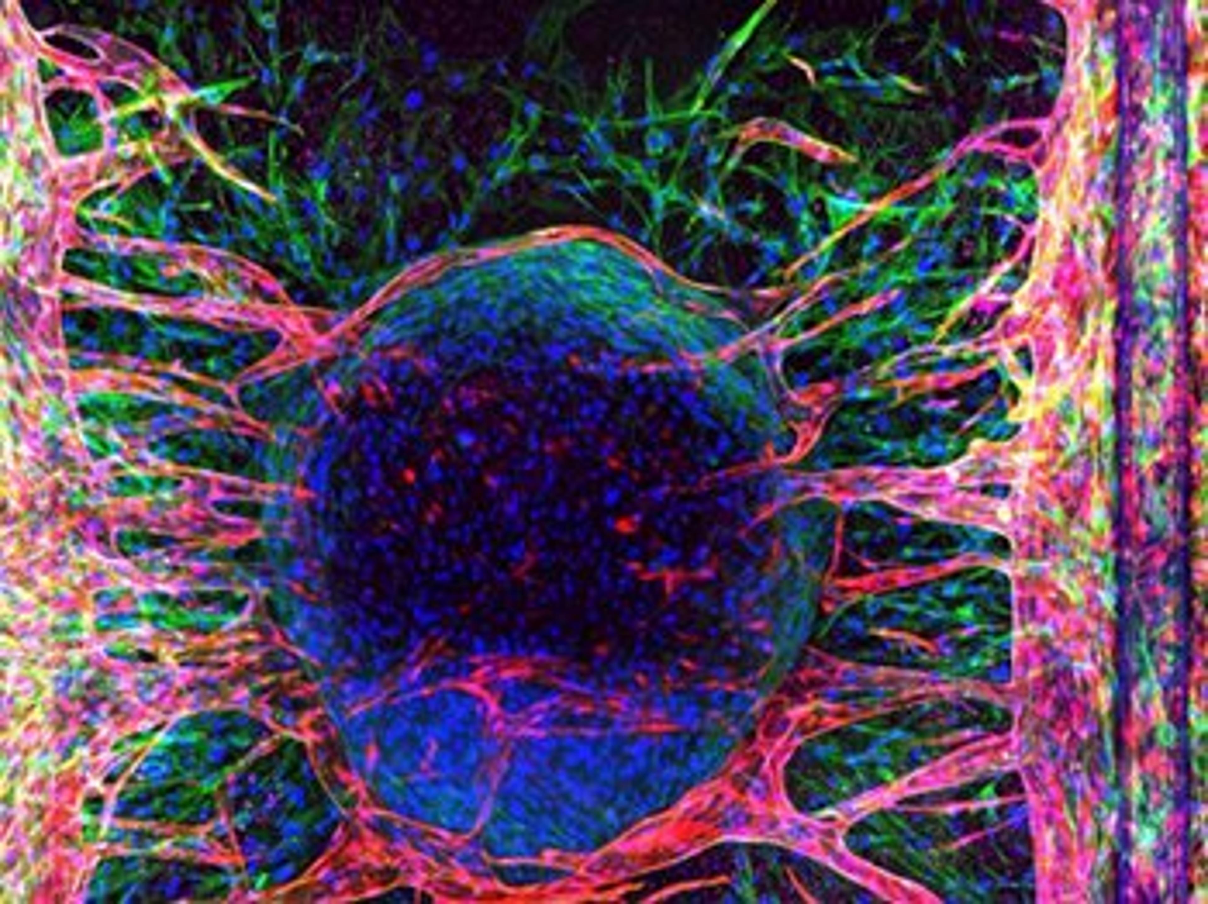
Angiogenesis Research: High-Content Imaging Systems Help Unlock the Full Potential of 3D Tissue Models
Angiogenesis is an important field of research and a focus for cancer therapeutics. In this interview, Dr. Bas Trietsch, CTO, MIMETAS, introduces a new solution for the study of angiogenesis; the OrganoPlate® Graft, an in vitro cell culture microplate platform that allows vascularization of 3D tissues. Hear how Molecular Devices’ ImageXpress Pico Automated Cell Imager and ImageXpress Micro Confocal High-Content Imaging System play a vital role in the development and analysis of 3D tissue models built on the OrganoPlate® Graft.
Video filmed at SLAS Europe 2019 – visit the SelectScience special feature for more videos from the event.
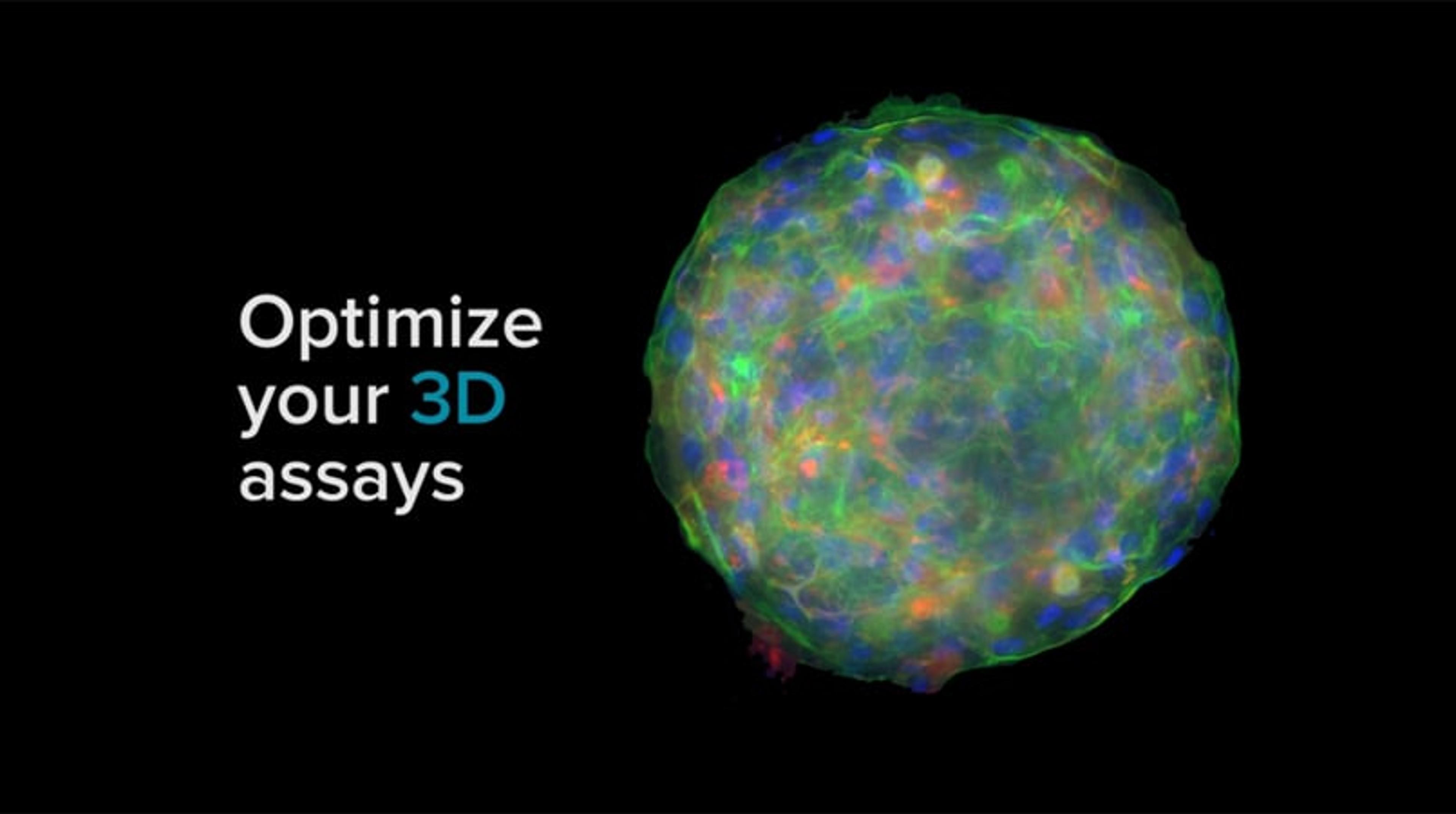
Optimize Your 3D Assays with ImageXpress
Optimize your 3D assay and make your next breakthrough in cellular imaging - with intelligent, innovative imaging solutions.
ImageXpress Micro Confocal Launch Video
This video introduces the main features and benefits of the ImageXpress Micro Confocal system from Molecular Devices. Explore new dimensions with the confocal system for your complex biology, faster, easier and with better results.

Meet the winners of the 2022 Scientists’ Choice Awards for Drug Discovery & Development
Scientific, technological, and communications excellence has been celebrated at SLAS2022 with MOBILion Systems, Agilent Technologies, and Wyatt Technology Corp. among those recognized

Discover the ultimate guide to live-cell imaging
Download our new eBook with all the resources and expert advice you need to choose the best microscopy equipment for your cell analysis applications

5 essential resources to streamline your drug discovery and development processes
Explore the latest application eBooks with top tips on biopharmaceutical characterization and high-content screening
Improving acquisition and analysis of 3D cell model assays with water immersion objectives
Sebastian Peck discusses an automated high-content imaging solution designed to deliver more precise results from complex biological assays
Molecular Devices' imaging system helps Recursion produce the world's largest imaging dataset for COVID-19 research
More than 305,000 high-resolution, multi-channel COVID-19 cellular images are now available to the scientific community
Revolutionizing early drug discovery for immuno-oncology and neurodegenerative disease modeling: High-throughput imaging of 3D models of disease
How Bioneer A/S is using state-of-the-art imaging technology to advance screening of 3D cancer spheroids and monitor neurite outgrowth
The importance of the cellular microenvironment in the development of more efficient drugs against complex diseases
Learn about the 3D imaging technologies advancing phenotypic screening at HCS Pharma
Harnessing the latest imaging technology to advance angiogenesis research
Learn how one biotechnology company is using cutting-edge imaging to improve the visualization of angiogenesis in its 3D tissue models
SLAS Europe 2019: the new technologies changing the future of drug discovery
Solutions for automation, assay development, high-throughput screening, imaging and more