Thermo Scientific™ HCS Studio Cell Analysis Software
Thermo Fisher ScientificIntuitive interfaces, intelligent design and powerful image analysis tools leading to meaningful dynamic measurements at the cell-, well- and field-level.
Intuitive interfaces, intelligent design and powerful image analysis tools leading to meaningful dynamic measurements at the cell-, well- and field-level.
Image processing, manual and automatic measurements over multiple channels, to segmentation and classification tools that help you transform images into quantitative data
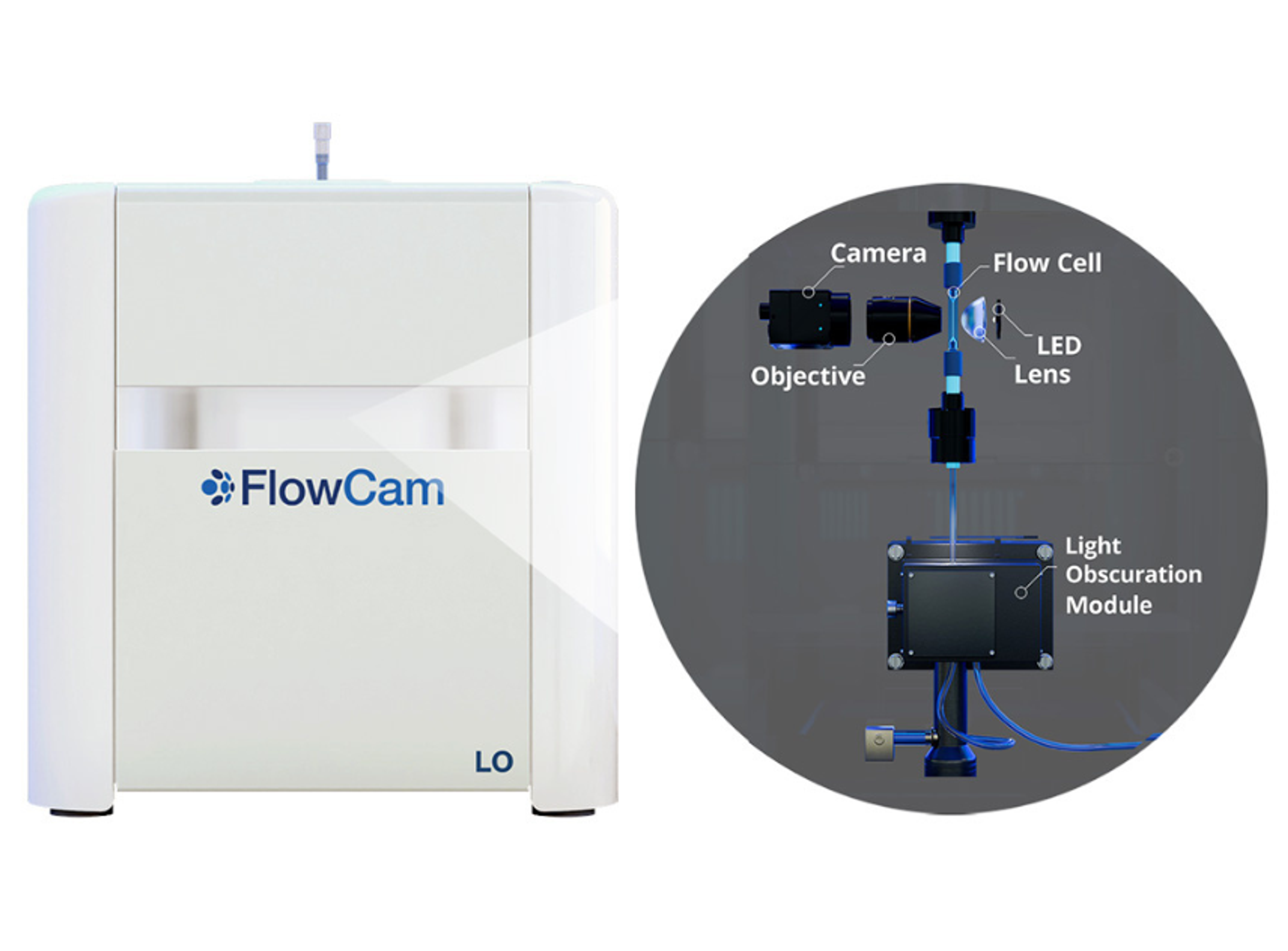
Innovative particle characterization with FlowCam® LO combines flow imaging microscopy (FIM) and light obscuration (LO) into a single analytical solution. Beyond the compendial light obscuration method to fulfill USP <787> and <788> requirements, flow imaging microscopy provides an orthogonal method for quality control of subvisible particulate matter.
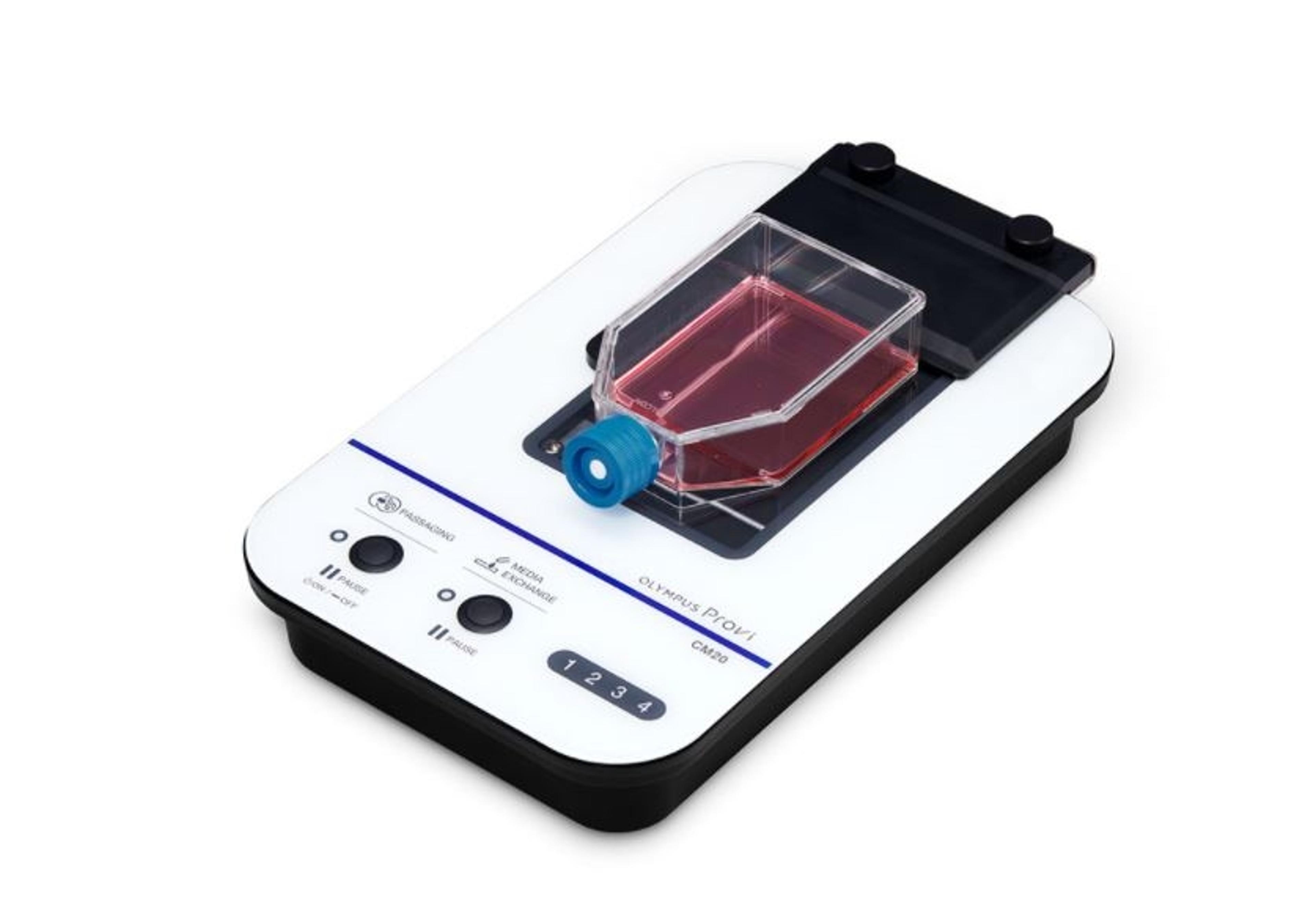
The CM20 system provides quantitative data remotely - place the head and your cell cultures in the incubator, and the system will periodically scan it, count the number of cells, and determine confluency. The data are wirelessly communicated to a PC or a tablet through an optional router, so you can monitor your cultures' progress without entering a clean room.
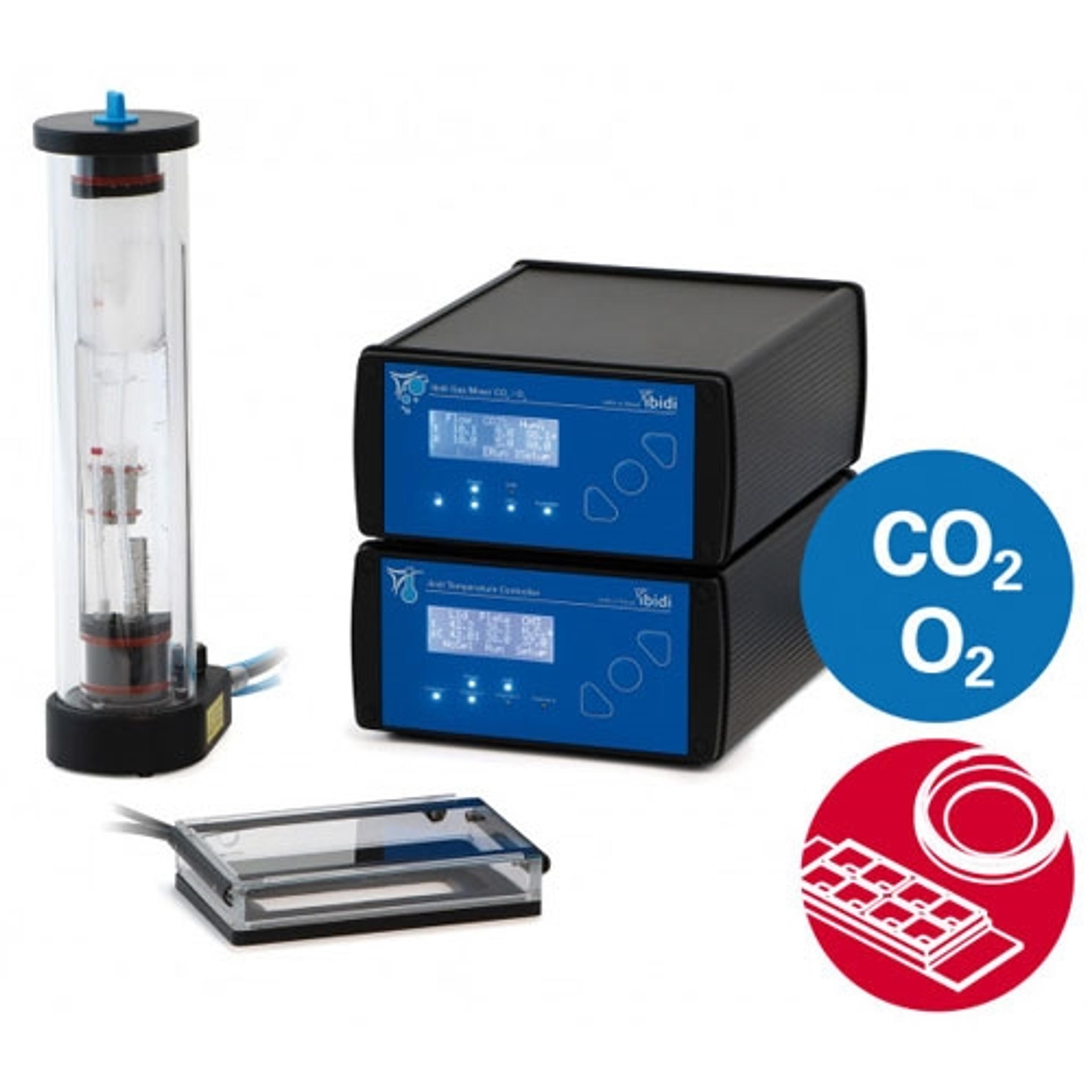
Easy setup directly on your microscope: perform live cell imaging in dishes, slides, or multiwell plates. The ibidi Stage Top Incubation Systems fit every standard inverted microscope and include CO 2 and O 2 control as well as actively controlled humidity. They are ideal for all live cell imaging applications and available for single slides and dishes as well as for multiwell plates.
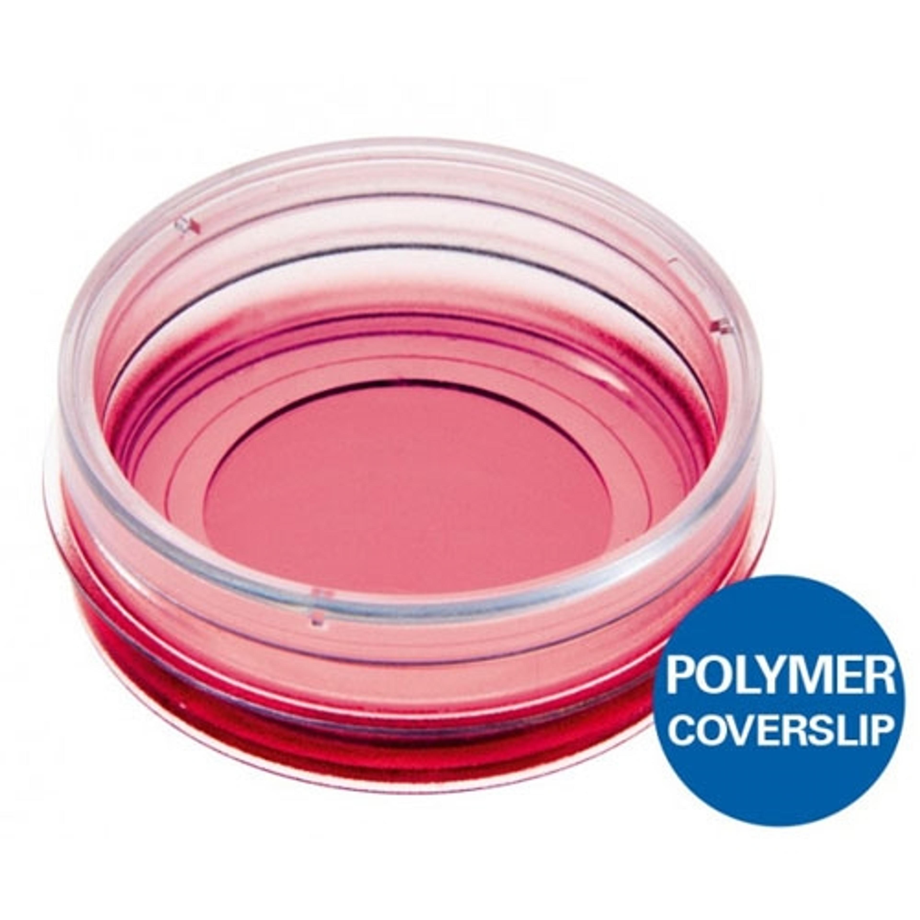
Use this versatile 35 mm imaging Dish for high-end microscopy through the #1.5 coverslip bottom.
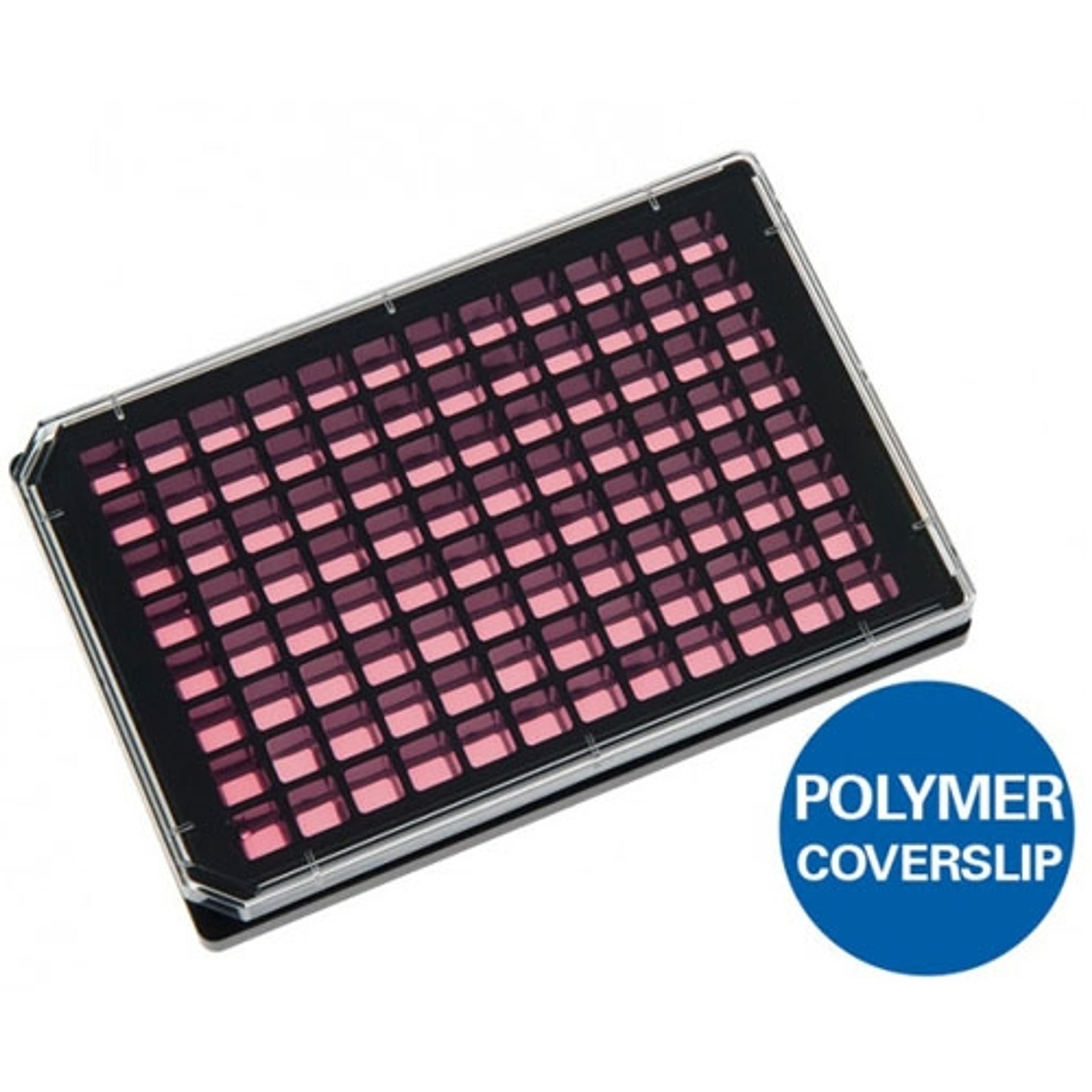
Use this Multiwell plate with black walls, square wells, and a flat and clear coverslip bottom (#1.5 ibidi Polymer Coverslip) for high-throughput applications in cell-based assays. Also available with 24 wells.
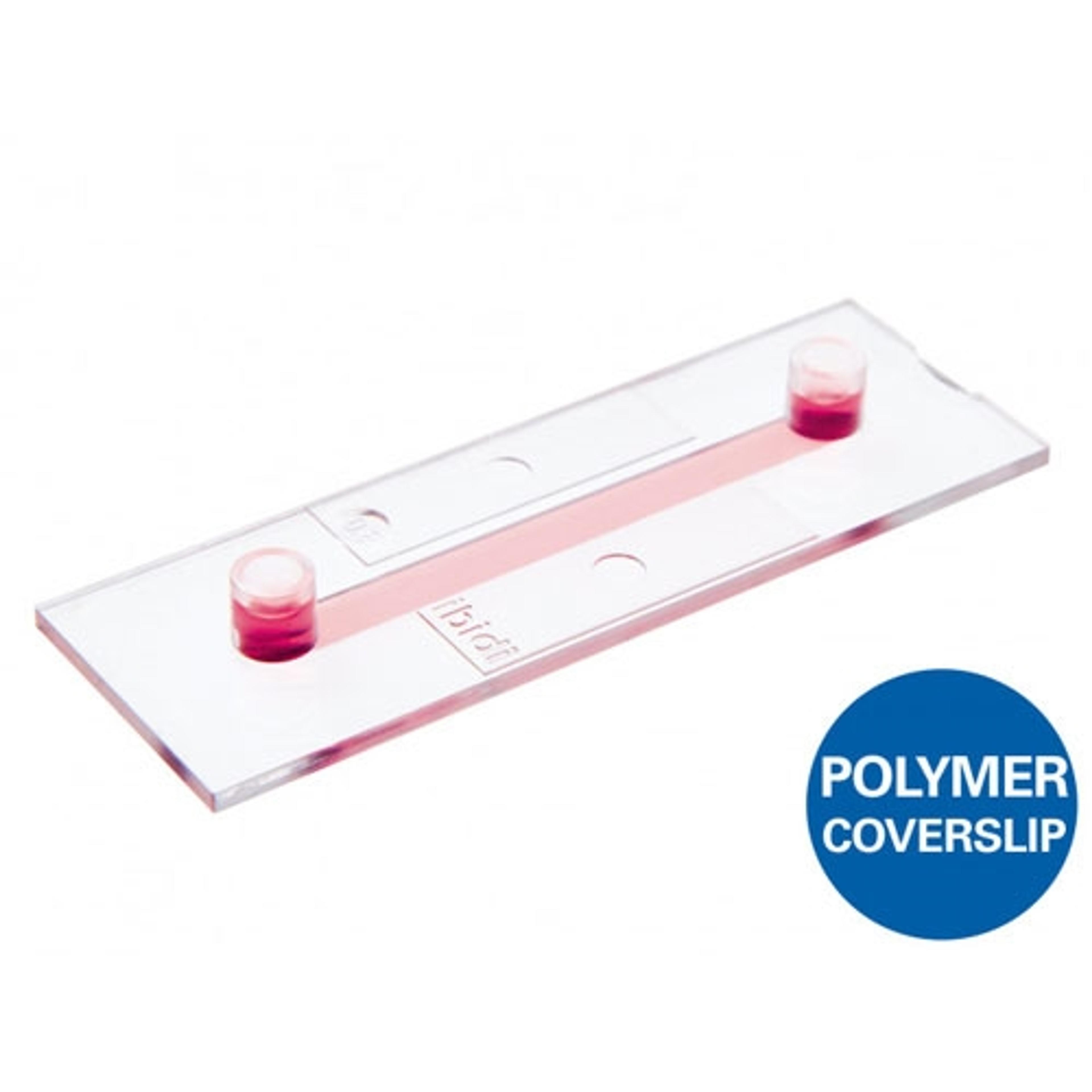
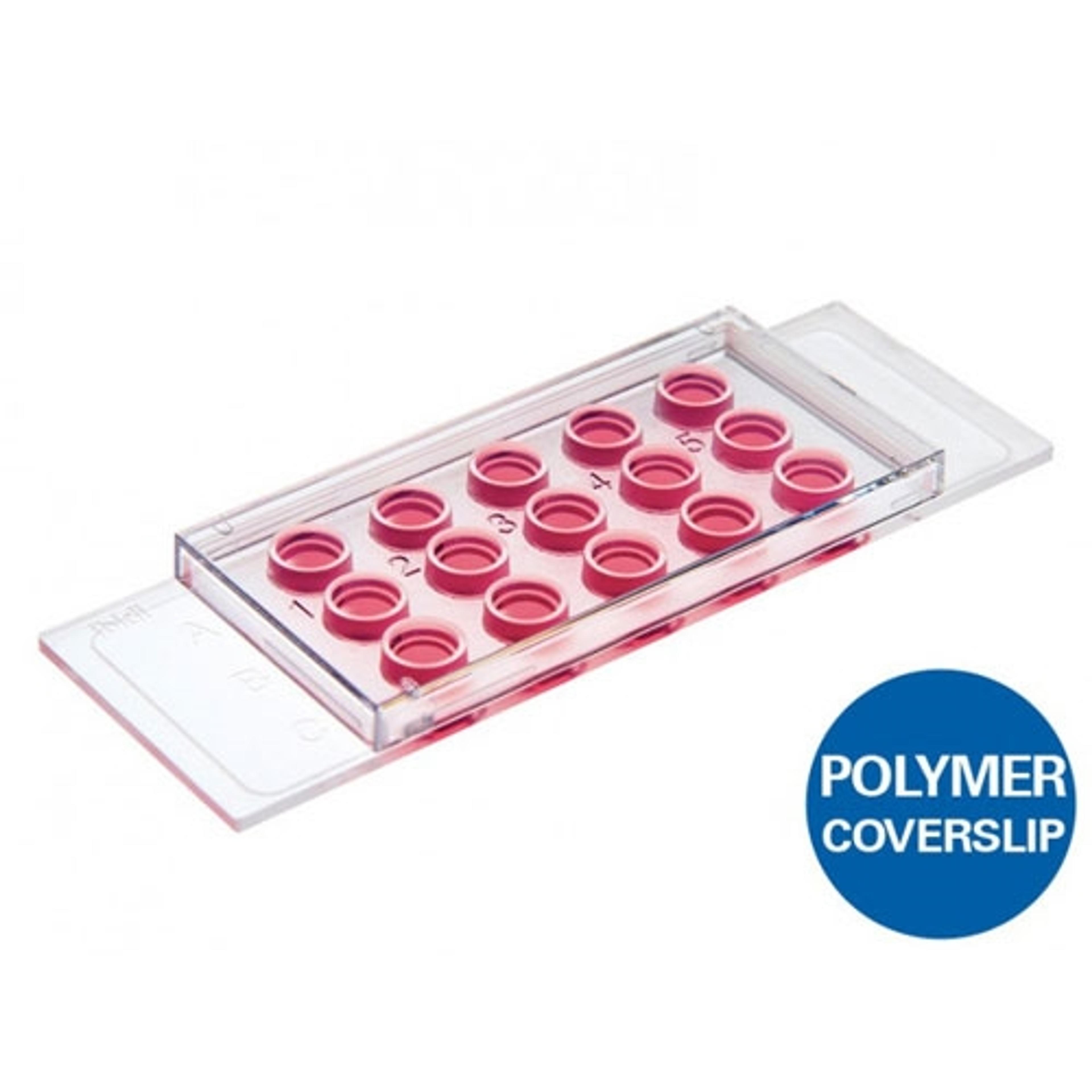
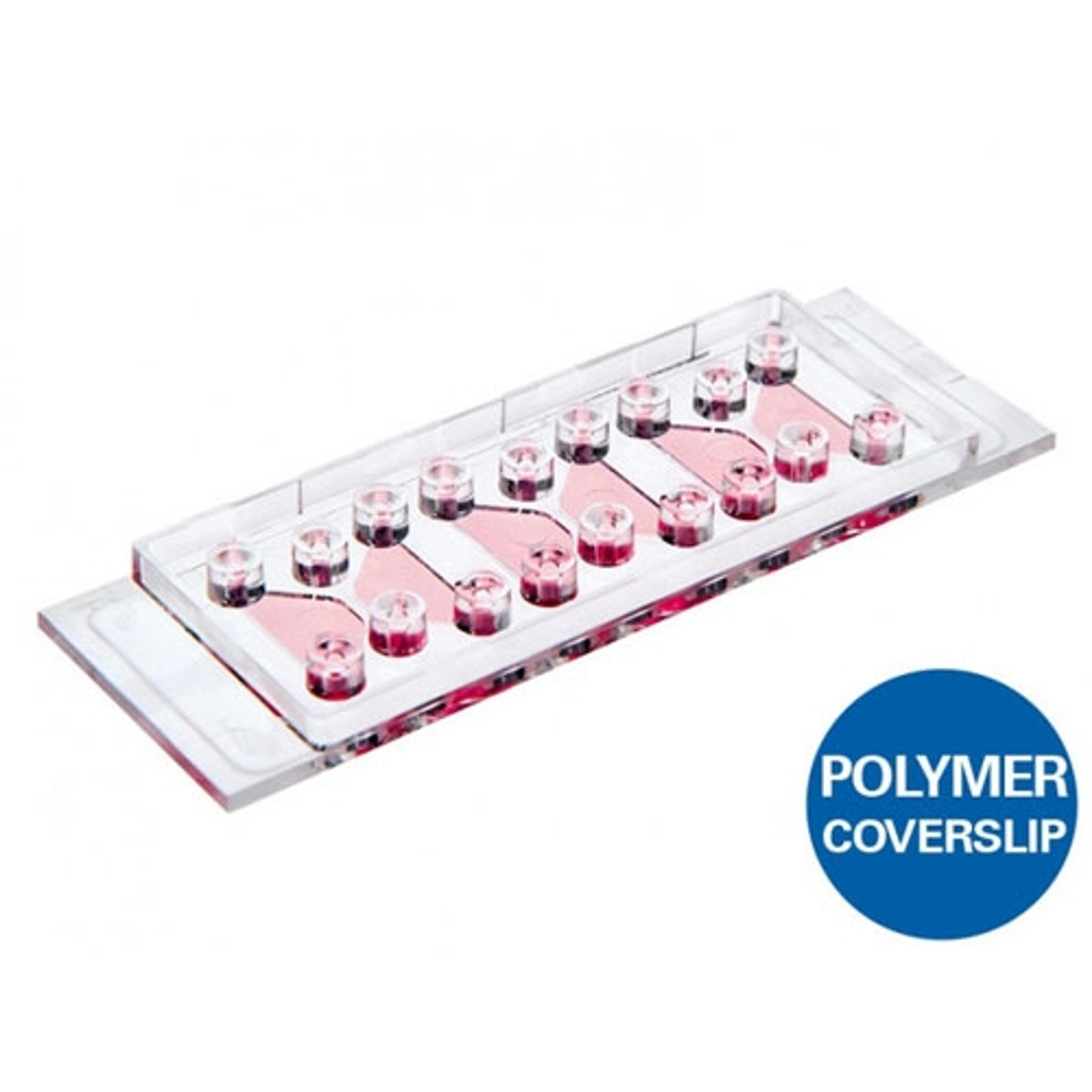
Use this channel microscopy slide with a coverslip bottom (#1.5 ibidi Polymer or #1.5H glass) for immunofluorescence, cell culture under flow, live cell imaging, and high resolution microscopy on inverted microscopes. Available in various channel heights.
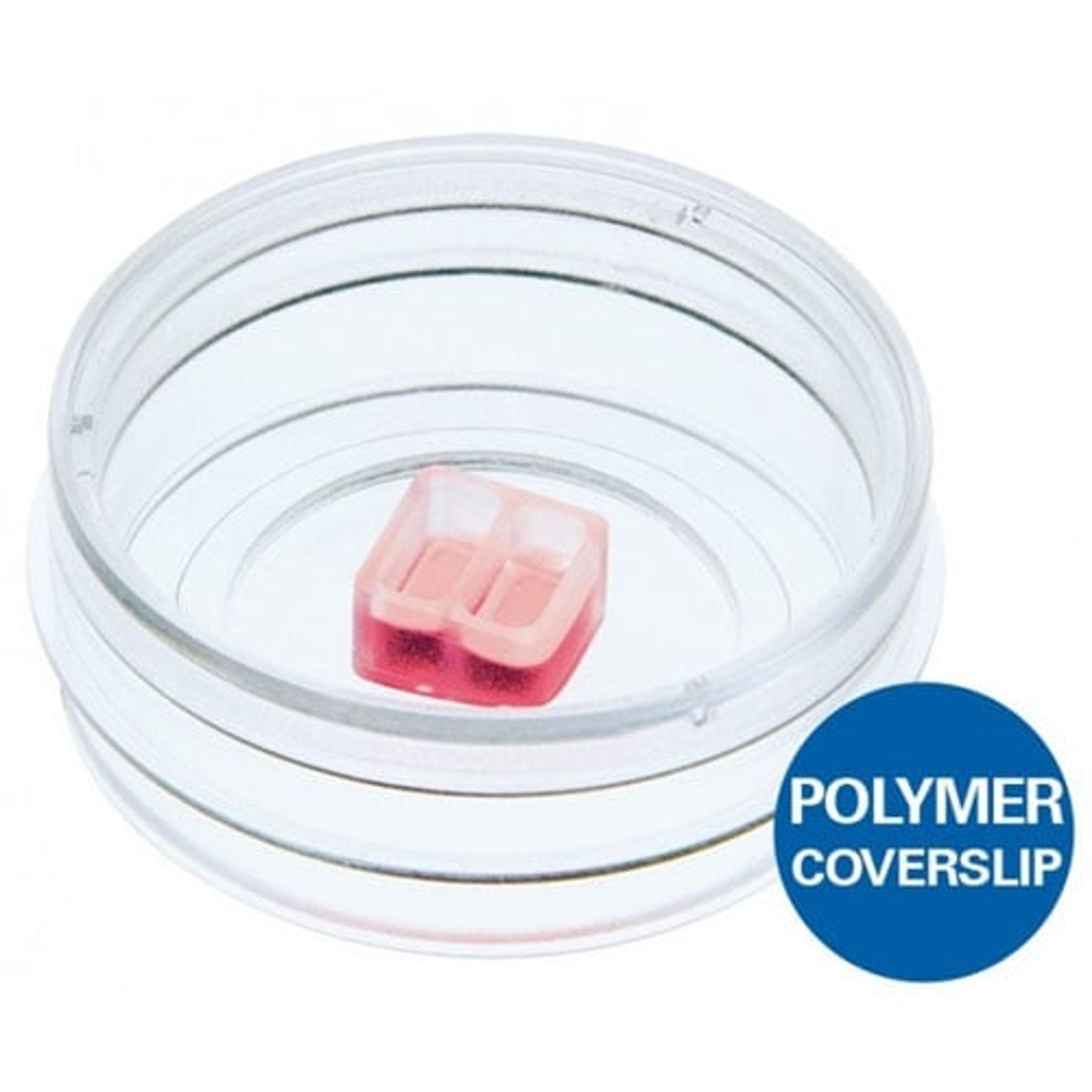
Use these silicone inserts with a defined cell-free gap for wound healing assays, migration assays, 2D invasion assays, and co-cultivation of cells. Available with 2, 3, or 4 wells.
Use this slide to investigate angiogenesis in tube formation assays. Also ideal for 3D cell culture and immunofluorescence staining. Also available in a 96 well format.
Investigate chemotaxis of fast or slow migrating adherent cells and non-adherent cells in 2D or 3D gel matrices.
Access the future of AI microscopy. Using state-of-the-art, AI-first software architecture, Aivia is a uniquely innovative and complete 2-to-5D image visualization, analysis and interpretation platform designed for the reliable processing and reconstruction of highly complex images in just minutes. Aivia provides powerful 2D & 3D spatial relational analysis for objects across diverse types, morphological complexities, and hie…
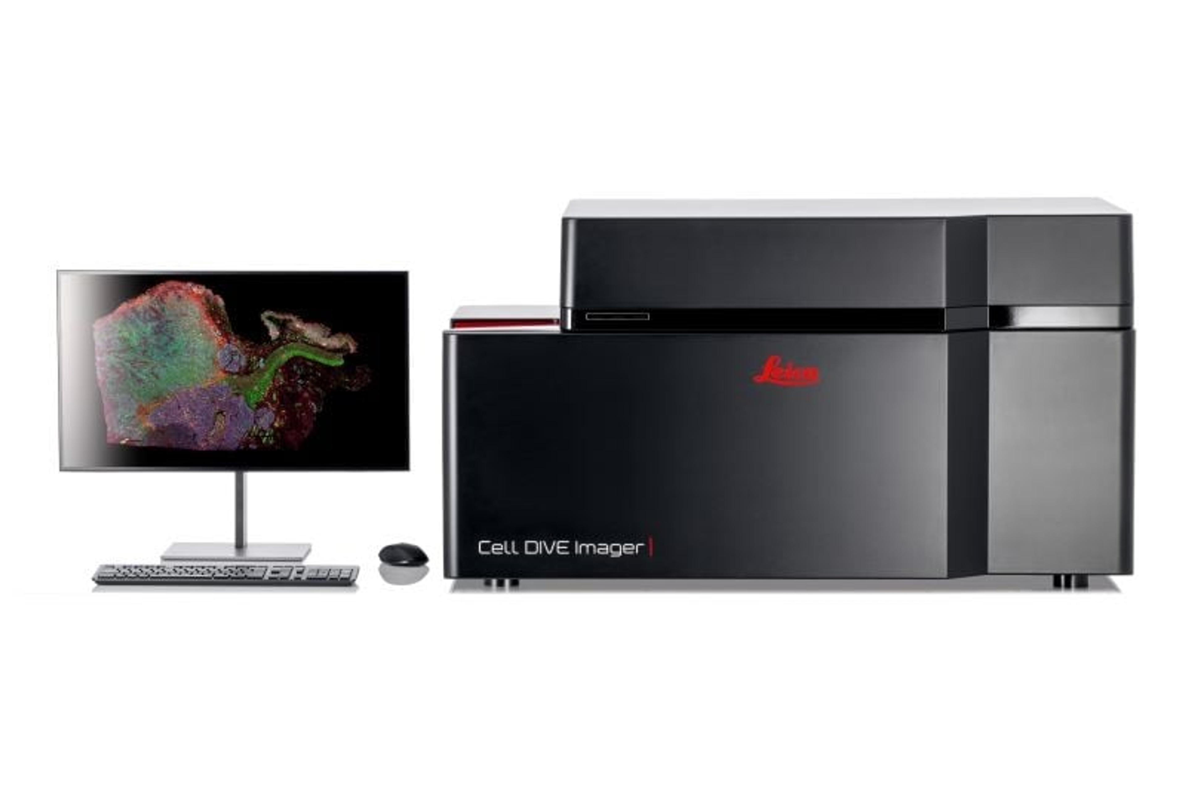
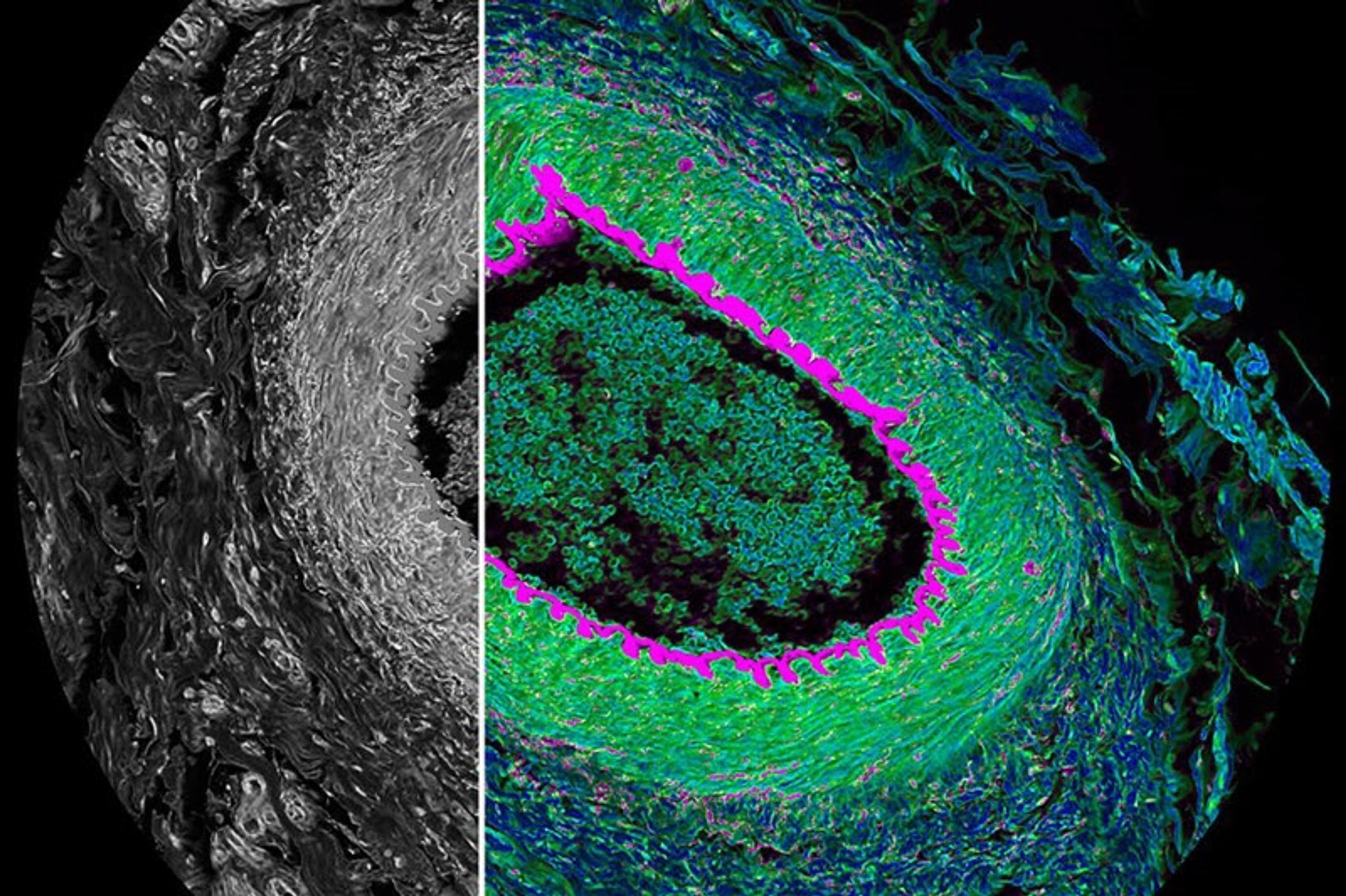
Cell DIVE multiplexed imaging is an antibody-based hyperplexed technique addressing spatial cell biology and function within the tumor microenvironment.
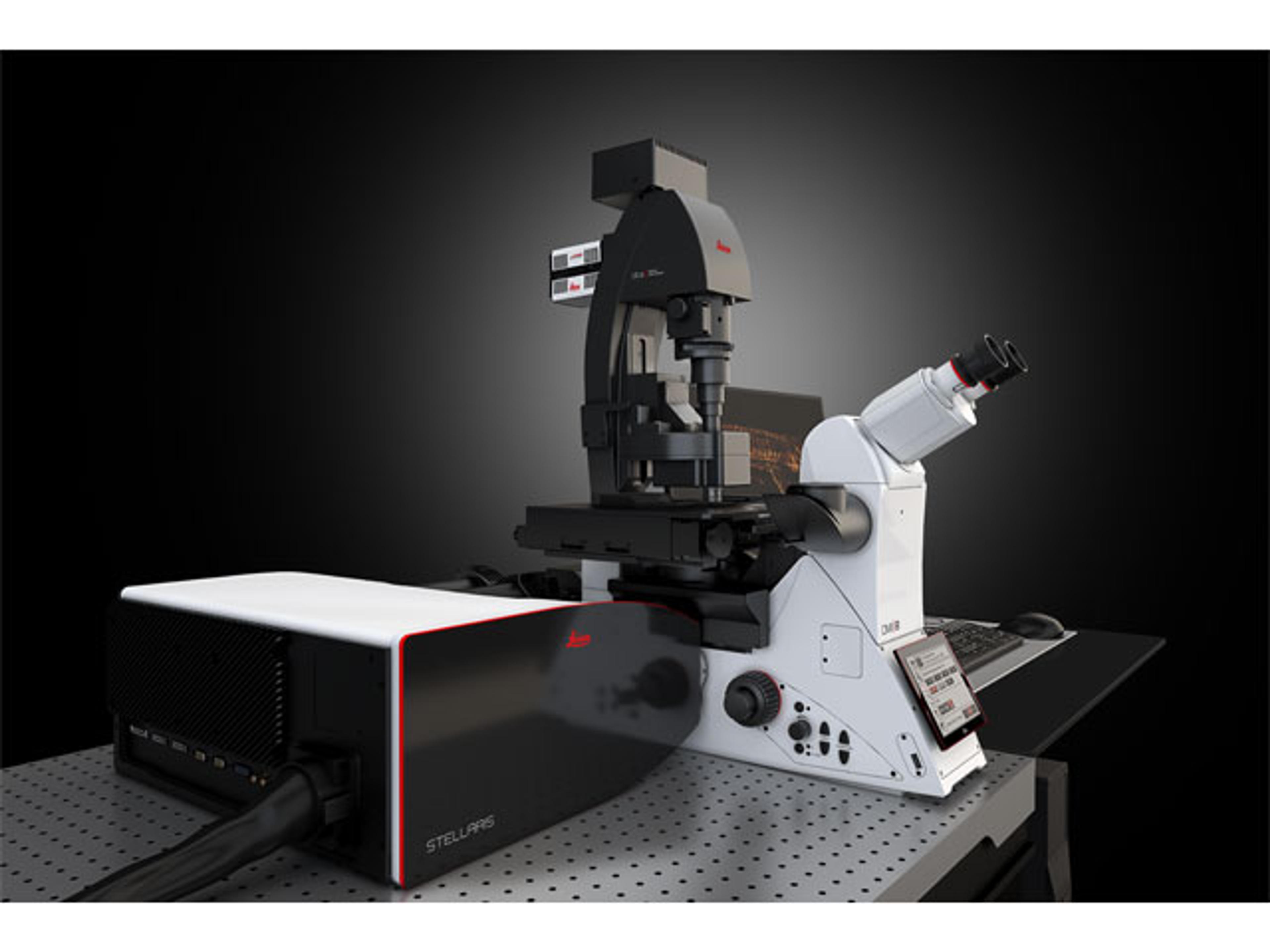
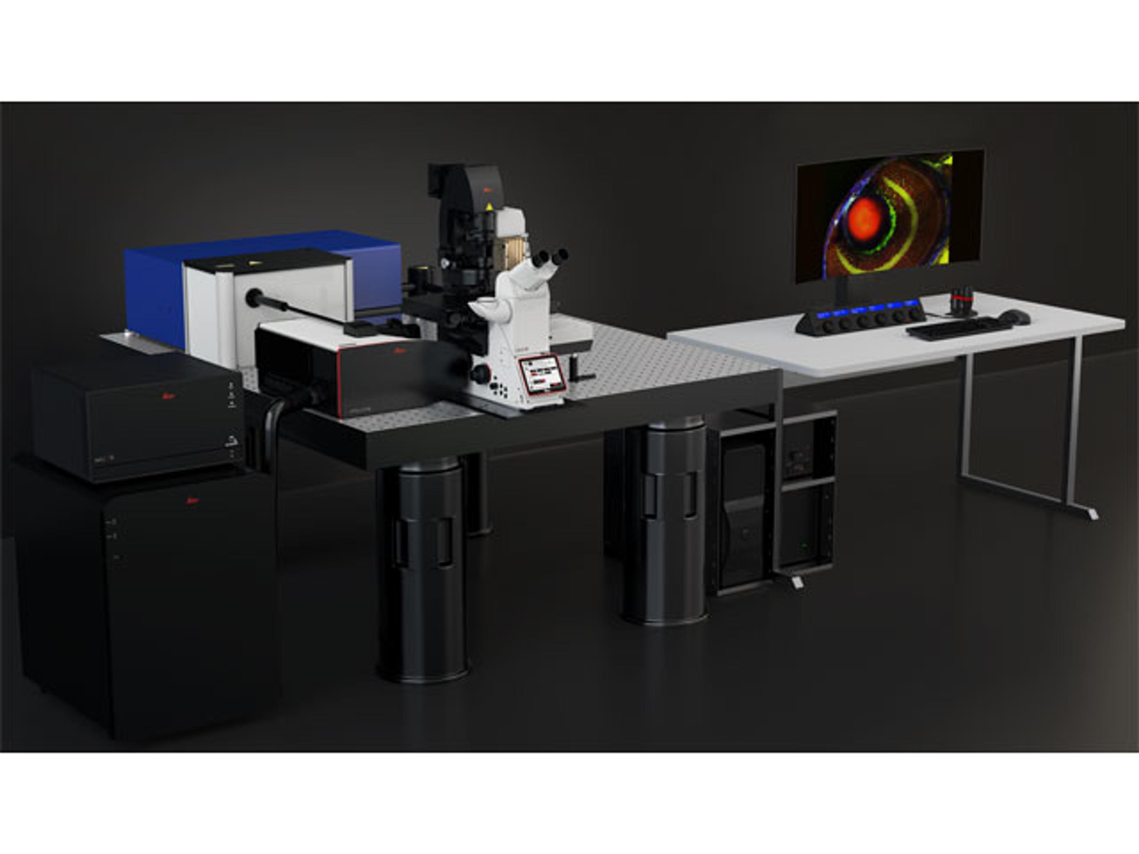
STELLARIS 8 FALCON (FAst Lifetime CONtrast) is the future of functional imaging. Harness the power of fluorescence lifetime to investigate cellular physiology and explore dynamics in living cells.
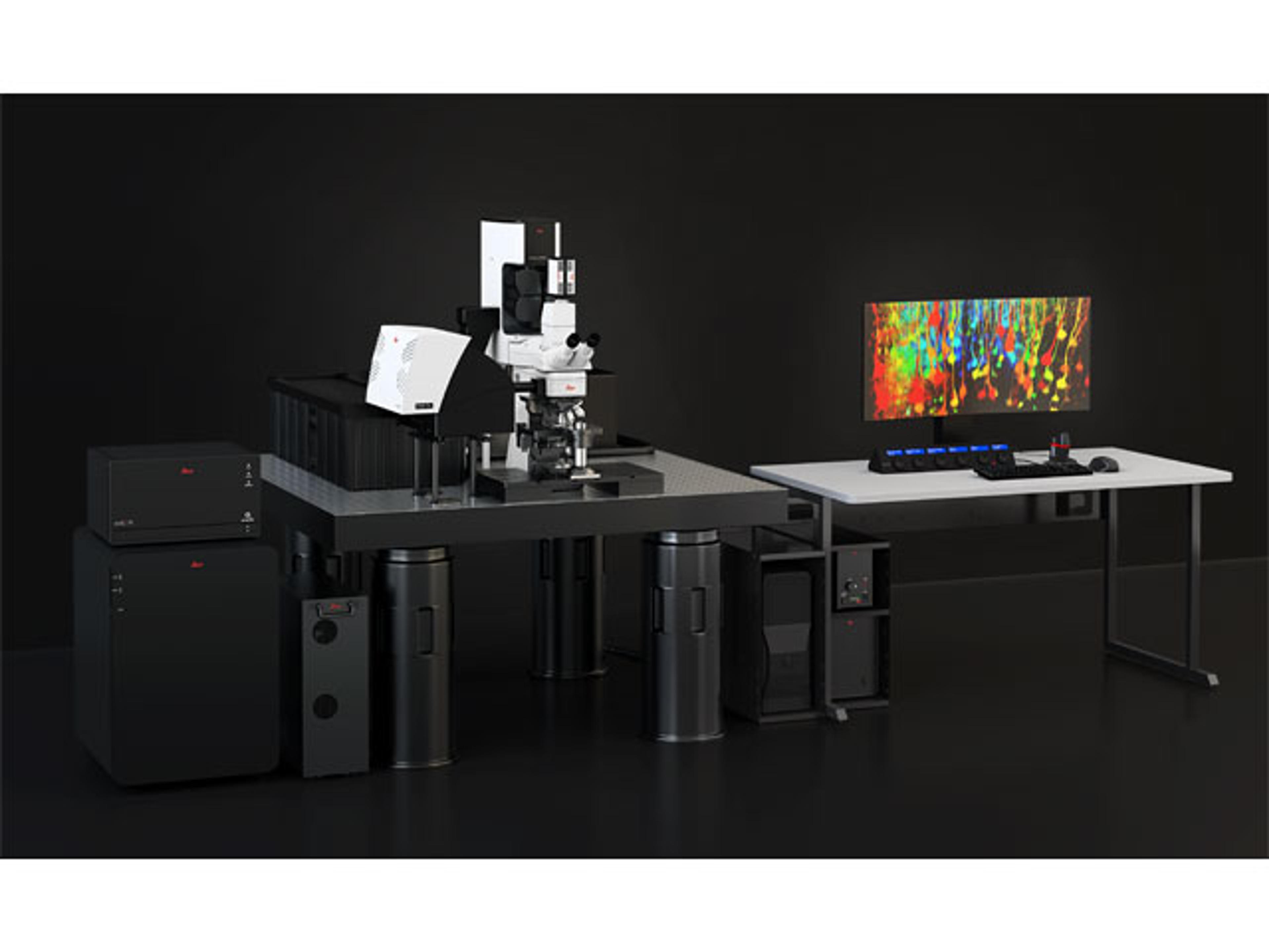
DIVE and STELLARIS have now merged to provide you with the power of flexible multicolor multiphoton imaging.
STELLARIS LightSheet (DLS) unites in one place a confocal system and a light sheet microscope – a unique combination aimed to make your research more versatile. DLS and STELLARIS enable even gentler imaging giving you the ability to perform fast and gentle volumetric light sheet imaging and to improve your live cell imaging applications.
When you need to study structures that cannot be visualized with traditional fluorescent microscopy methods, the STELLARIS 8 Coherent Raman Scattering (CRS) microscope enables you to implement label-free chemical imaging into your workflow to answer those challenging research questions.
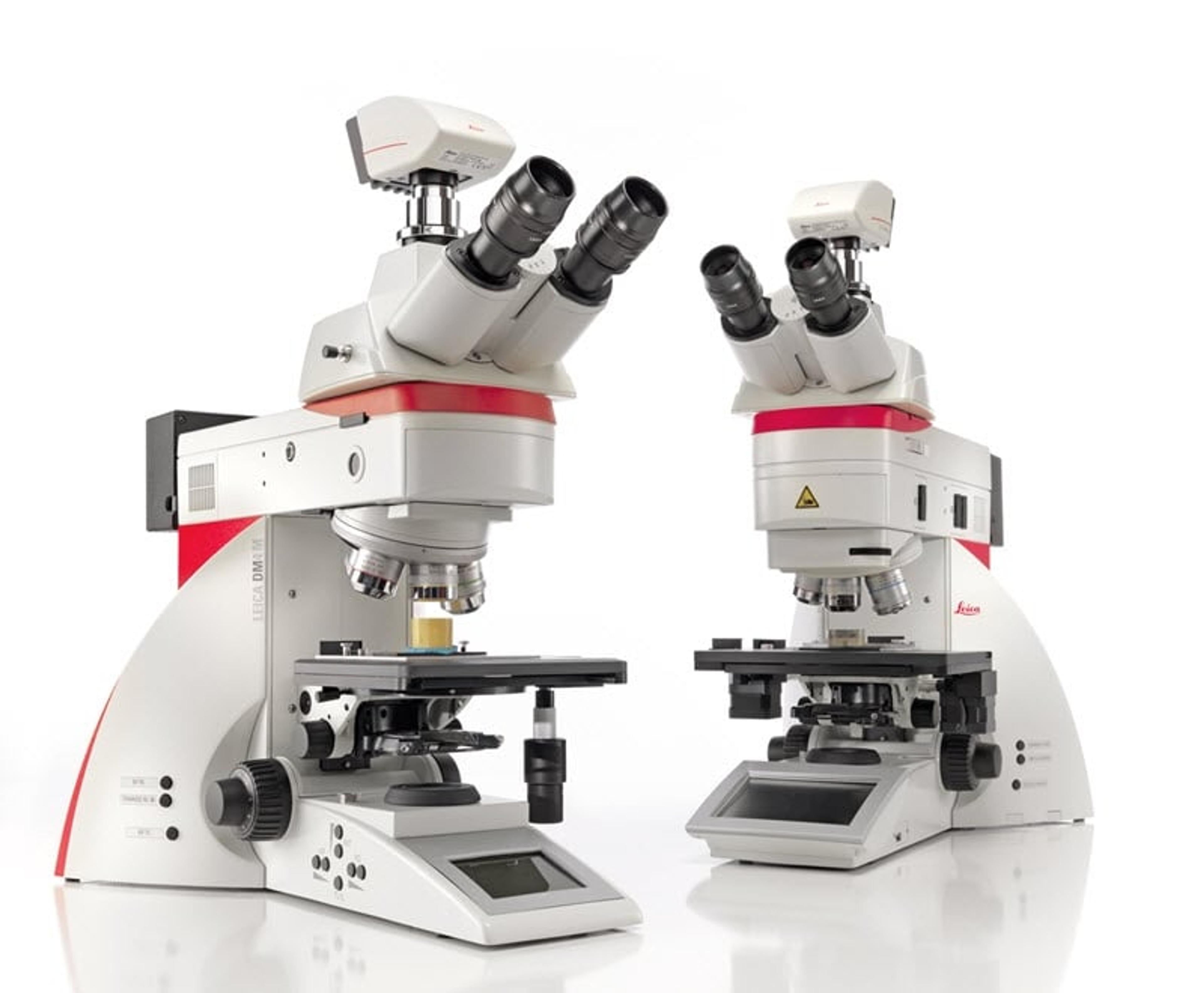
Do you need to image, measure, and analyze similar features across many samples and materials science and analysis? The Leica DM4 M and Leica DM6 M are the Industrial Microscopes for you, whether microscopy novice or professional.
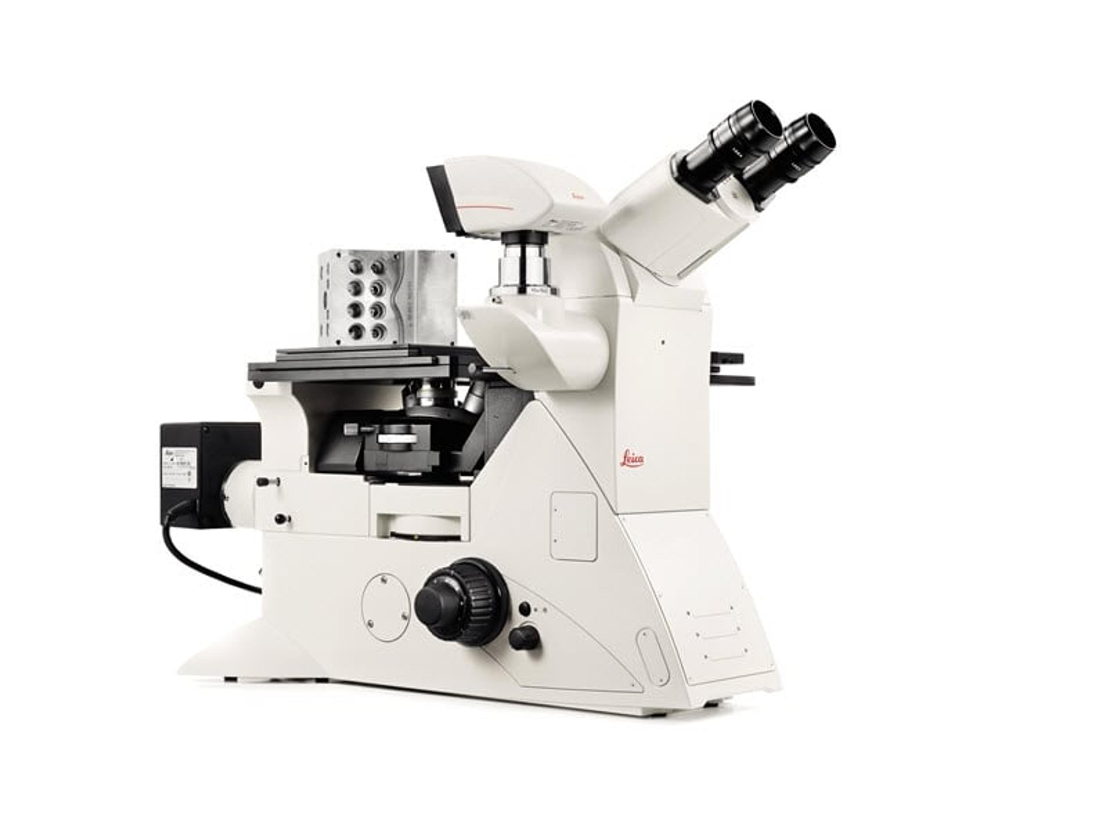
Inverted Microscopes for Industry
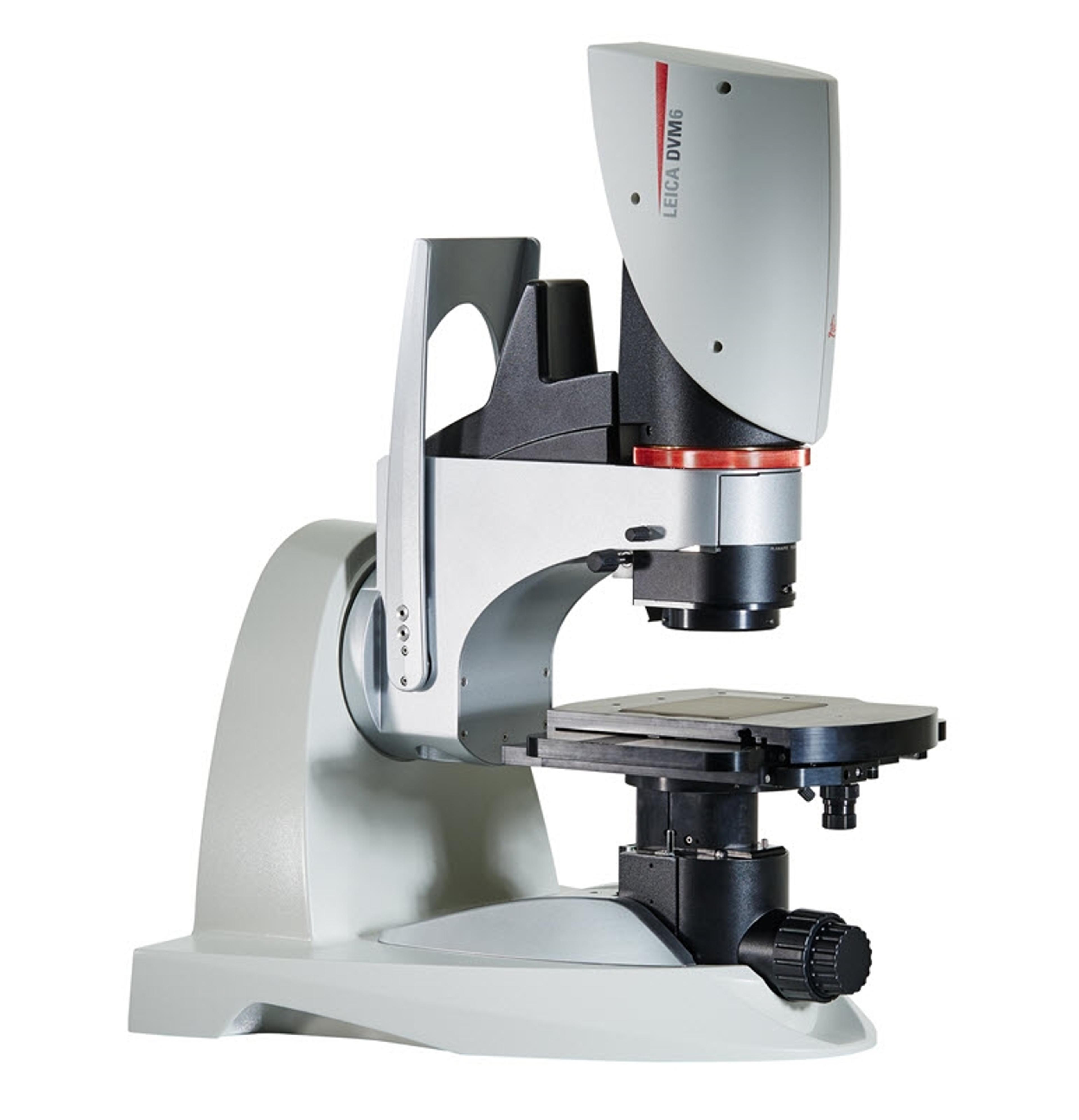
If you work in quality control/assurance, failure analysis, research and development, or in forensics, searching for the detail can take up a lot of your time in microscopy. The DVM6 digital microscope is a fast, reliable and easy to use solution that combines outstanding optics, intuitive operation, and smart software to save you time.
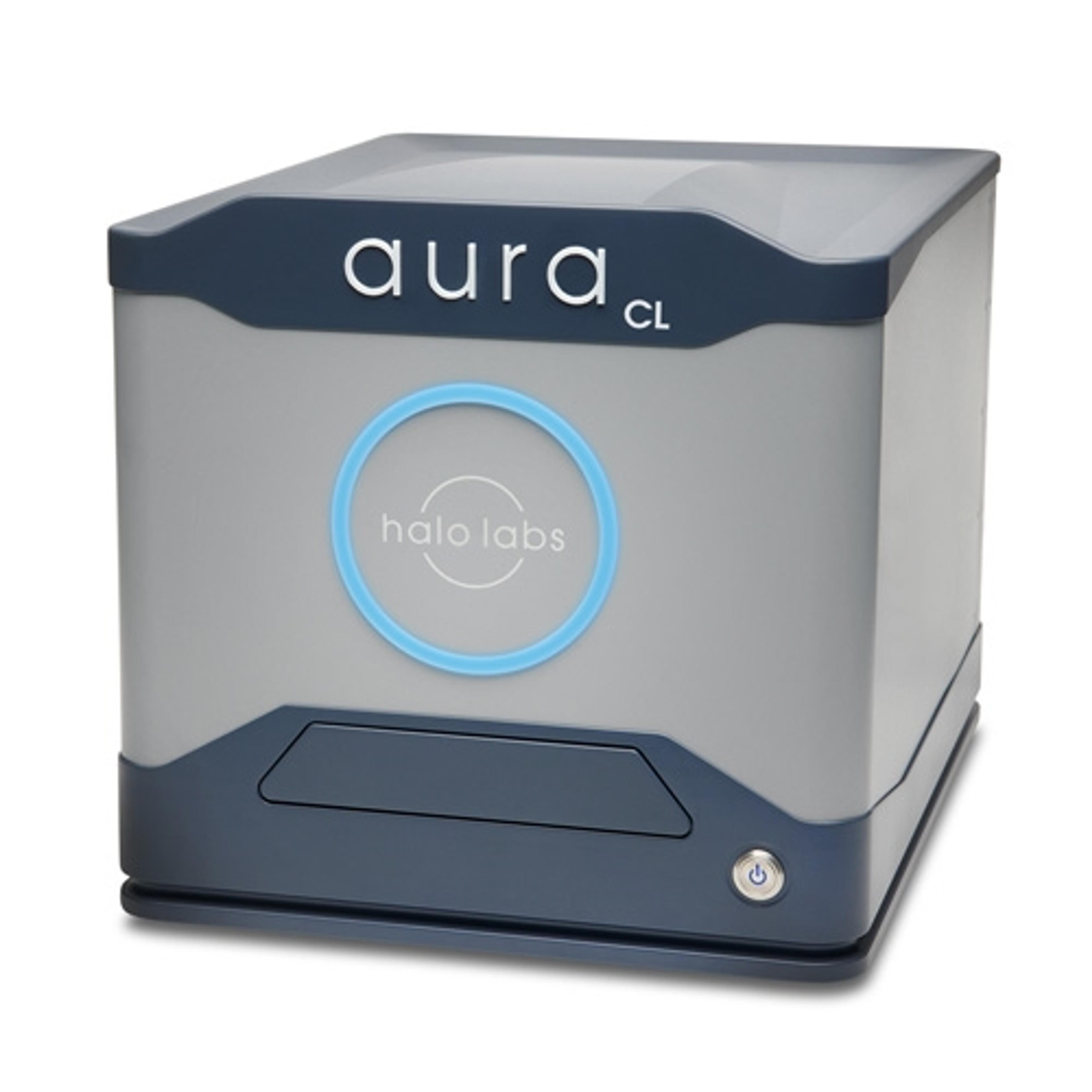
Aura® CL systems couples Fluorescent Membrane Microscopy and Backgrounded Membrane Imaging to help you understand what’s in your cell therapy. It detects, counts, sizes, and IDs cellular aggregates and subvisible particles and ID’s cellular from other particles without any machine learning so you can maximize the purity, safety, and efficacy of your therapeutic.
Aura® is the particle and aggregate detection system that combines Backgrounded Membrane Imaging which images 100% of your sample to give you count, size, and morphological information, with up to 2 channels of Fluorescence Membrane Microscopy. Identify if particles are protein or not with the first FMM channel and determine if your sample contains lipids, hydrophobic entities, or other aggregates of your choice with a second…
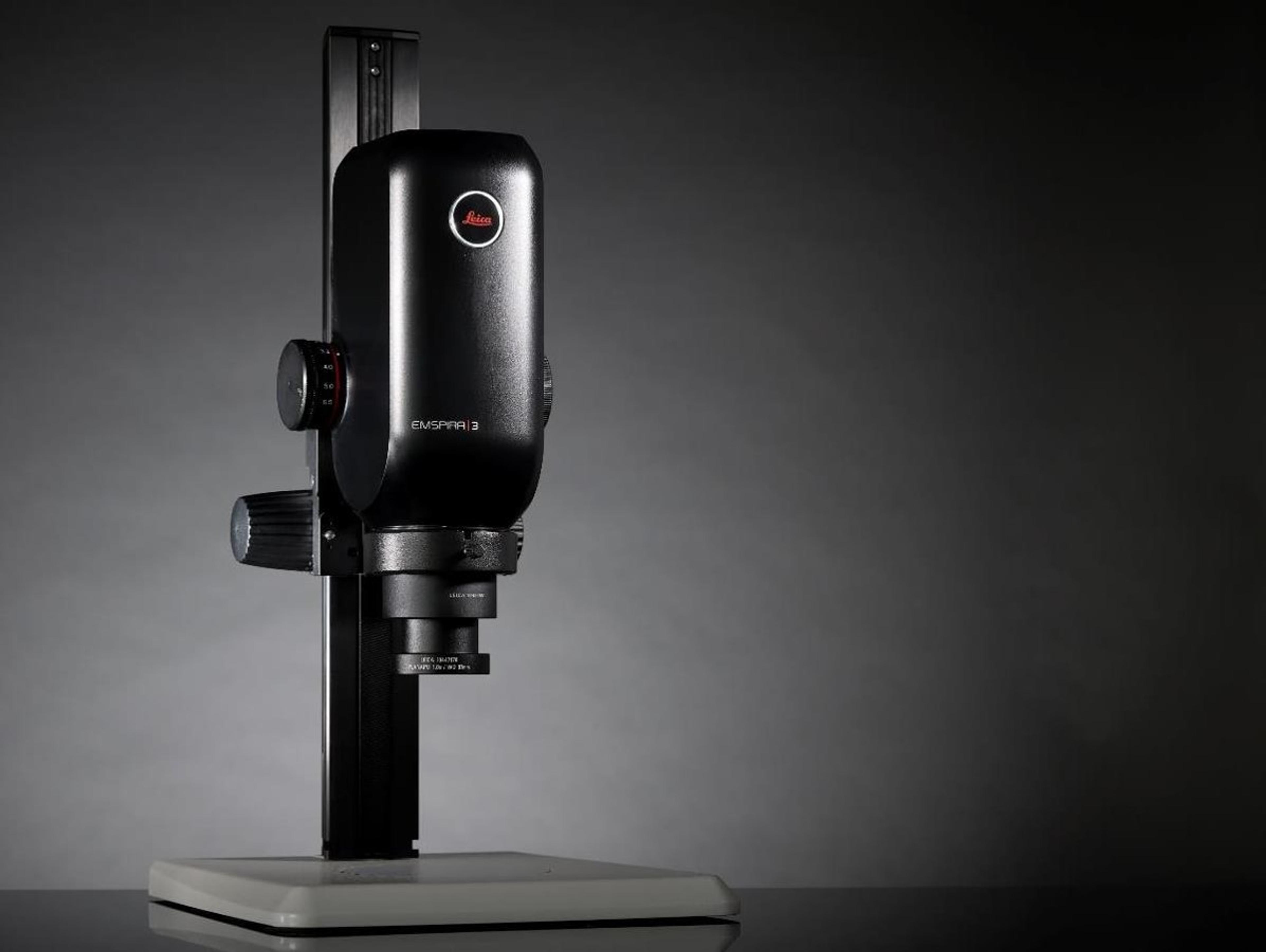
An all-in-one digital inspection solution from Leica Microsystems. The Emspira 3 digital microscope empowers users to streamline inspection processes, cover inspection needs flexibly, and work in a confident and reliable way, with a single system.
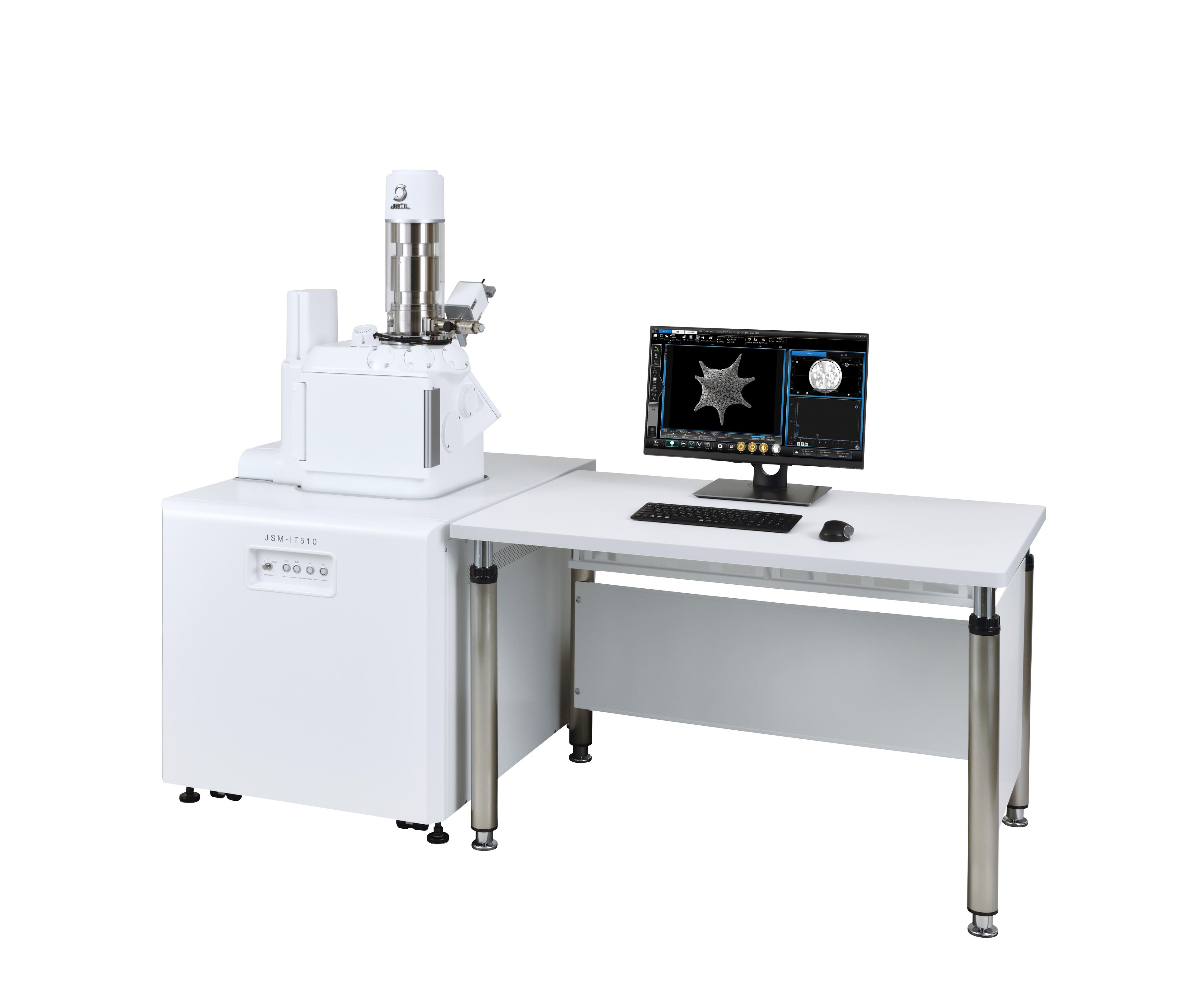
The newest generation InTouchScope™ SEM enhances productivity with Simple SEM automation. Automated image collection at multiple locations and conditions simplifies workflow for the most routine tasks. Embedded Signal Depth display enhances understanding of analytical spatial resolution. Seamless navigation from optical to SEM imaging, Live EDS (both spectrum and X-ray maps) and auto functions from alignment to focus produce…