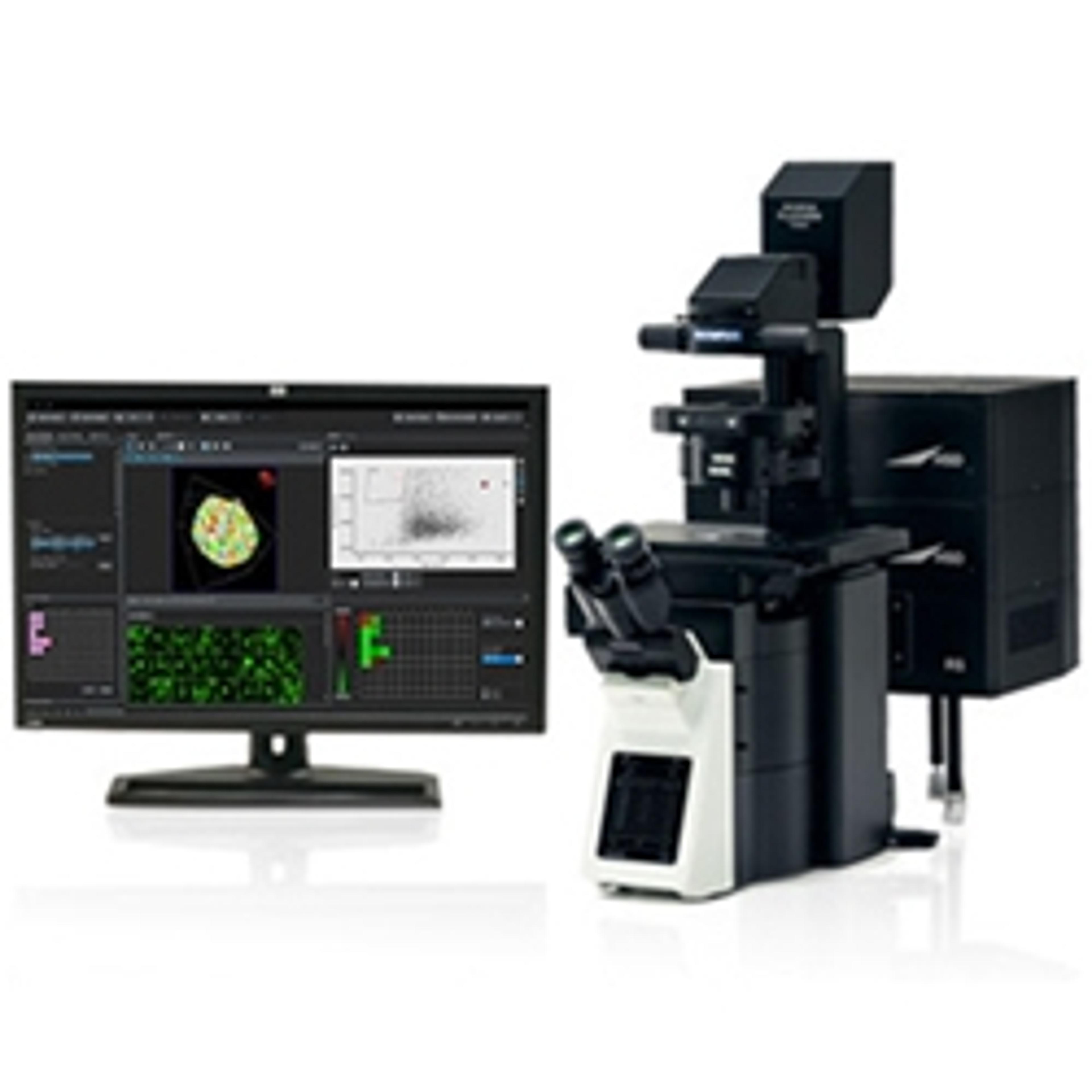
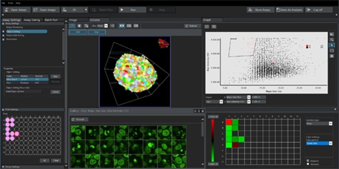
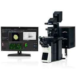
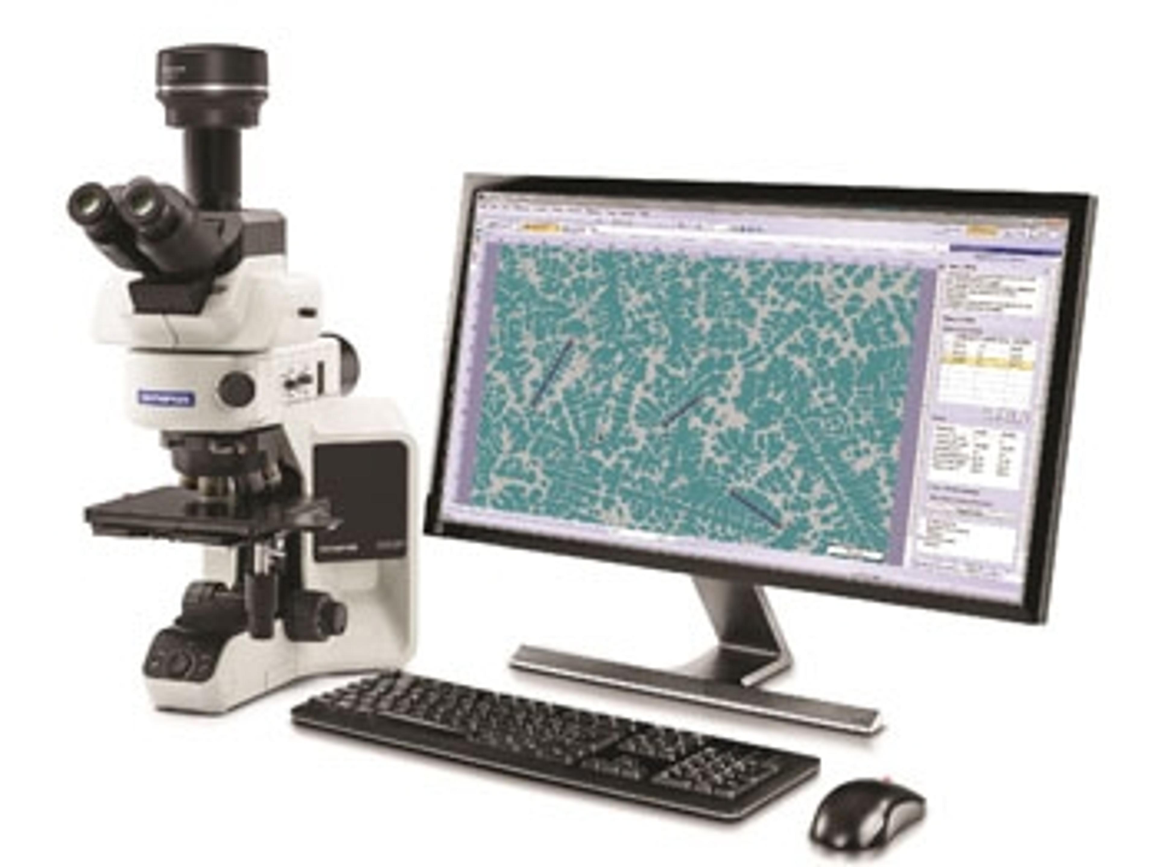
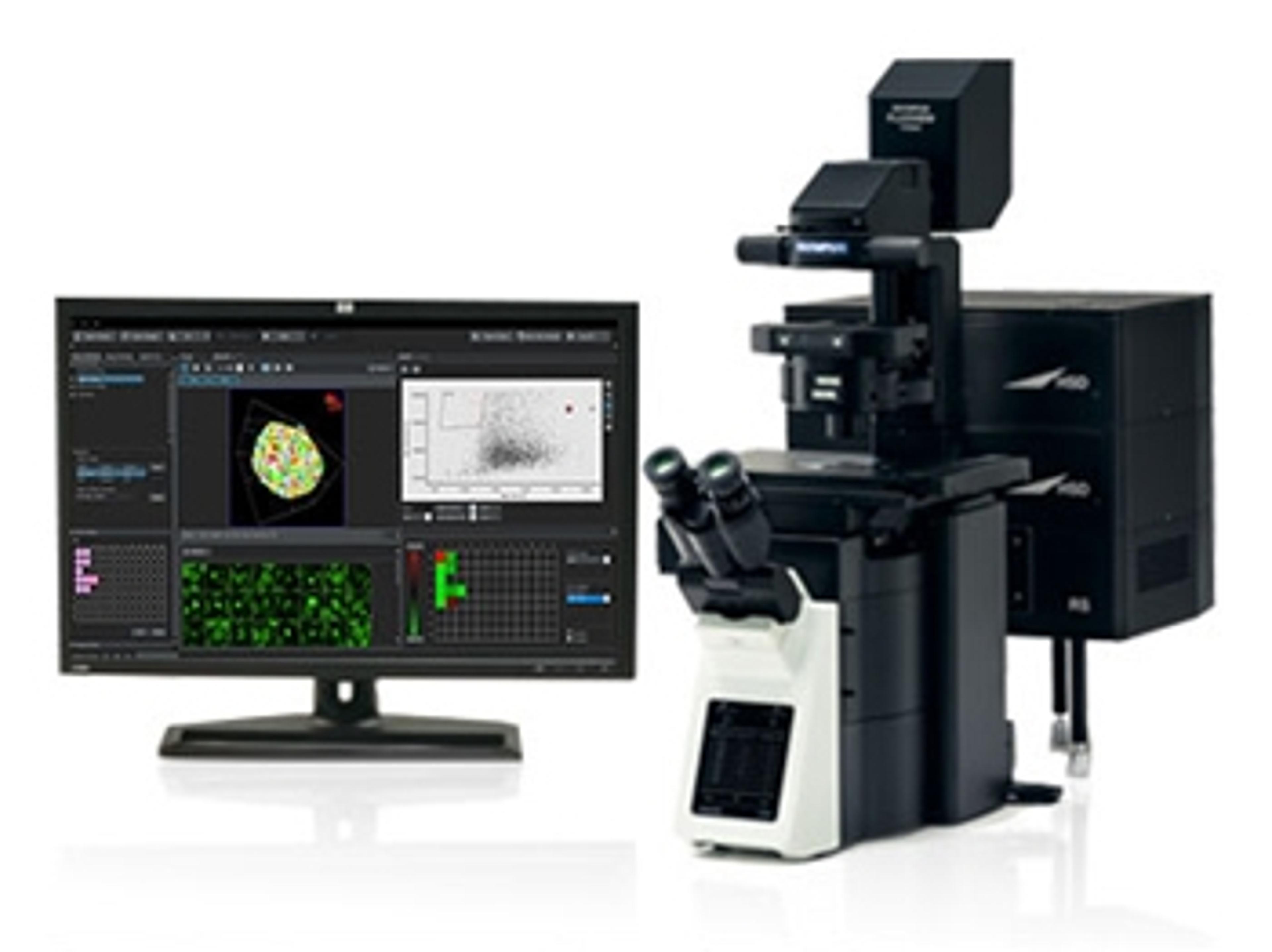
NoviSight™ 3D High Content Analysis Software
Improve the speed of your discovery


The supplier does not provide quotations for this product through SelectScience. You can search for similar products in our Product Directory.
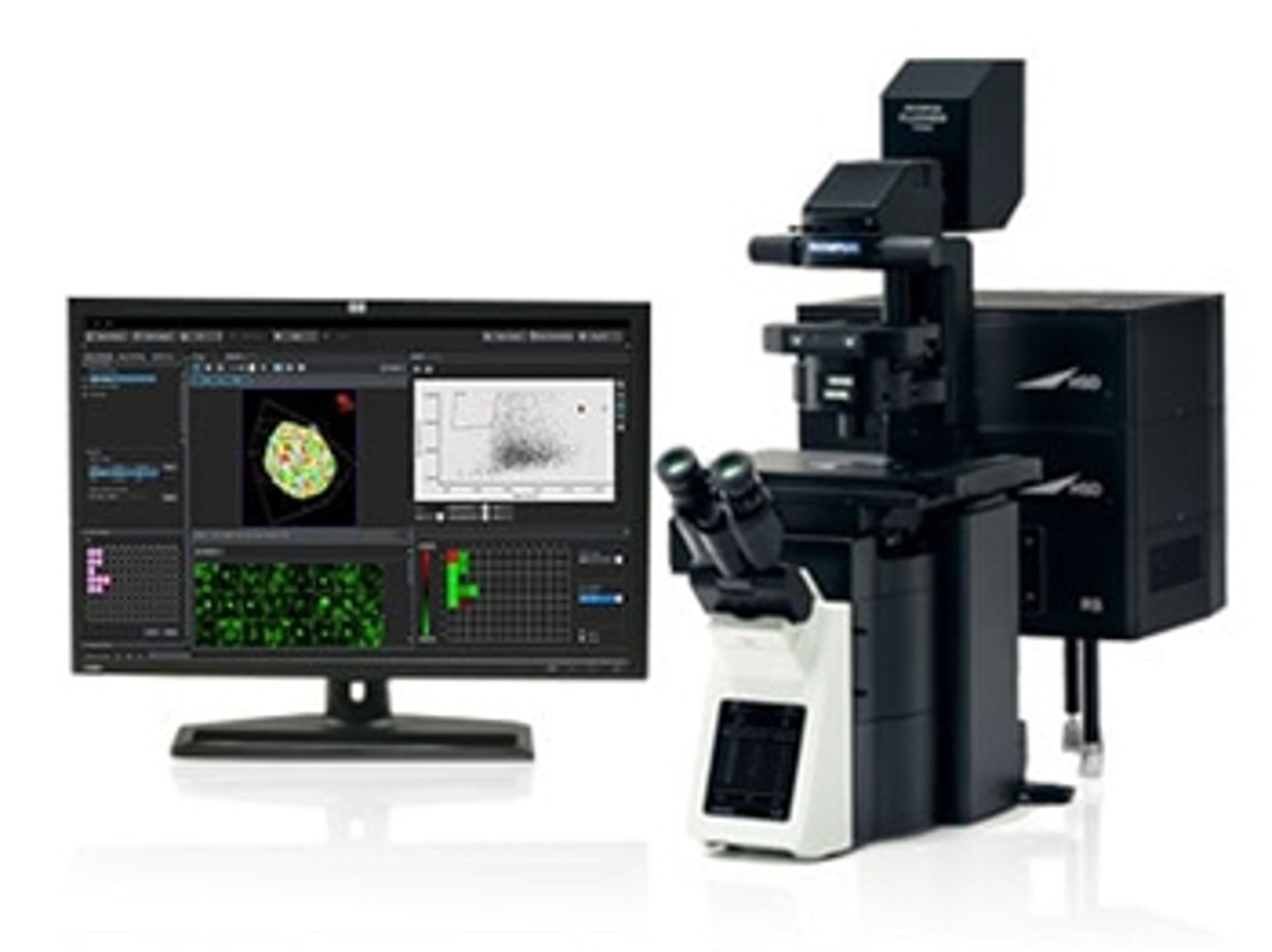
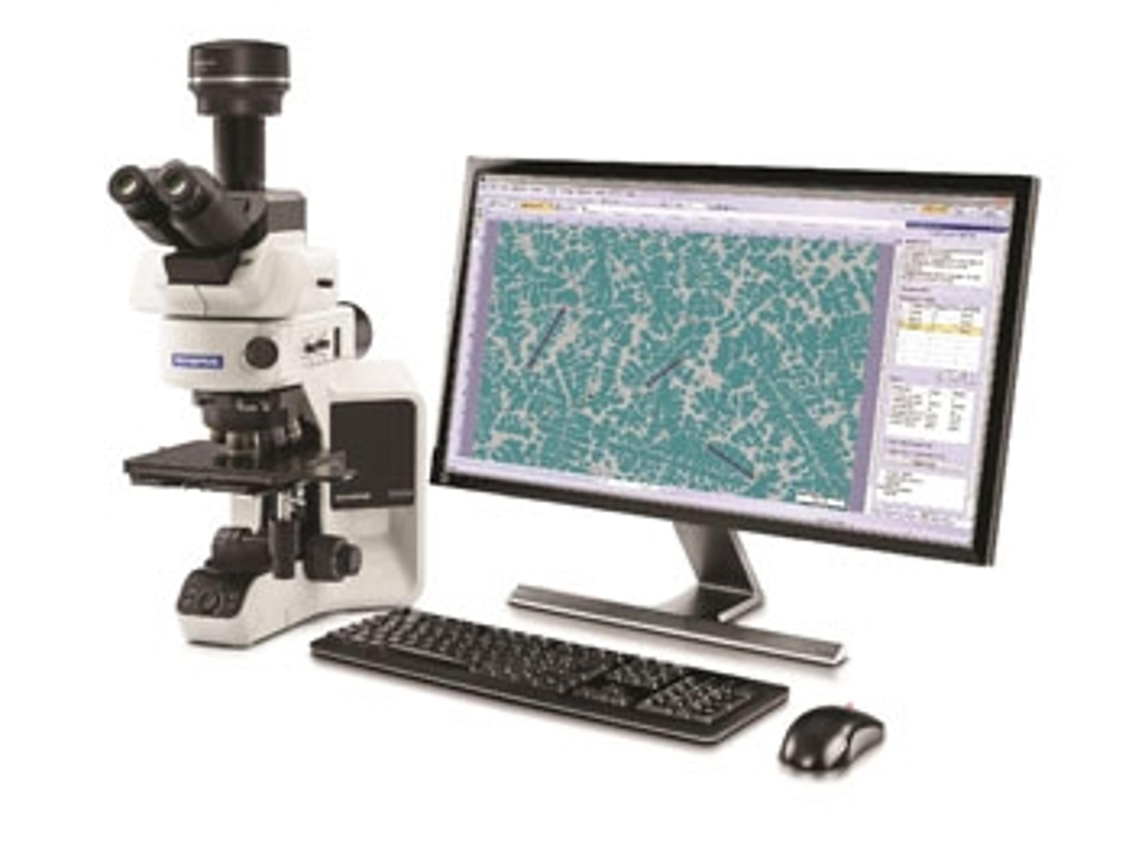
Olympus' NoviSight™ software with True 3D cell analysis technology enables accurate analysis of 3D cell cultures down to the nuclei, improving the reliability and accuracy of the entire drug discovery process.
Designed for Olympus laser scanning confocal microscopes, NoviSight software facilitates data interpretation and validation through its practical features:
- Recognition, analysis, and statistical results are displayed on one screen
- Switch between 3D and 2D views
- Scatterplot, heat map, and graph display options
- Clicking points in these display options shows the corresponding images
- Export data as CSV and FCS files for further analysis
The biologist’s guide to fluorescent live cell imaging: Explore a microscopic world
New technological developments and discoveries in the field of microscopy have enabled us to observe, explore, and analyze the microscopic world like never before. The ability to measure dynamic processes inside living cells, and observe biological entities, such as bacteria, in real-time, has opened up new avenues of research, allowing scientists to routinely make new insights and breakthroughs.
In this eBook, explore the fascinating world of microscopy, including time-lapse imaging, the important relationship between microscopy and genomics, and dynamic volumetric imaging. Plus, discover how to increase microscopy efficiency, so you no longer need to invest hours manually searching for samples.
This guide will also explain:
- How to take macro-to-micro imaging to the next level
- How to effectively combine microscopy with genomics
- How to capture cell motility and proliferation

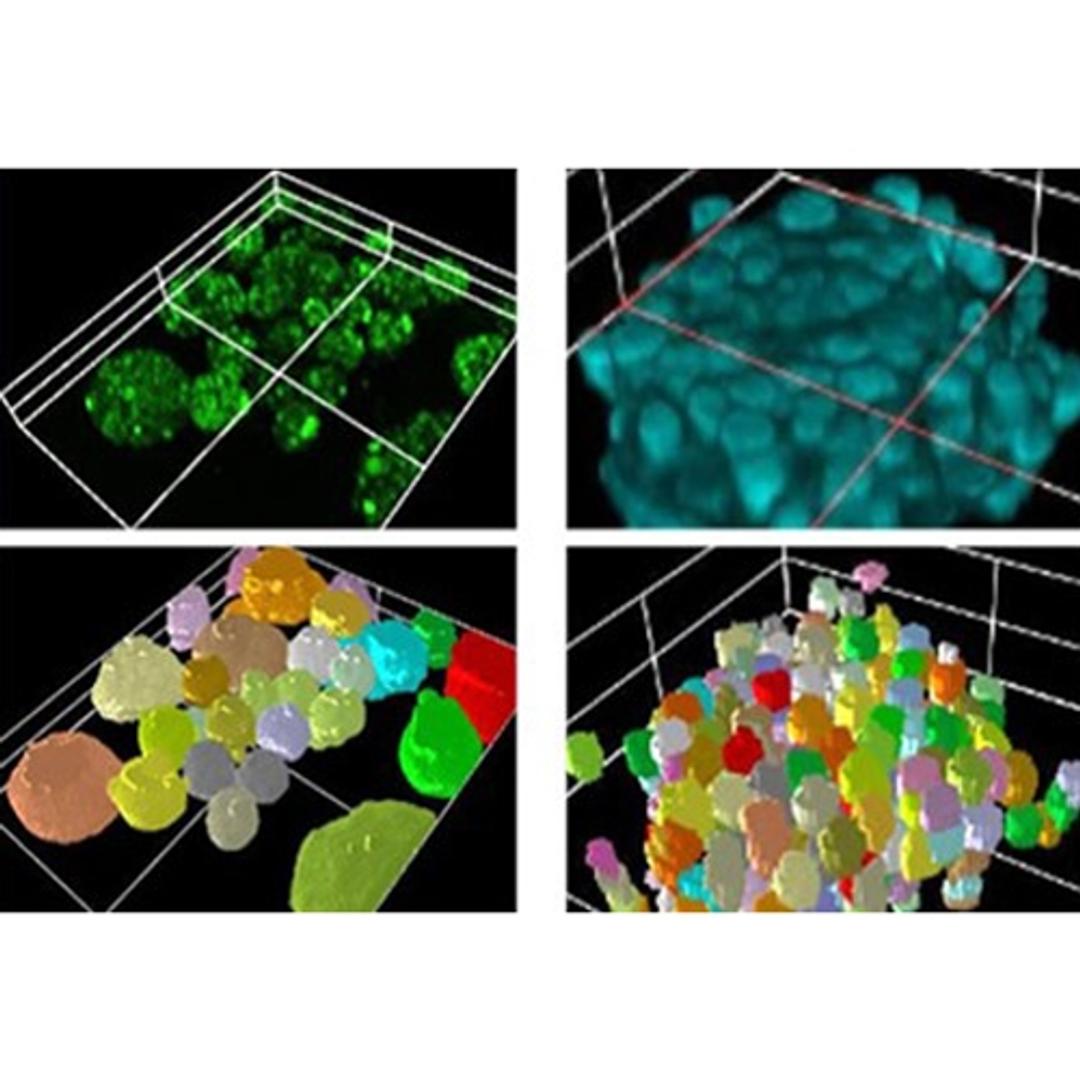
3D analysis of co-culture cancer spheroids
In this case study, confocal images of co-culture spheroids are quantitatively analyzed using NoviSight™ software to evaluate their cell population and determine drug sensitivity in each cell. Assessing the performance of a drug using three-dimensional cancer spheroids is important because the spheroids reflect the complicated in vitro microenvironment of the cancer. This enables researchers to evaluate a drug’s effectiveness under parameters that more closely resemble a tumor’s natural environment.
The importance of tissue clearing and objective selection in 3D analysis of spheroids
In this application note, Olympus introduce a one-step spheroid clearing protocal using SCALEVIEW-S4 tissue clearing reagent. This application note describes the importance of refractive index (RI) matching between cleared spheroids and the objective lens.
3D analysis of co-culture cancer spheroids using NoviSight™ software
In this application note, Olympus, uses NoviSight 3D analysis software and the FLUOVIEW® FV3000 confocal laser scanning microscope to classify the types of cells contained in co-culture cancer spheroids and analyze cell-type-specific drug responsiveness.
3D analysis of patient-derived tumor organoids
In this application note, Olympus’ FLUOVIEW® FV3000 confocal laser scanning microscope and NoviSight™ 3D software were used to quantitatively analyze patient-derived organoids (PDOs), in order to evaluate their morphological features and antibody binding of molecular targeted drugs. The combination of the high-resolution images of the FV3000 microscope, with the high recognition accuracy of NoviSight software meant that more reliable results of drug distribution and morphology were obtained.
3D spatial analysis of high-throughput drug screening
In this application note, Olympus Life Sciences highlights how to perform a homogeneous cell viability assay in a 384-well plate without liquid exchange. Here, high-speed images were taken using the FV3000RS confocal microscope’s resonant scanner and the images were analyzed with NoviSight 3D software. This spatial analysis technique promises more advanced drug evaluation for multiple samples.
True 3D analysis of an intracellular autophagic pathway in a 3D spheroid model
3D culture models have been used for predicting drug efficacy or toxicity. In this application note, we focus on an intracellular autophagic pathway. First, we confirmed the usefulness of the EGFP-LC3 probe for monitoring the models’ autophagic status. Then, Olympus’ true 3D analysis workflow using the FLUOVIEW™ FV3000 confocal microscope and NoviSight™ software was used to quantitatively evaluate the effect of a drug candidate called chloroquine. We demonstrated that this analysis workflow offers an alternative to profiling drug candidates using in vivo studies.
3D analysis of patient-derived tumor organoids using NoviSight™ analysis software
In this whitepaper, Olympus reveals how the NoviSight™ 3D software can perform quantitative analysis of patient-derived tumor organoids (PDOs), morphologically and pharmacologically, using images captured by Olympus' FV3000 confocal laser scanning microscope.
Optical Sectioning Solutions for 3D Cancer Cell Analysis – Your Application Guide
In this eBook, we present several cancer-based examples of non-damaging, high-resolution, live-cell and fixed-cell assays, using some of the most innovative optical sectioning technologies available. These include the analysis of antibody-dependent cell-mediated toxicity (ADCC), 3D in-gel invasion, DNA damage signaling, mitochondrial uncoupling, and drug efficacy in multicellular models.
Fluorescent Image Analysis – Cell Division in Spheroids
Cell division inside spheroids can be visualized by staining cell nuclei and microtubules. The number of cells in mitosis in spheroids can be quantitatively evaluated using images captured by a laser confocal microscope and NoviSight™ software's counting module.
Using 3D cell culture analysis to support drug discovery
In this video, discover how the use of 3D cell cultures is enabling more effective and safer drugs to be discovered early on in the development process. Here, Olympus Life Science also outlines how the Olympus NoviSight™ 3D High Content Analysis Software is designed to help support these processes.
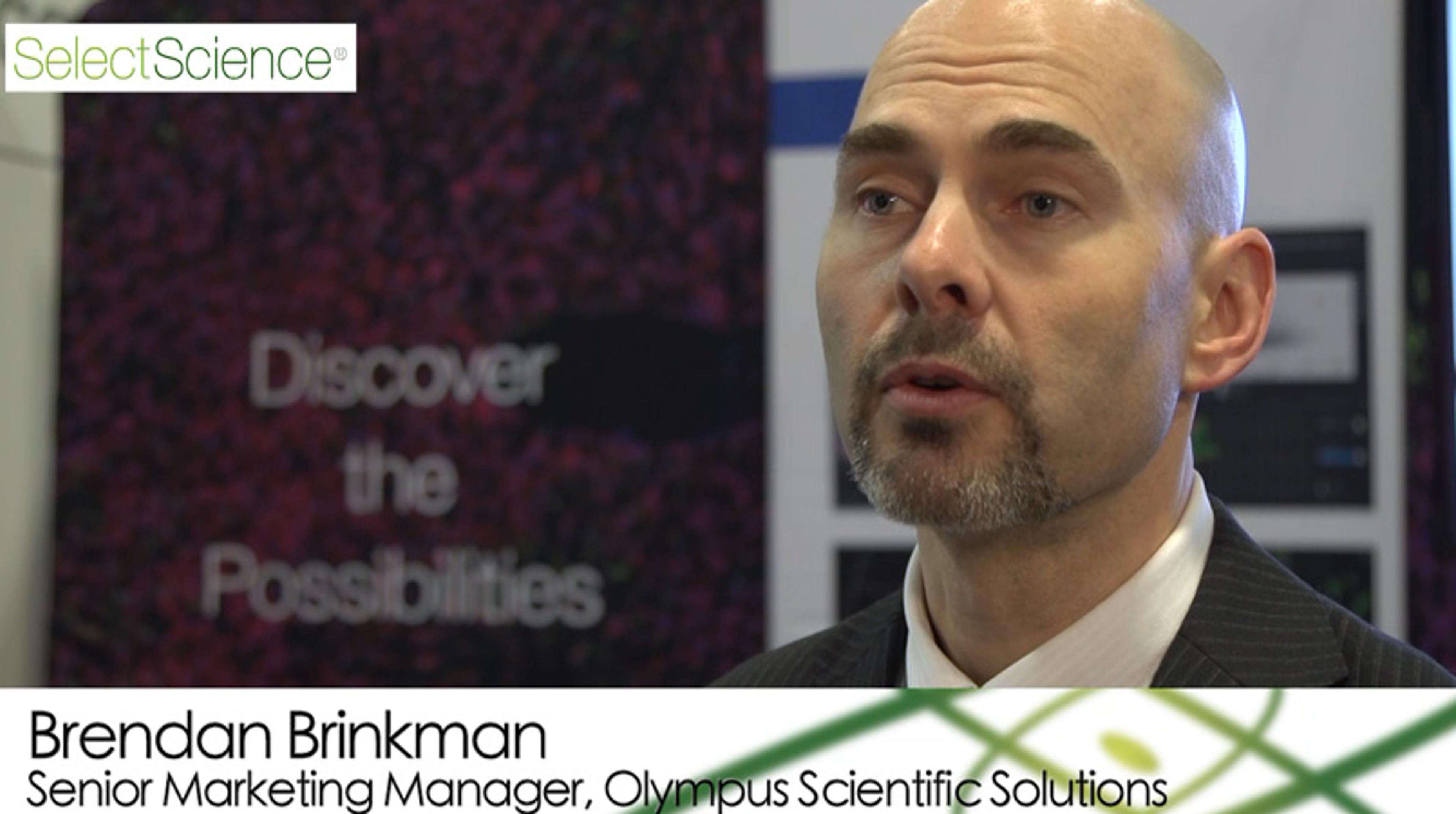
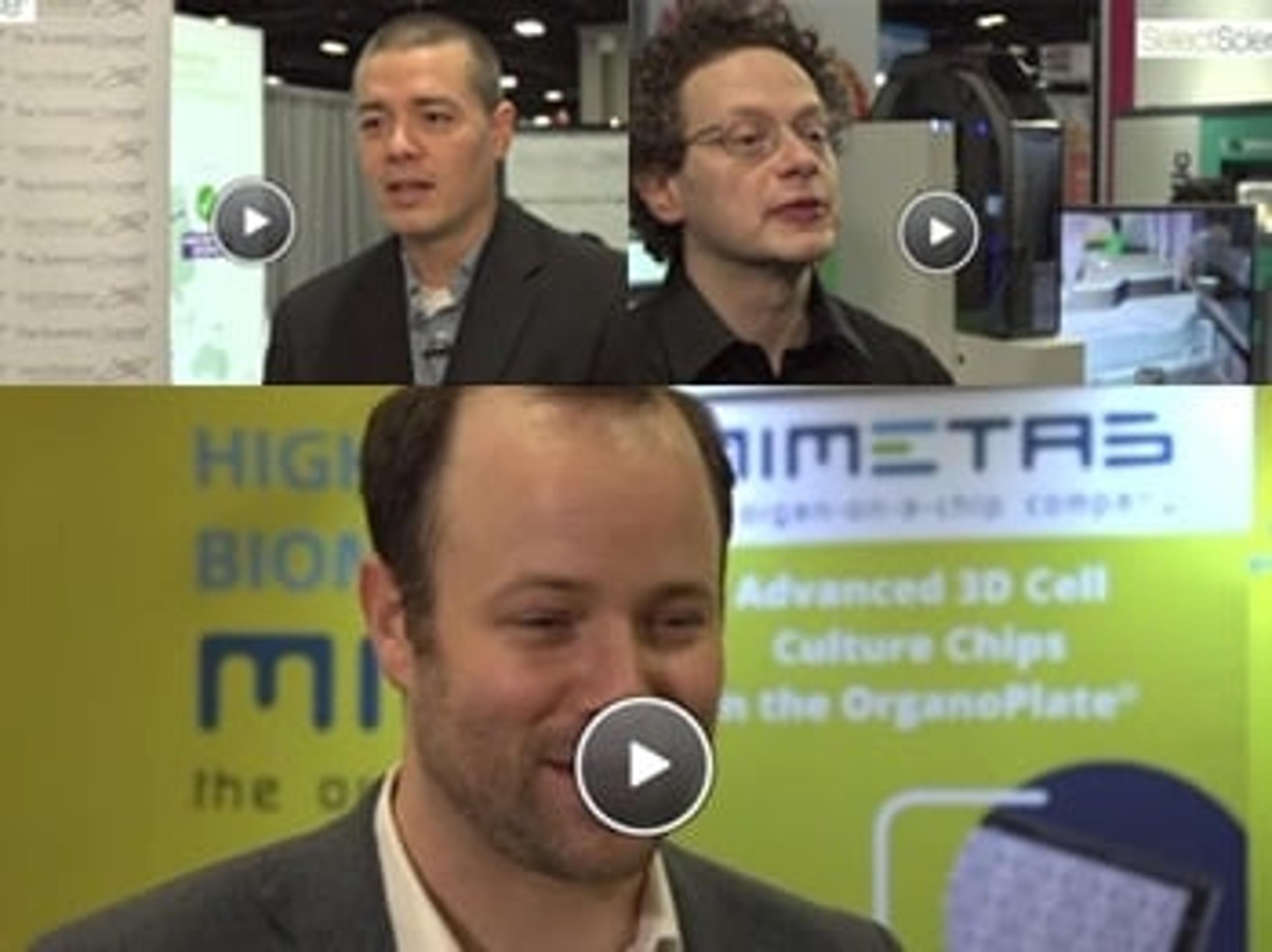
Make the Most of Your 3D Cell Cultures with NoviSight 3D Cell Analysis Software
The use of physiologically relevant 3D cell models is becoming increasingly important within the drug discovery and development community. In this video interview, Brendan Brinkman, from Olympus Scientific Solutions, explains how the NoviSight 3D Cell Analysis software offers true 3D high-content analysis, to improve the accuracy of drug discovery research and screening.
This interview was filmed as part of our coverage of SLAS2019 - see other video highlights here >>

Helping scientists succeed: A revolution in scientific collaboration
Dr. James Evans explains how his expert team help other drug discovery companies grow and succeed, and highlights the power behind scientific collaboration, sharing ideas, and teaming up to win
OLYMPUS Stream software version 2.4.2 offers an improved image analysis method for steel quality control
The update adds device support and new capabilities to determine the non-metallic inclusion content in steel
Latest Advances in Cell-Based Assays: SelectScience Special Feature
From rapid multiplexed analysis, 3D organoid imaging software, and automated plate processing, discover the top technologies, techniques and best practices for cell-based assay research

3D high-content analysis: An emerging solution for drug discovery
Learn about Olympus’ new solution for 3D high-content analysis and how patient-derived organoids can be utilized for more effective drug candidate profiling

How to Evaluate the Drug Response in Patient-Derived Organoids Using True 3D HCA
Join this expert webinar to find out how imaging and true 3D high-content analysis can benefit drug studies based on patient-derived organoids
3D Cell Culture Methods and Protocols
From 3D Culture Systems to Spheroids – break into three-dimensional (3D) cell culture with the latest methods and resources
Olympus Stream Software: Inspection Without Limitation
Conduct fast and precise observations on a large variety of samples while maintaining data security and measurement reliability
Drug Discovery Video Playlist: 6 Experts Share Research & Technology Insights
Watch exclusive highlights from the show floor at SLAS2019 as we find out about the latest technologies innovating life sciences discovery
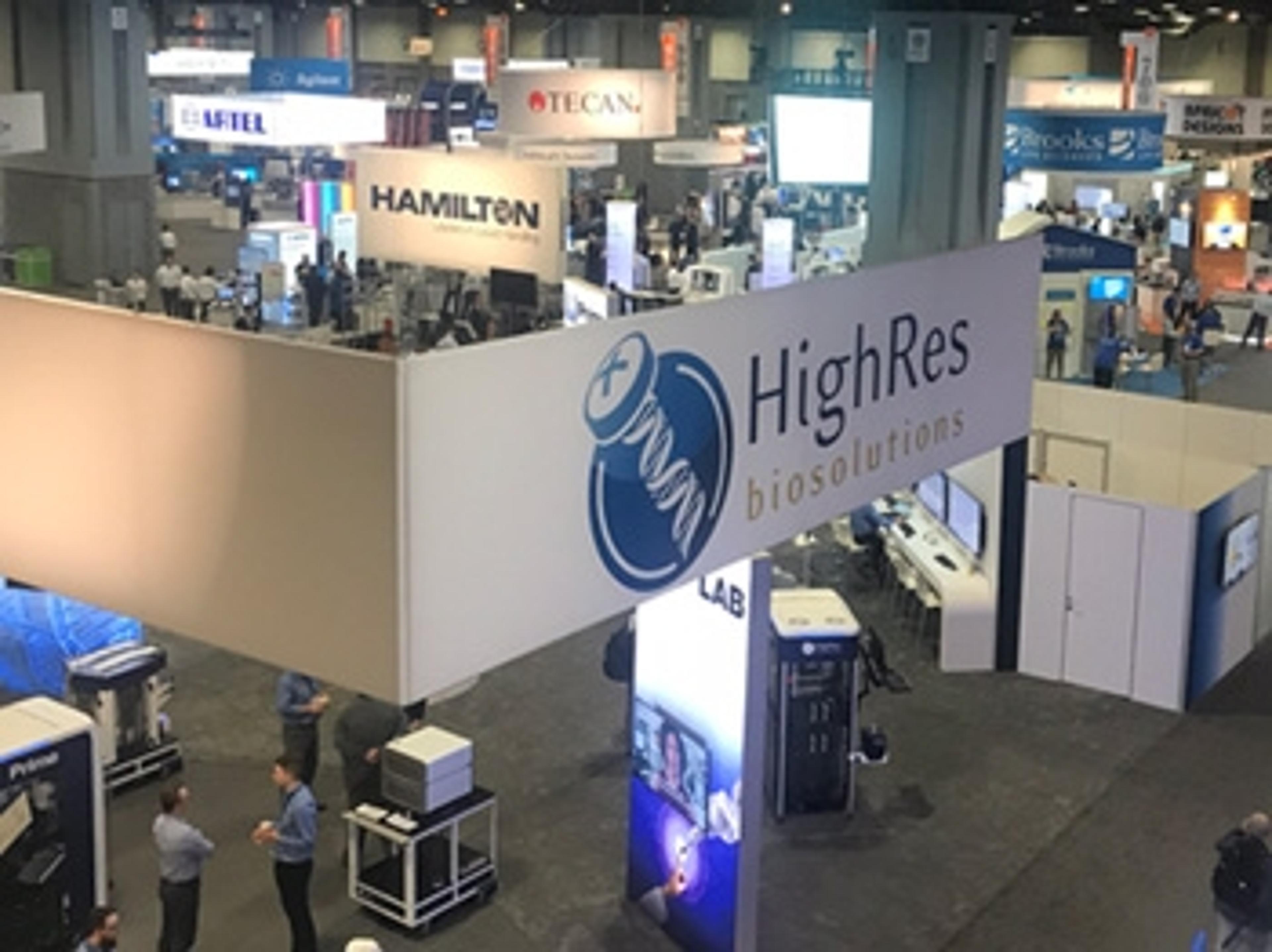
Spotted at SLAS2019: 9 Top Technologies from the Exhibition Floor
SelectScience editors reflect on the most exciting technologies showcased at the 2019 Society for Laboratory Automation and Screening conference
Increase Efficiency and Accelerate Drug Discovery Research with Olympus NoviSight™ 3D Cell Analysis Software
3D imaging technology provides images of the cell cluster down to the nuclei while mitigating drug discovery risks