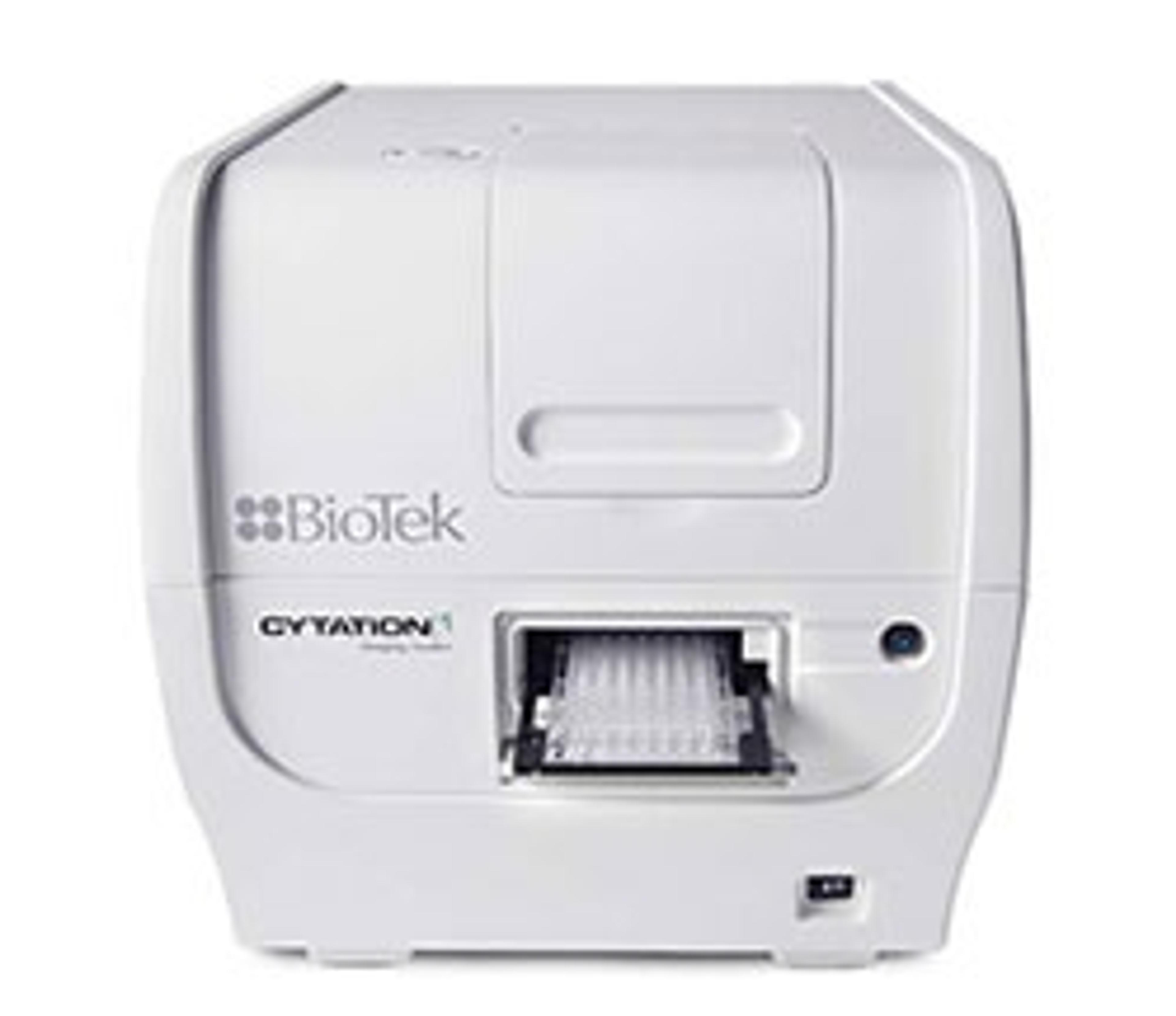
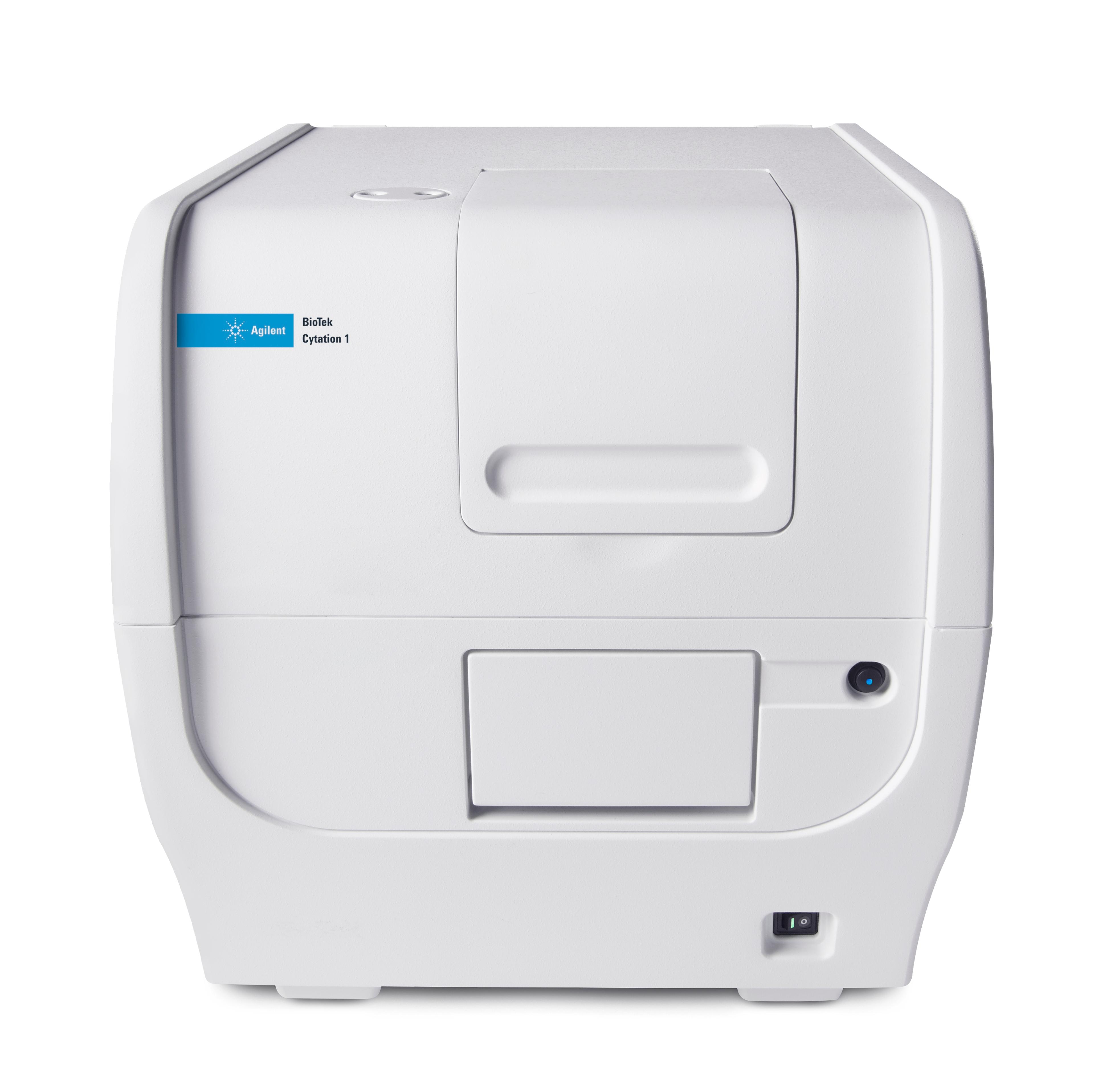
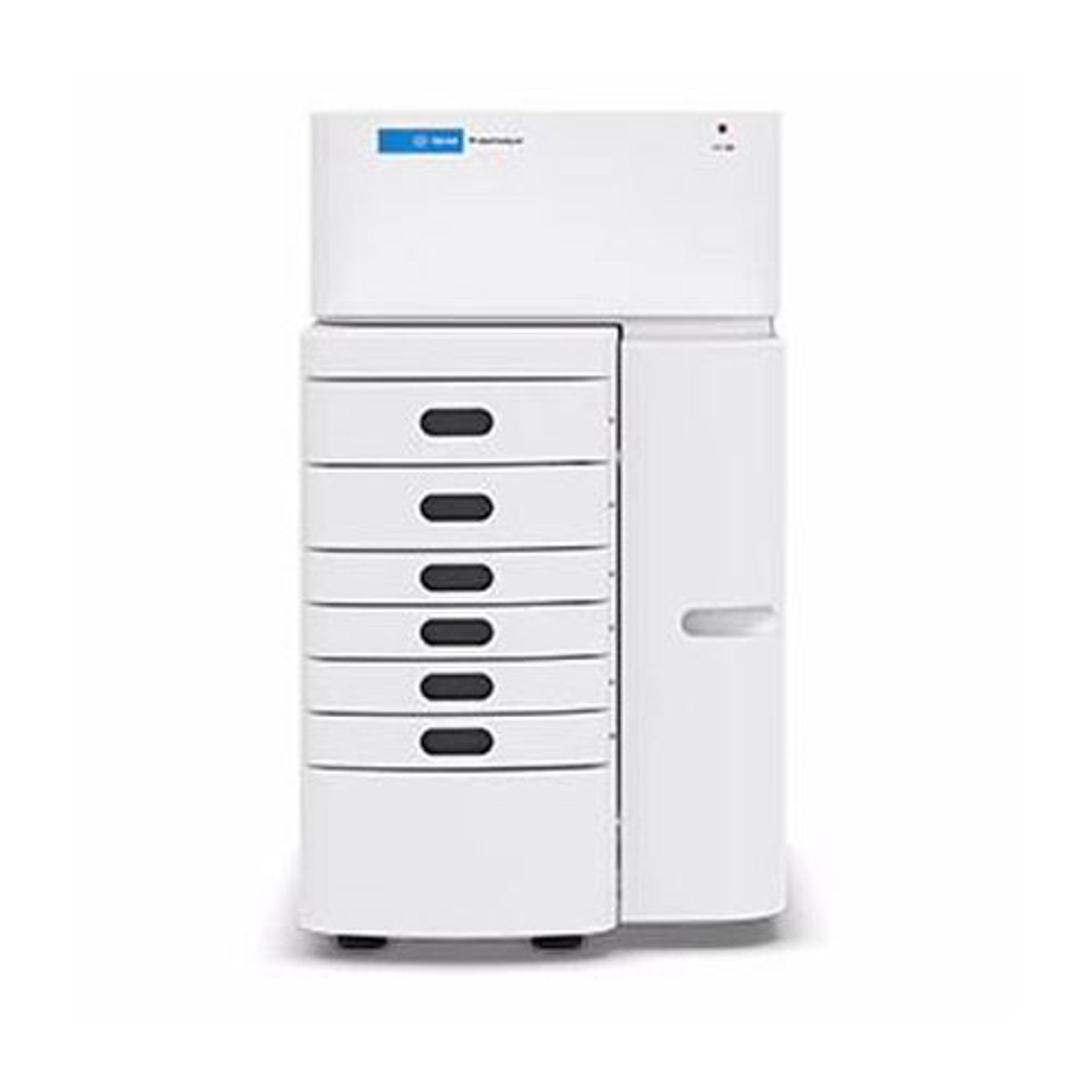
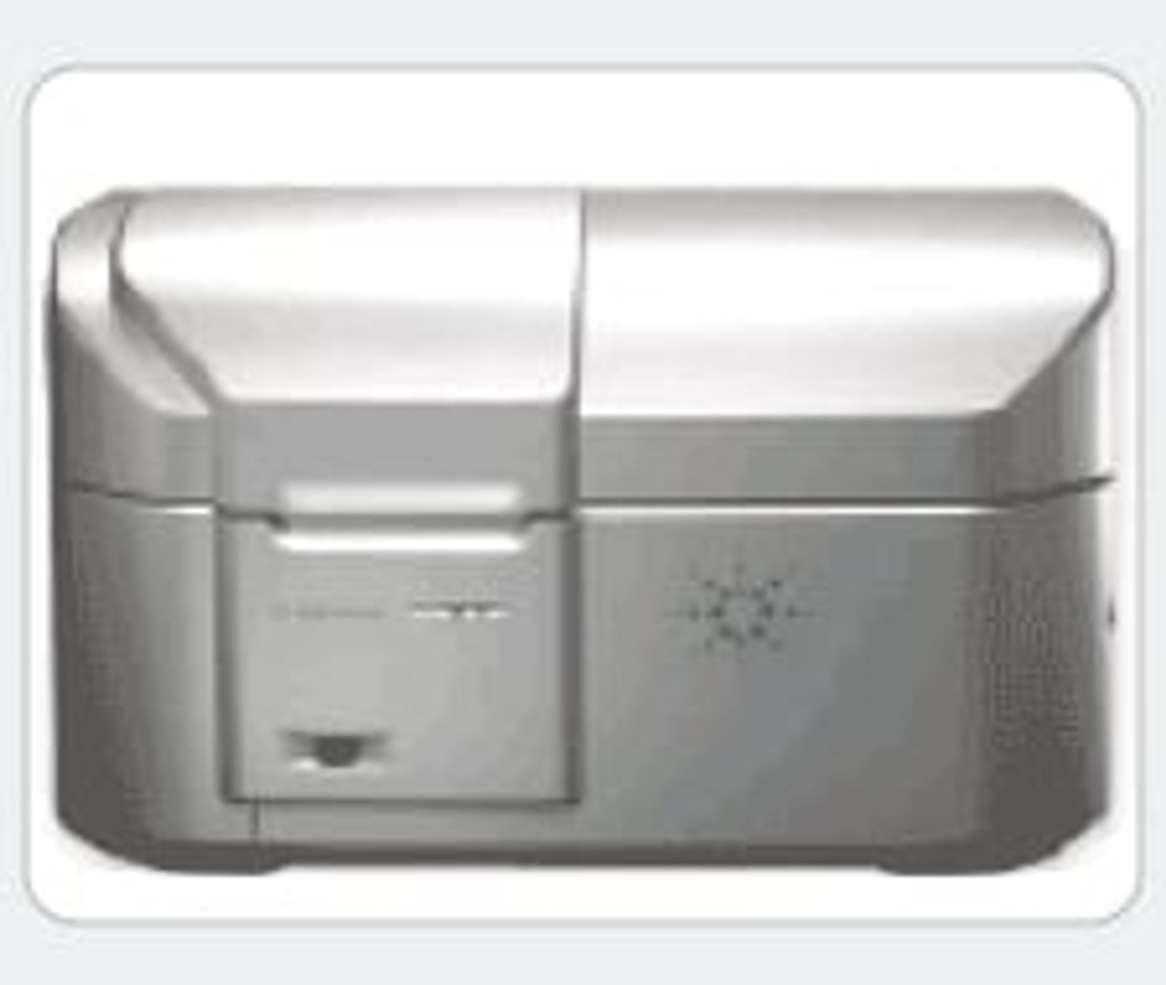
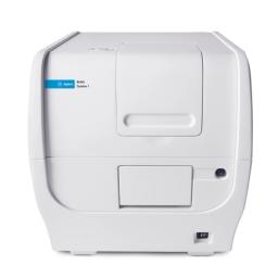
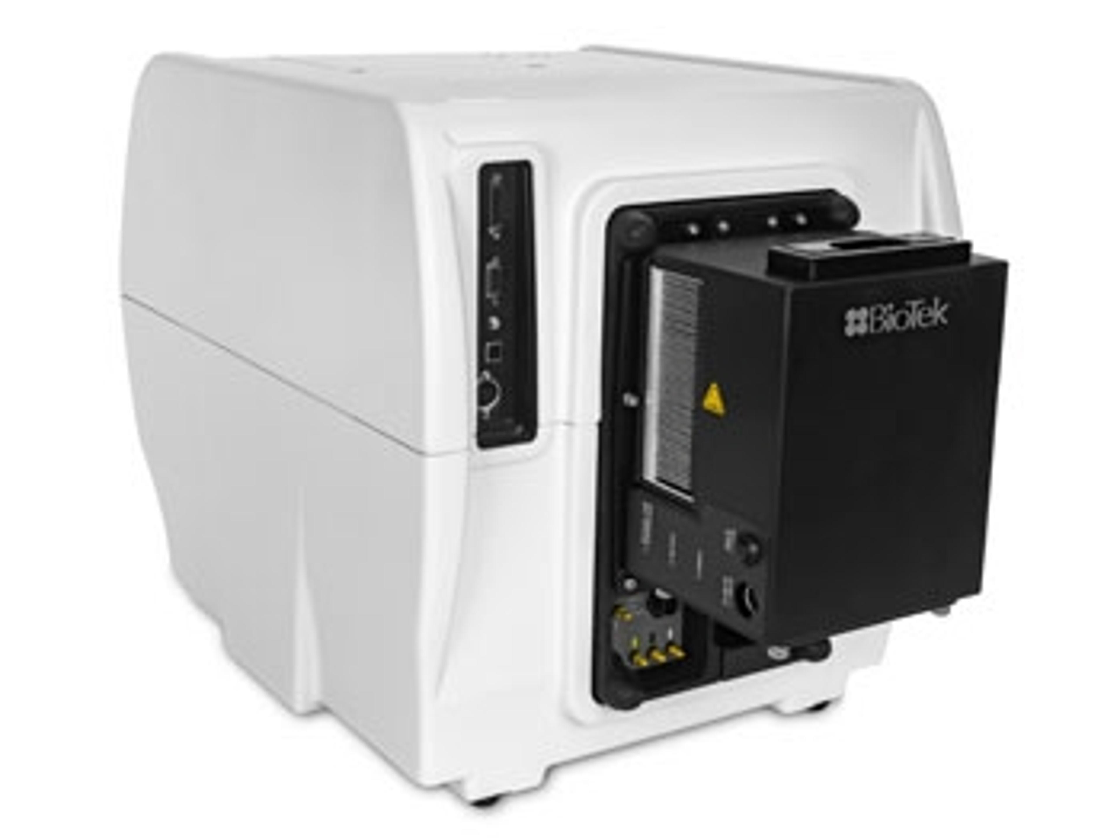
Agilent BioTek Cytation 1 Cell Imaging Multimode Reader
Cell Imaging Multi-Mode Reader


Receive your quote directly from the manufacturer.
Great instrument, and super helpful customer support!
Analyze in vitro growth assays
While this instrument and corresponding analysis software is pretty user-friendly and easy to use, we have ran into a few questions when setting up new assays. The customer support has been amazingly helpful, from both our local sales representative to the regional customer support team. Both of them have been very prompt in responding to our questions, and really make you feel like they want you to get the most out of their instrument.
Review Date: 26 Apr 2018 | Agilent Technologies
The Cytation 1 is a must have for an R&D lab.
High throughput screening of cells for viral titer
The Cytation has been a wonderful addition to our lab. It allows us to generate a lot of data in a short amount of time. We are constantly adding new assays based on the Cytation's capabilities.
Review Date: 26 Apr 2018 | Agilent Technologies
Very good instrument. Expensive but will be worth the money in the long run.
Antimicrobial assay, anticancer assay, enzyme inhibition assays, phytochemical profiling
The product is very useful for what we want to do. The learning curve is simple which makes its use easy for undergraduate students. Having the imaging capabilities has moved our research and teaching labs to the next level. We expect that the modularity of the instrument we benefit us as we can add new functionalities as future needs develop.
Review Date: 26 Apr 2018 | Agilent Technologies
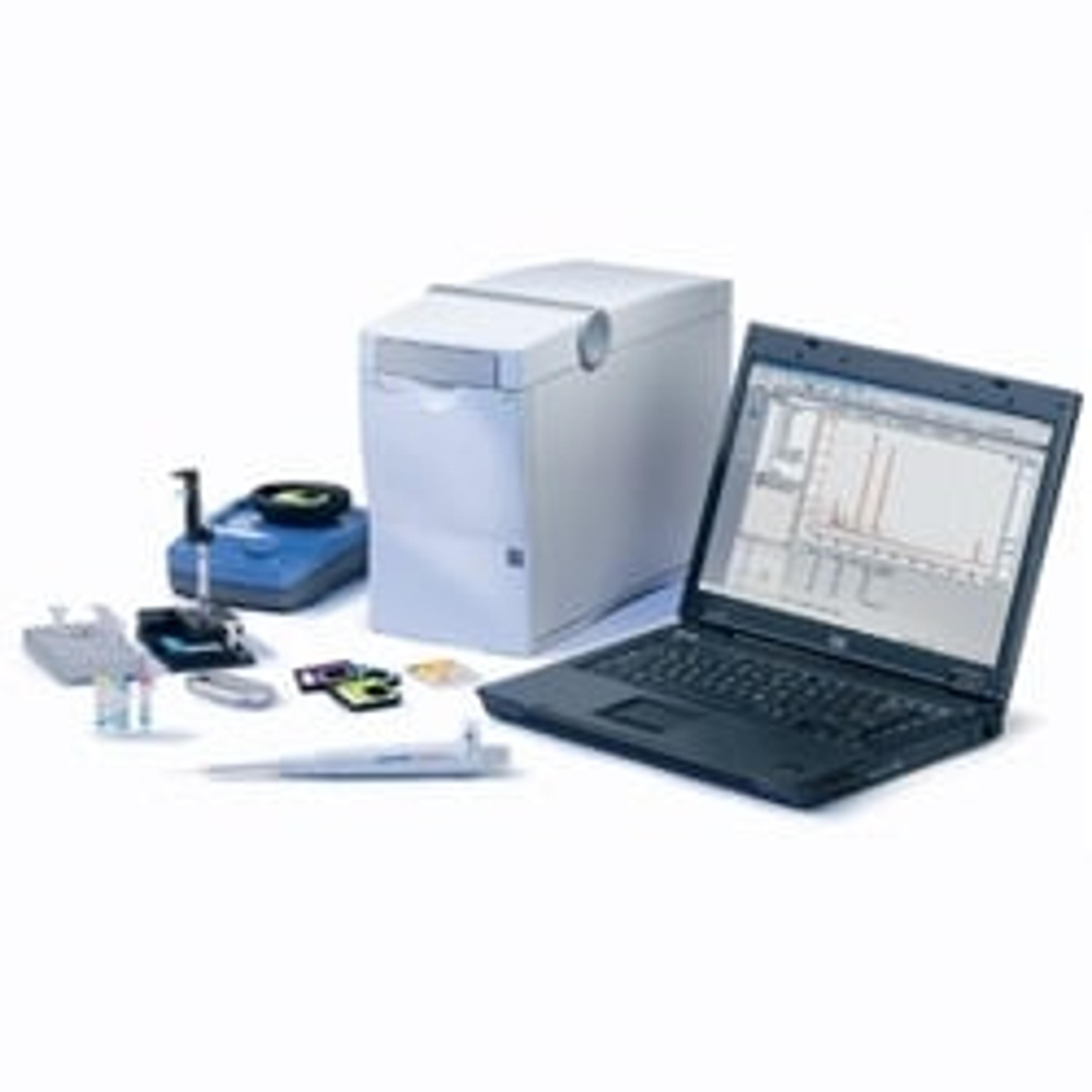
The Agilent BioTek Cytation 1 cell imaging multimode reader combines fluorescence and high contrast brightfield imaging with conventional multimode detection. This affordable imaging reader delivers performance and economy that is not typically available in other digital microscopy systems.
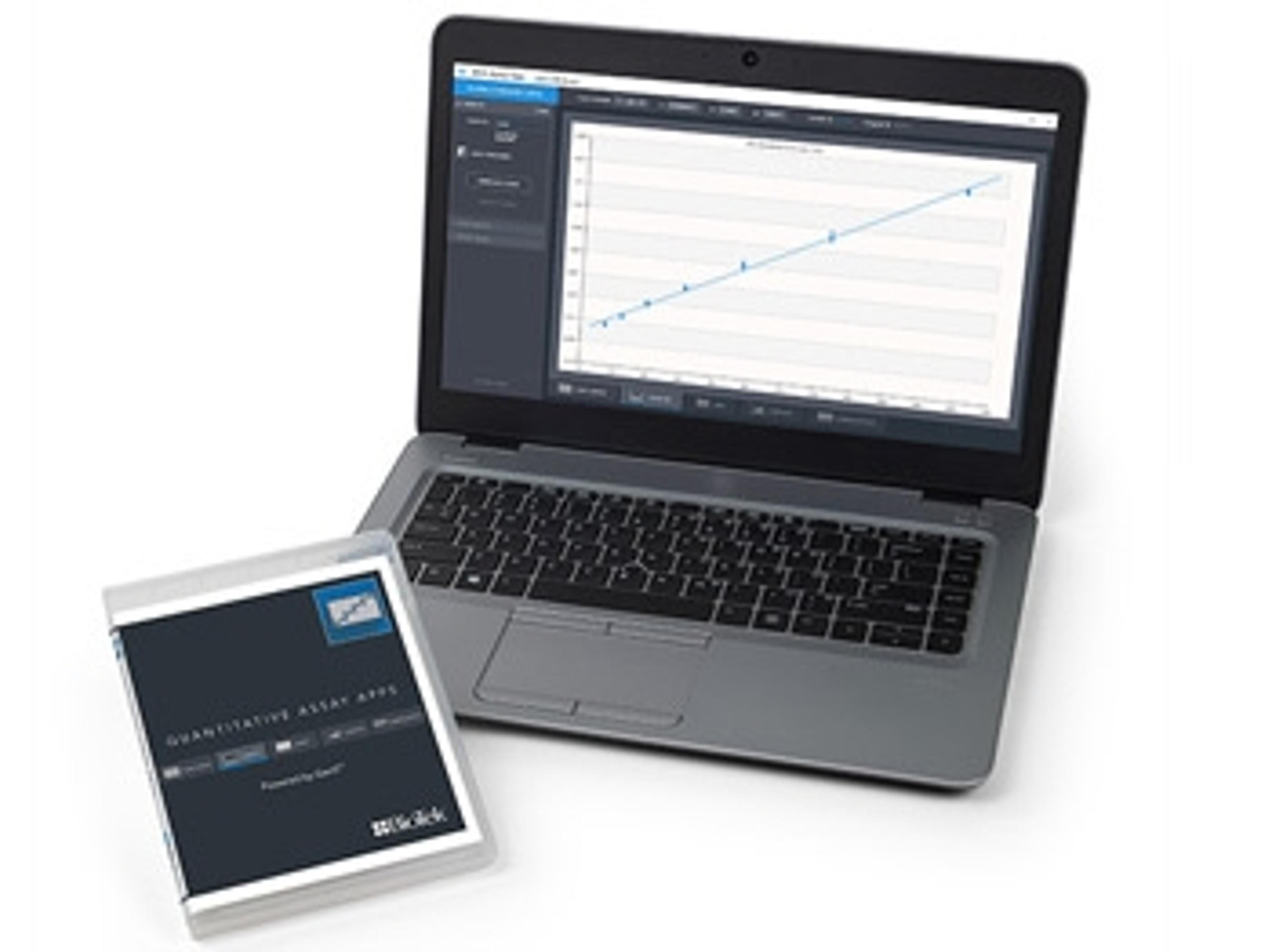
The BioTek Cytation 1 design provides both quantitative phenotypic cellular data and well-based quantitative data. The multimode detection module has high sensitivity filter-based fluorescence and a monochromator for UV-Vis absorbance. Temperature control and shaking are standard. CO2/O2 control and reagent injectors are available. Gen5 software simplifies image capture and plate reading.

Long-Term 3D Spheroid Culture & Analysis - An Essential Guide
Interest has intensified recently in the use of 3D models in life sciences research, to improve the understanding of basic cellular processes and reduce drug attrition rate. In the past, however, 3D models have proven difficult to reliably cultivate long enough to perform multiple dosing studies as well as to monitor chronic toxicity, long-term kinetic analyses, and stem cell differentiation. With a focus on four key applications, this application eBook speaks to these challenges - demonstrating robust workflows for high-throughput spheroid assembly and long-term quantitative analysis.
An Image-Based Method to Detect and Quantify T Cell Mediated Cytotoxicity of 2D and 3D Target Cell Models
T Cell mediated cytotoxicity is an important part of new methods being developed to fight cancer. This application note demonstrates a methodolgy for automated monitoring and measurement of CTL-mediated cytotoxicity .
Cost-Effective Automated Hepatotoxicity Testing Using 3D Bio-Printing
To meet the demand for increased hepatotoxicity testing, automation has been incorporated to streamline the procedure and reduce the need for large-scale manual manipulations. Typically, liquid handling systems have been large, expensive, and required placement into clean rooms for sterile processing. While this type of solution is suitable for larger core facilities, the size and cost can be prohibitive to the typical academic research lab. This application note demonstrates the ability to combine liquid handling with a novel cell imaging multi-mode reader to perform automated 3D hepatotoxicity studies, which will drastically reduce the overall costs for academic researchers.
Automation of Non-Alcoholic Fatty Liver Disease Model Assay
Non-alcoholic fatty liver disease is a common malady whose major feature is fat accumulation in the liver, which can lead to liver damage and cirrhosis. One phenotypic marker for this disease is the presence of intracellular neutral lipid droplets. An in vitro model for this phenotype uses the exposure of cells to free fatty acids to elicit the response. Here we describe the automation of the assay workflow using a BioSpa™ 8 Automated Incubator linked to a Cytation™ 5 Cell Imaging Multi-Mode Reader and a MultiFlo™ FX Microplate Dispenser.
Introducing Cytation 1: A Dual Microplate Analysis and Automated Imaging System
Xavier Amouretti, head of product management, Biotek, describes the Cytation 1, a third generation microplate detection and imaging platform. This innovative system enables cellular assay multiplexing and high-resolution imaging of cells in real time.
BioTek’s Cytation™ Cell Imaging Multi-Mode Reader Accelerates Cancer Drug Discovery
The Cytation™ Cell Imaging Multi-Mode Reader by BioTek Instruments, Inc. has been an invaluable instrument within the Gunning laboratory in Toronto, Canada, helping to accelerate drug discovery by enabling the identification of many promising small molecules in pre-clinical trials. Watch this video to find out more.

Trending in drug discovery and biopharma - the best of 2019
From CRISPR screening in primary T cells to photodynamic therapy for curing cancer, we take a look back at research highlights and emerging technologies from the past year

Optimizing Conditions for Temperature Sensitive Assays
New Peltier cooling module by BioTek prevents temperature fluctuations in multimode cell imagers


Major Drug Discovery Target Offers Promising New Cancer Treatment
Professor Patrick Gunning discusses the multidisciplinary approach that is helping his team to fast-track the development of small molecules for treating cancer