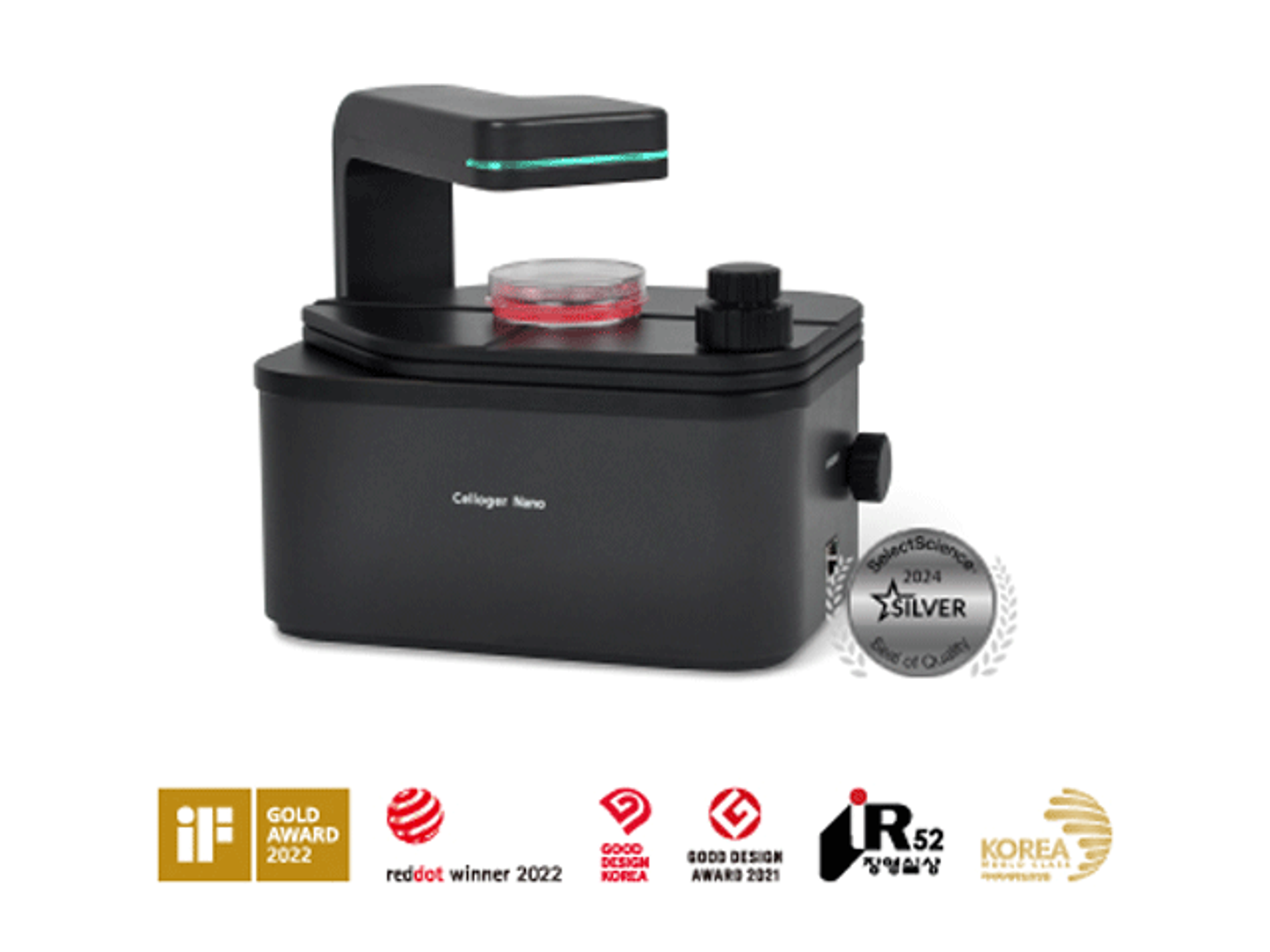
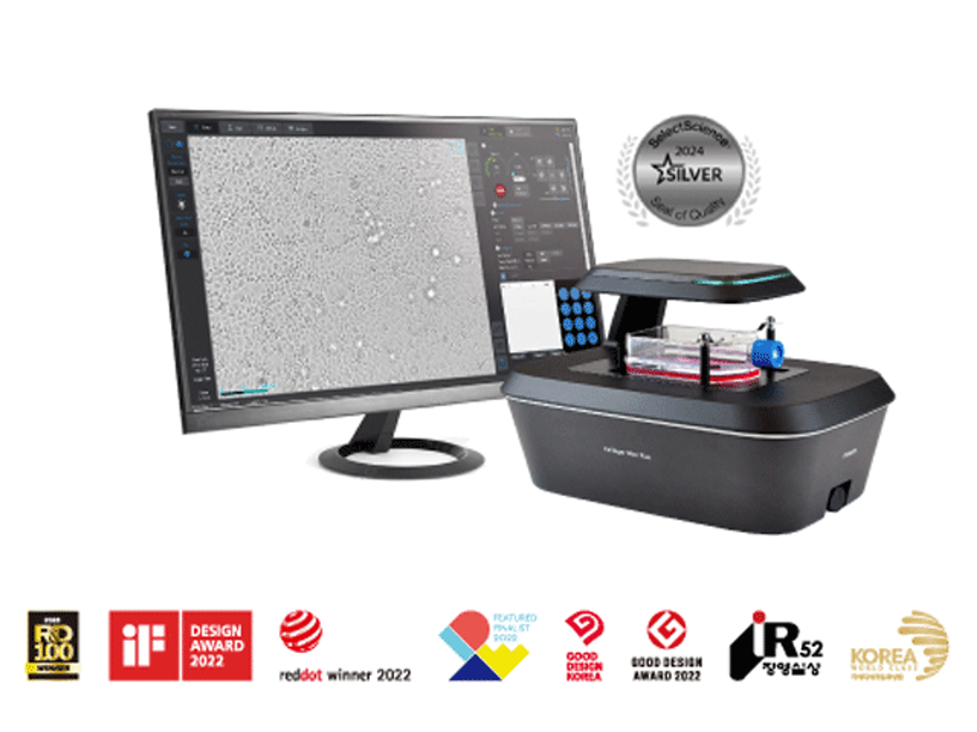
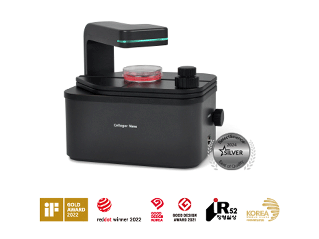
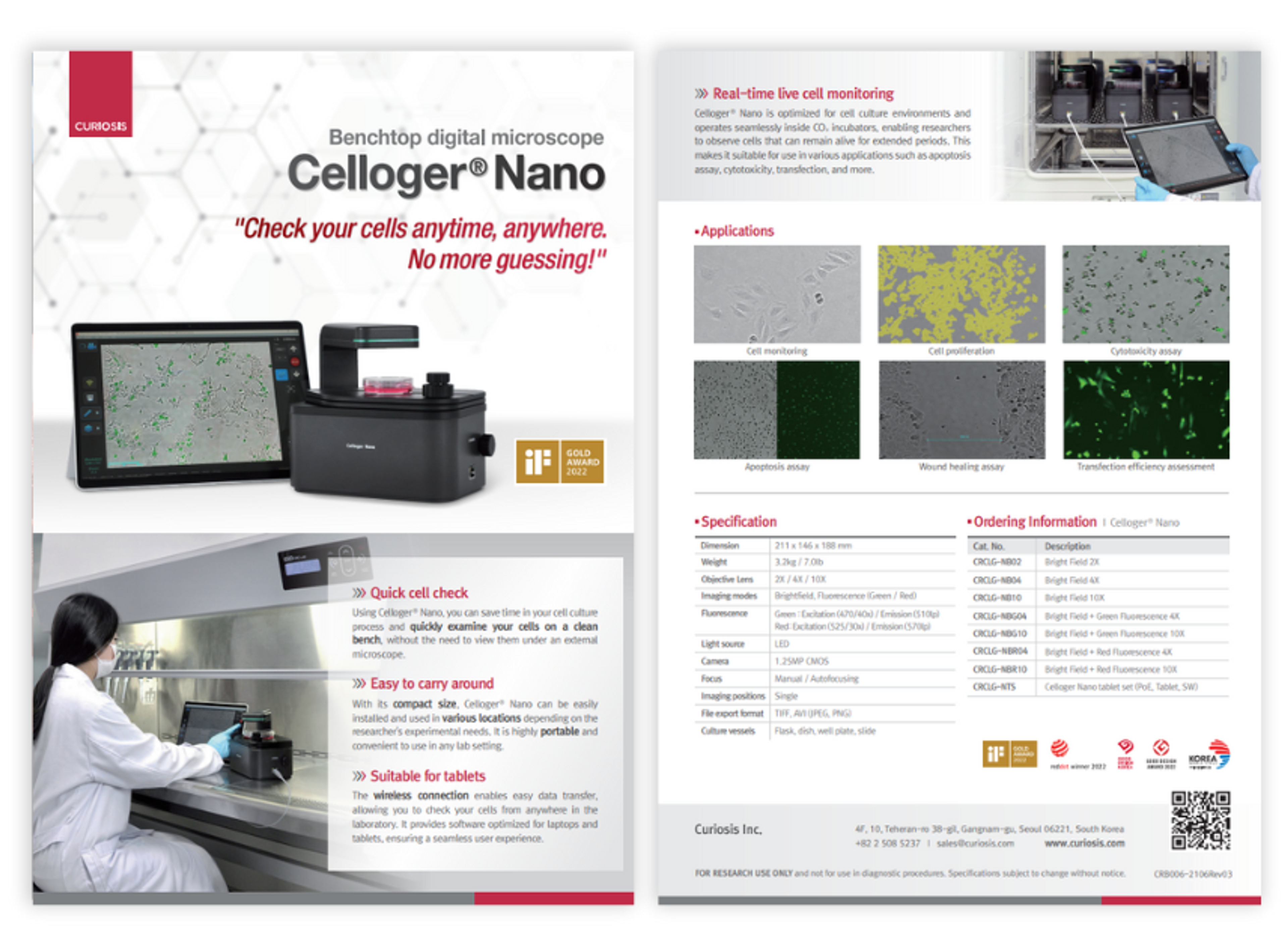
Celloger® Nano, Benchtop Digital Microscope from Curiosis
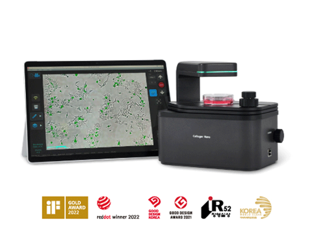
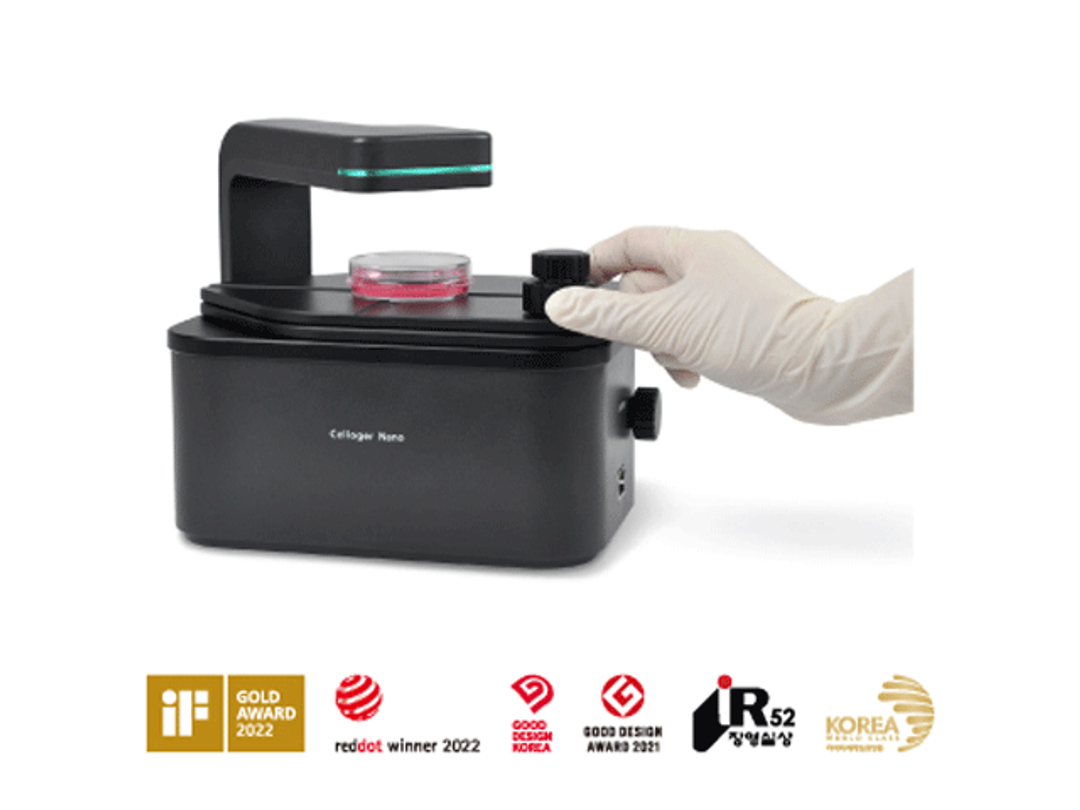
Celloger® Nano is a benchtop digital microscope equipped with a wireless connection, enabling you to check the state of your cells in real-time from any location within your laboratory. With all the necessary functions to check the cells, you can quickly assess the condition of your cells.


Receive your quote directly from the manufacturer.
The super compact benchtop digital microscope, Celloger® Nano, can be placed inside a clean bench and incubator without the risk of contamination of cells, greatly enhancing the efficiency of daily cell culture works through a simple and easy operation.
Key features
- Quick cell check
- Suitable for tablet
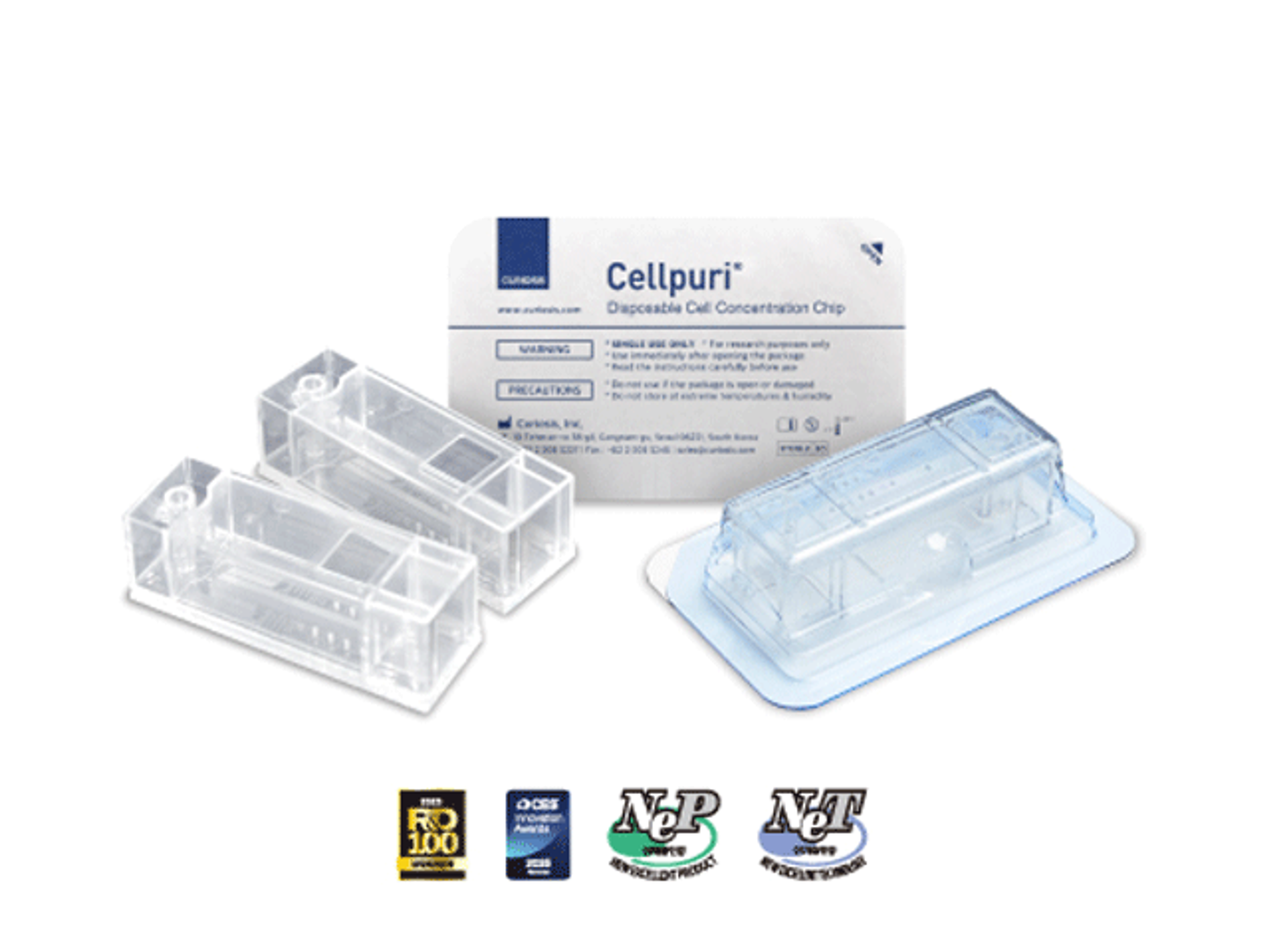
- Multiple vessel types
- User-friendly function(Precise stage controller, Preview record)
Applications
- Wound healing assay
- Cell migration
- Cell morphology
- Cell confluency
- Cell proliferation
- Cytotoxicity assay
- Transfection efficiency assessment
- Coculture monitoring
Specification
- Dimension: 211 x 146 x188mm
- Weight: 3.2kg / 7.0lb
- Objective: 2X / 4X / 10X
- Imaging modes: Brightfield, Fluorescence (Green/Red)
- Fluorescence: Green - Excitation (470/40x), Emission (510lp) / Red - Excitation (525/30x), Emission (570lp)
- Camera: 1.25MP CMOS
- Focus: Manual / Autofocusing
- Imaging positions: Single
Brochures
Celloger Nano product brochure
In this product brochure, CURIOSIS introduces the Celloger® Nano, a convenient solution for quick cell checks. With this device, researchers can save time by examining cells directly on a clean bench without the need for an external microscope. The compact and portable design of Celloger Nano allows for easy installation and usage in various locations, catering to the specific experimental needs of researchers.
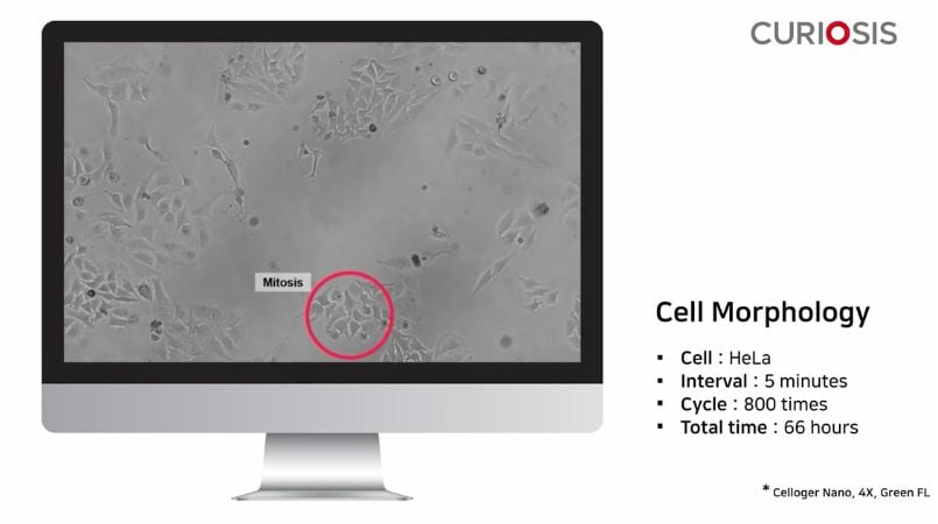
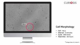
Celloger Nano application: Cell morphology
The changes in cell morphology occur at every major point in cell cycle and monitoring these changes in appearance of cells in real-time is very important. This video from CURIOSIS observes HeLa cell morphology and shows how its Celloger® Nano can be utilized here. The images were taken every 5 minutes for 66 hours using Celloger® Nano 4X objective.
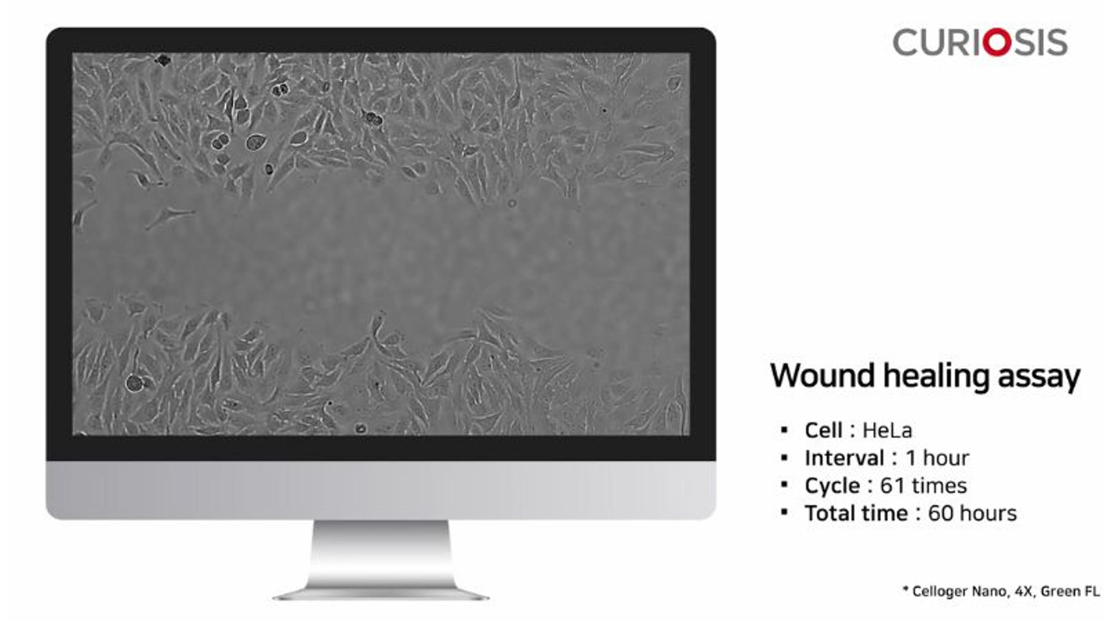
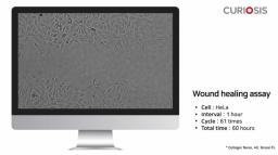
Celloger Nano application: Wound healing assay
Here, CURIOSIS shows how its Celloger® Nano can be utilized in wound healing assays. This time-lapse video shows the scratch wound closure of HeLa cells taken every hour for 60 hours using the Celloger® Nano 4X objective.
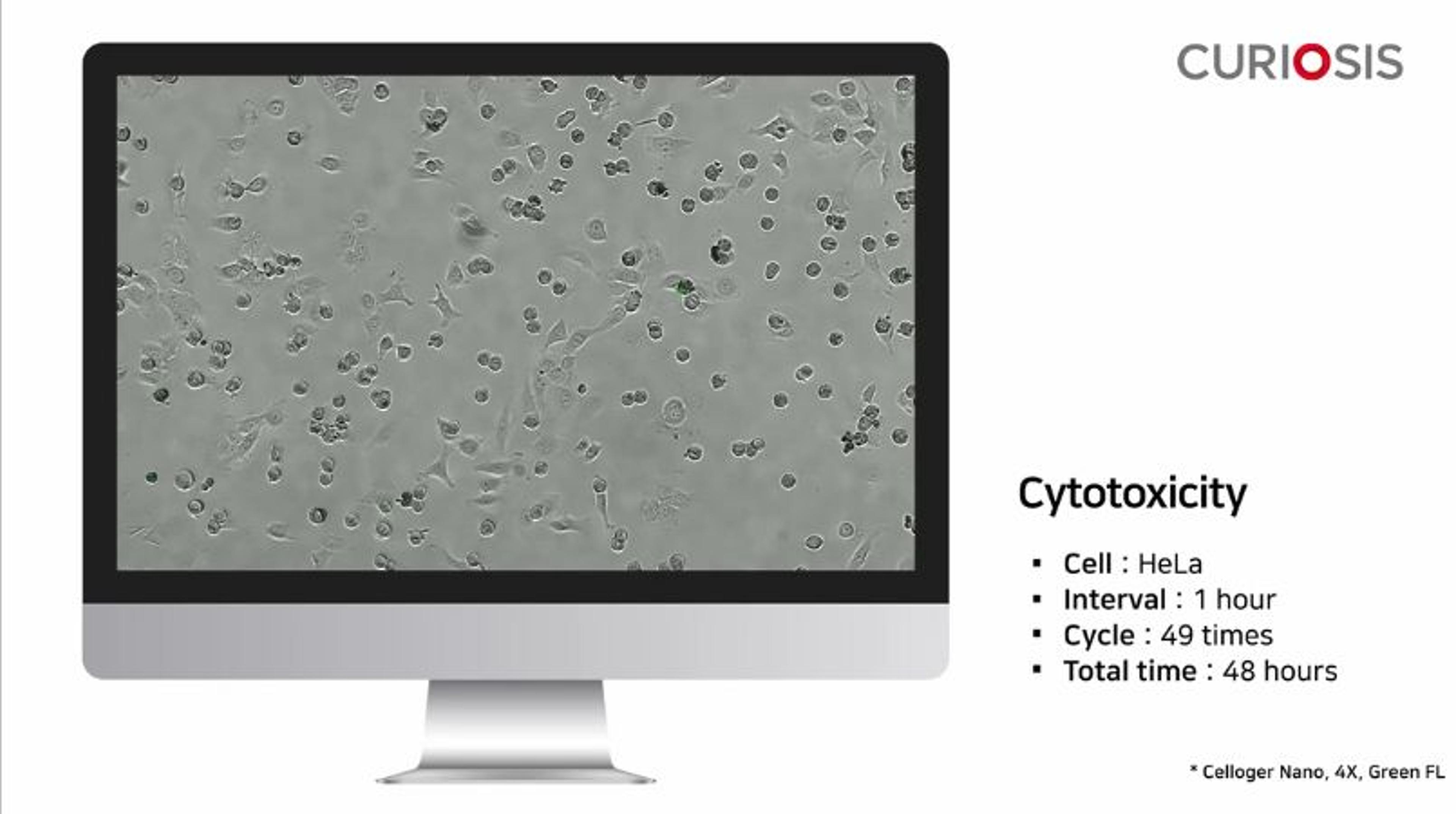
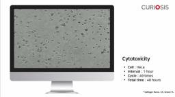
Celloger Nano application: Cytotoxicty
This video from CURIOSIS shows how its Celloger® Nano can be utilized for investigating the levels of cytotoxicity of HeLa cells when treated with either nocodazole or staurosporine. To measure the cytotoxicity of nocodazole, dead cells were stained with green fluorescent CellTox™ dye. Images of HeLa cells were taken every hour for a total of 48 hours using Celloger® Nano 4X objective, green fluorescence. To measure the cytotoxicity of staurosporine, dead cells were stained with red fluorescent Propidium iodide dye. Images of HeLa cells were taken every hour for a total of 71 hours using Celloger® Nano 4X objective, red fluorescence.
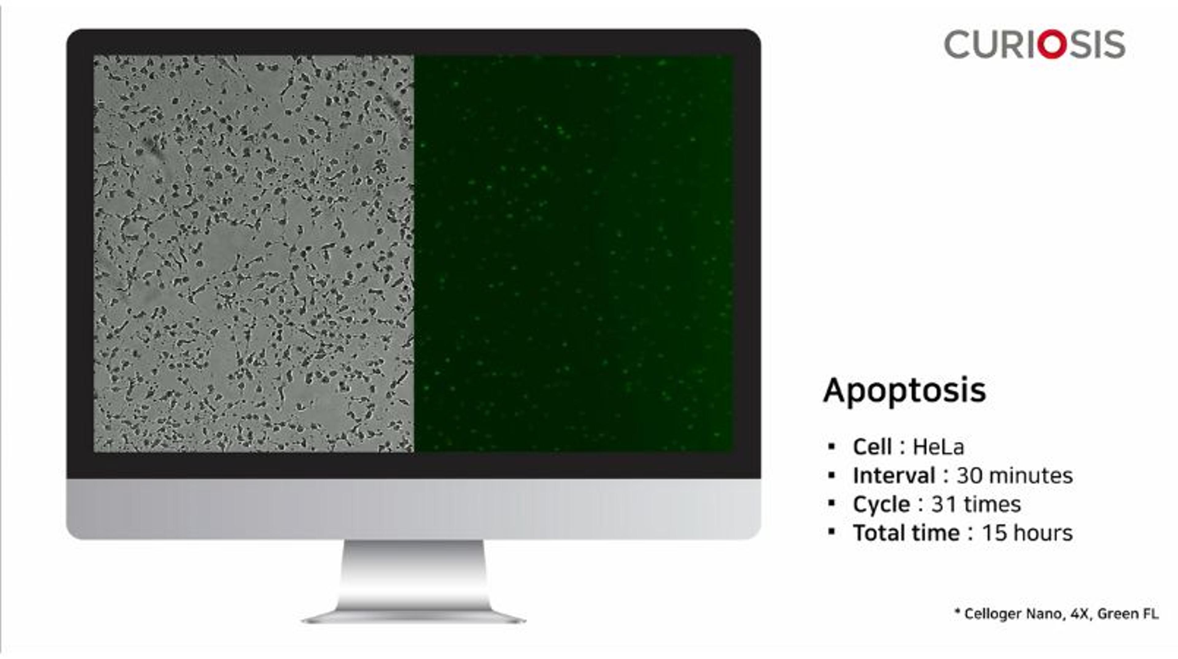
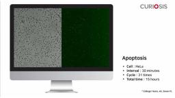
Celloger Nano application: Apoptosis
Apoptosis is the process of programmed cell death. Here, CURIOSIS shows how its Celloger® Nano can be utilized when investigating apoptosis. This time-lapse video analyzes the apoptotic effect of staurosporine treatment on HeLa cells, using the Celloger® Nano 4X objective, observed every 30 minutes for 15 hours.
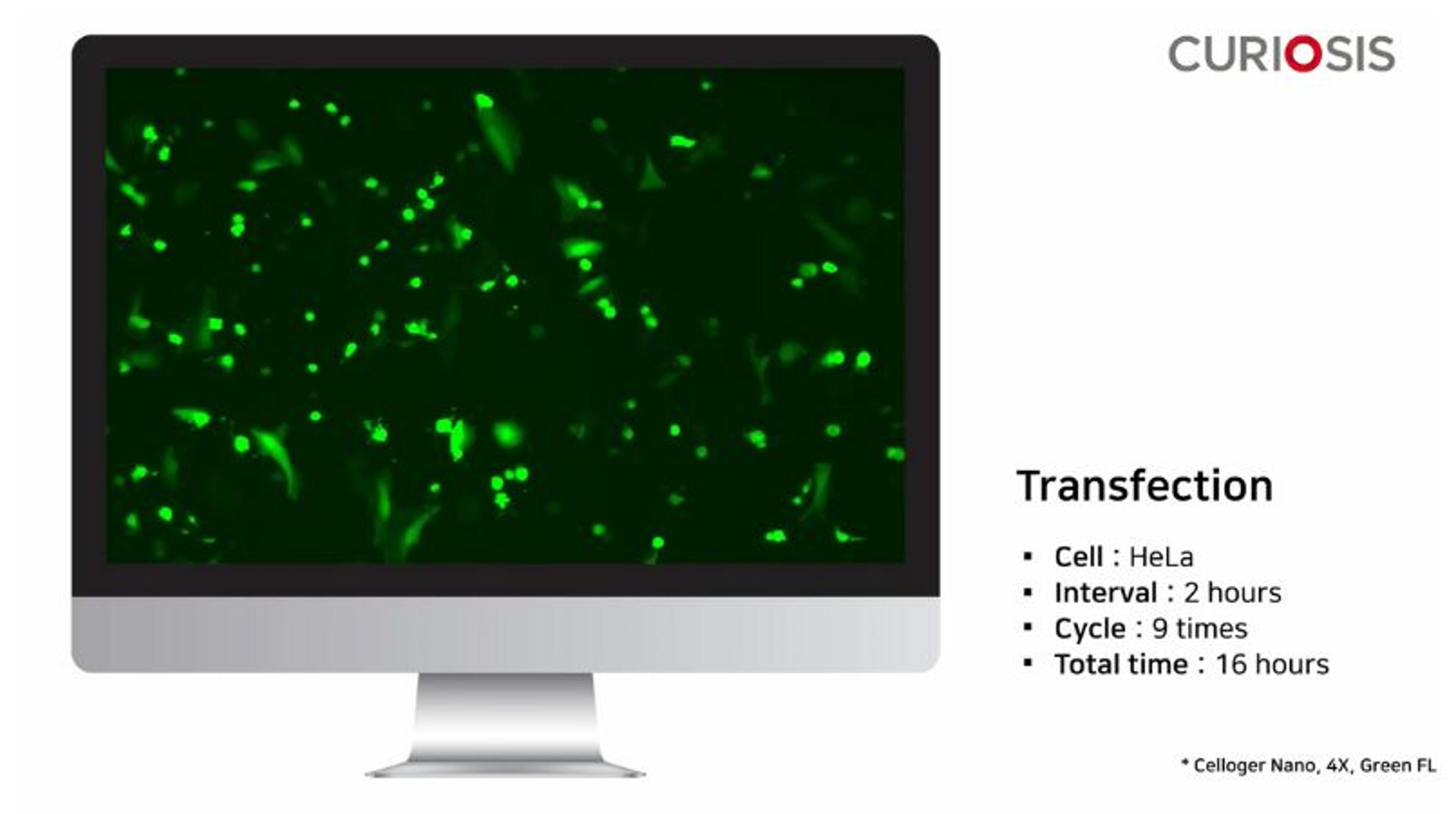
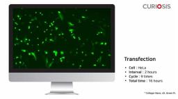
Celloger Nano application: Transfection
In this video, CURIOSIS shows how its Celloger® Nano can be utilized in transfection protocols. The expression of green fluorescence protein in the pCMV-GFP vector, which was transfected into cells, was observed every 2 hours through the Celloger® Nano.
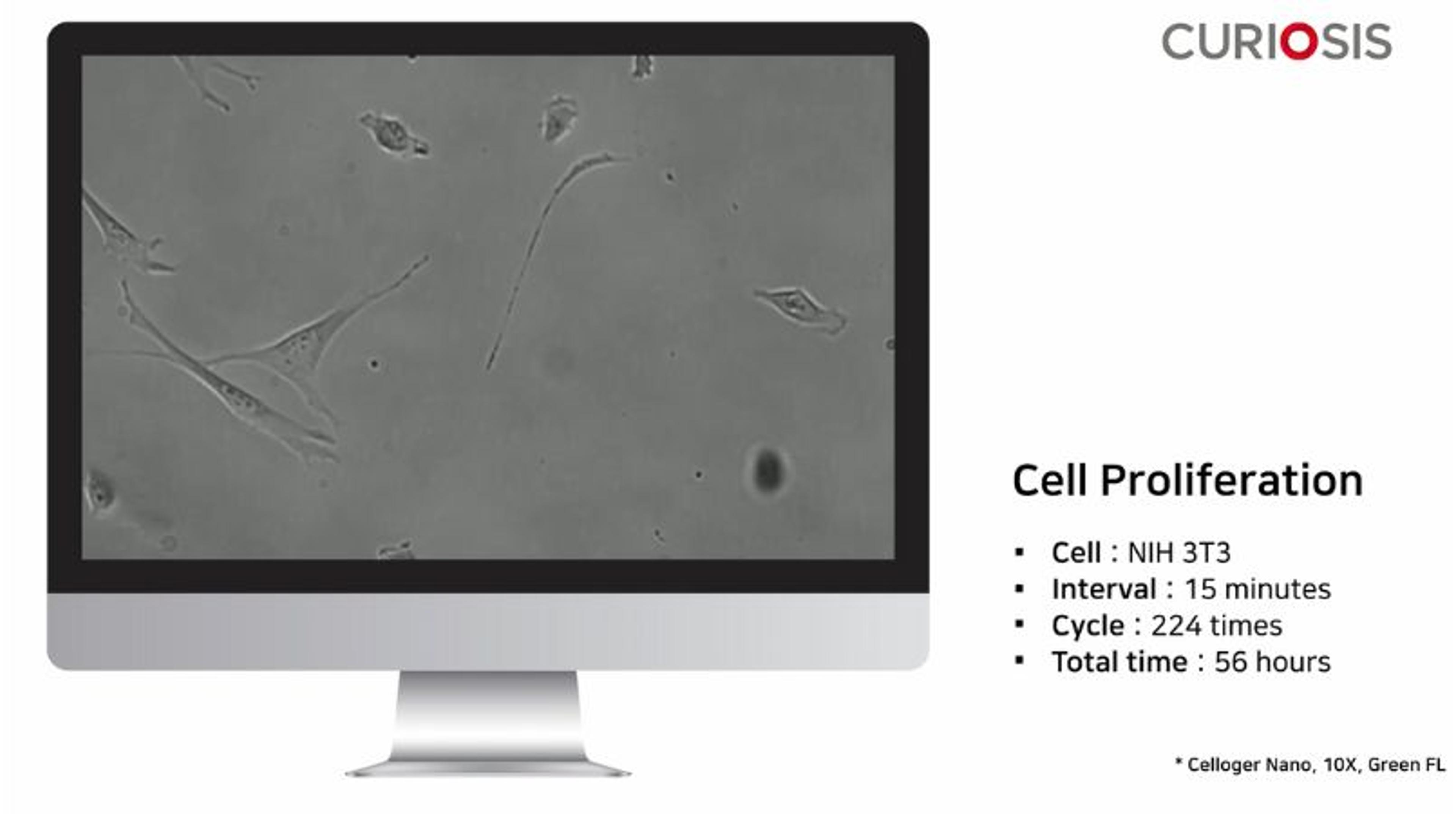
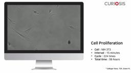
Celloger Nano application: Cell proliferation
Here, CURIOSIS shows how its Celloger® Nano can be utilized when studying cell proliferation. This time-lapse movie observes NIH 3T3 cell proliferation and the images were taken every 15 minutes for a total time of 56 hours using the Celloger® Nano 10X objective.
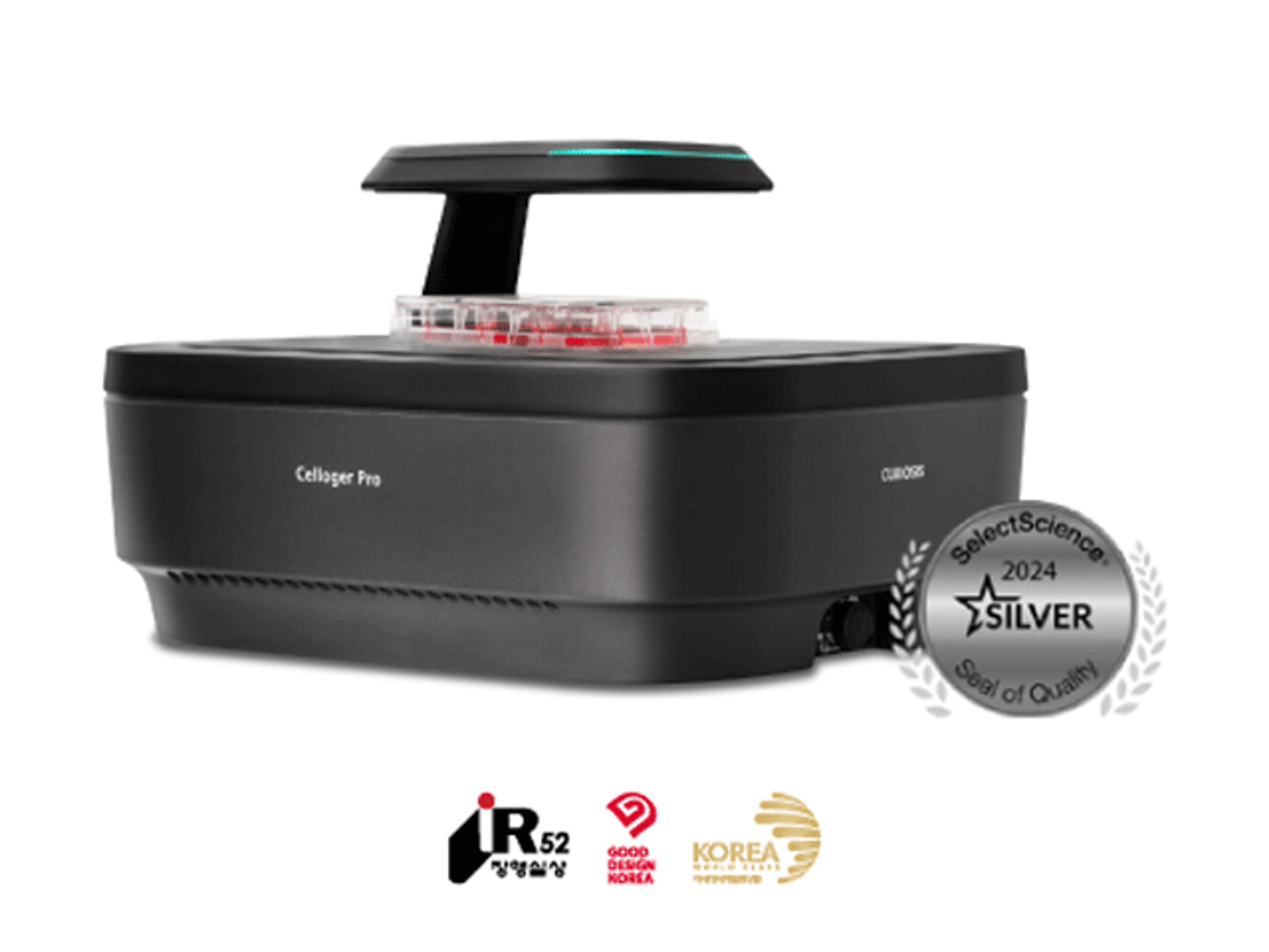
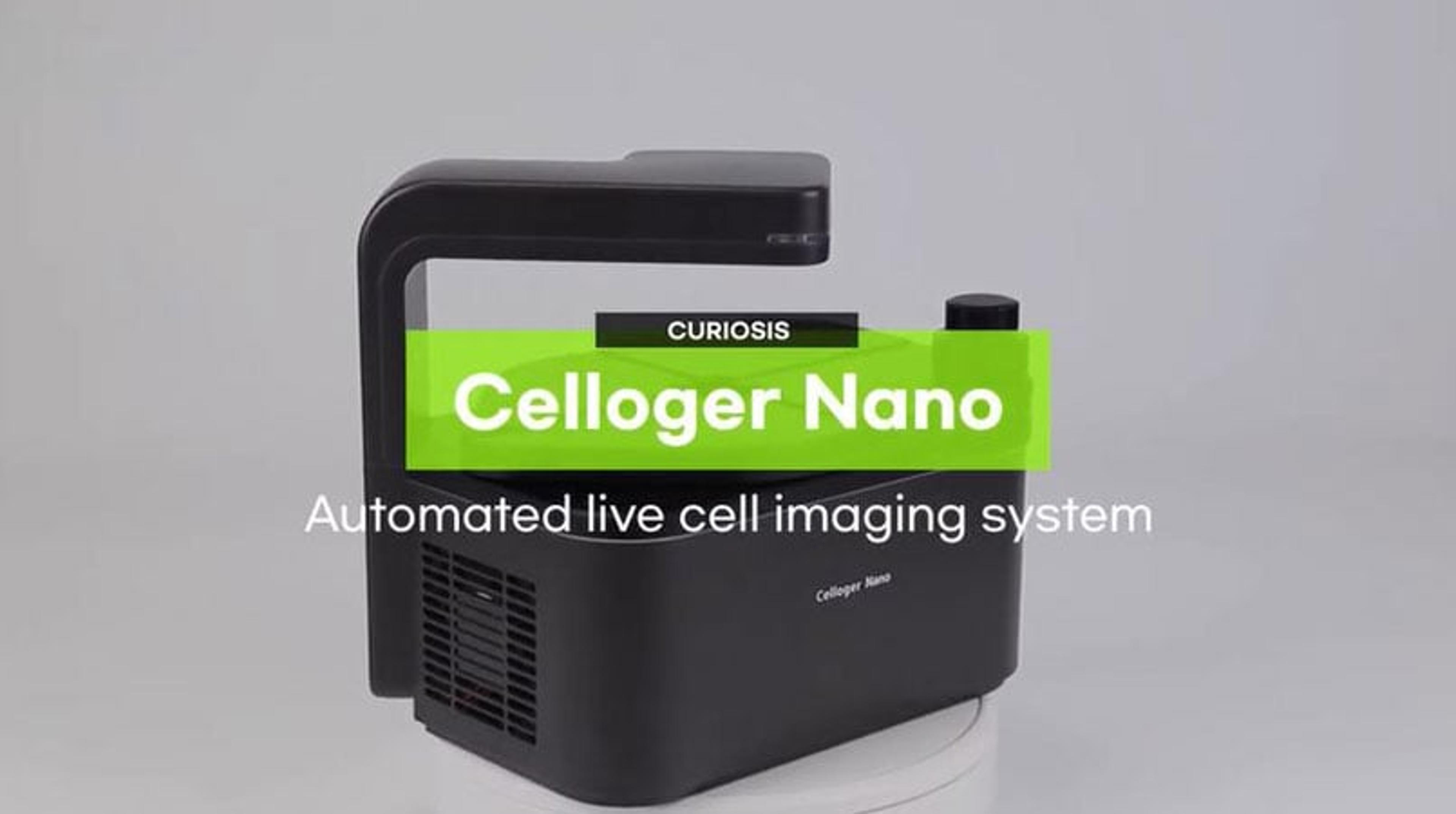
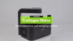
Celloger Nano, Automated Live Cell Imaging System from Curiosis
In this video, Curiosis introduces the Celloger Nano, which provides real time live-cell imaging and analysis within an incubator and is equipped with exceptional fluorescence and auto-focusing technology, and user-friendly software to accelerate your cell-based research works.
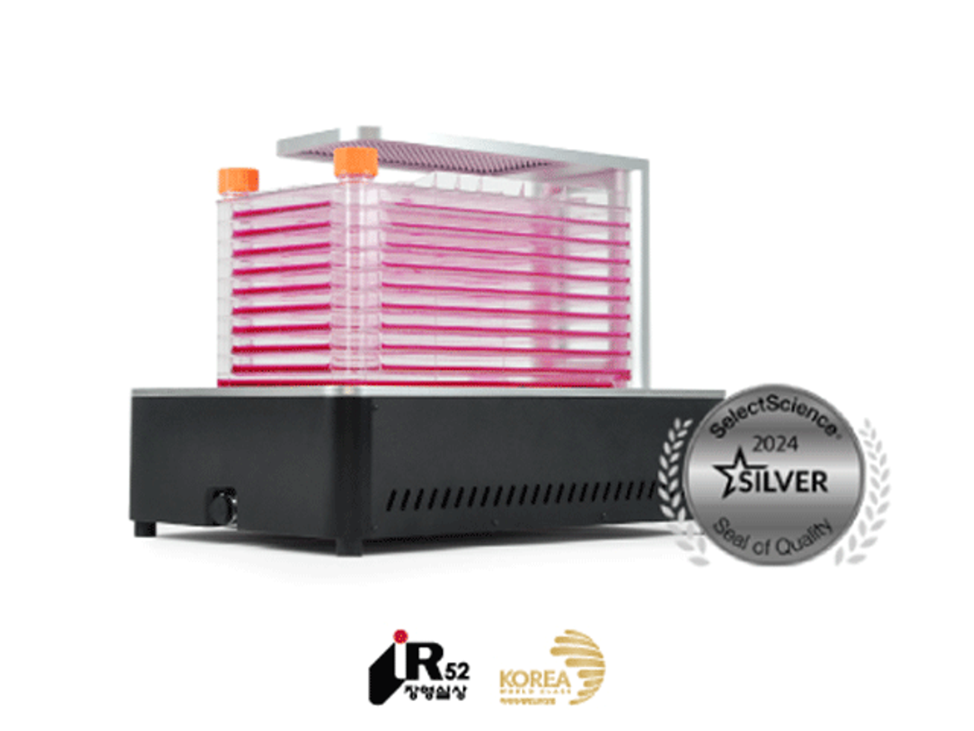
Looking for a compact microscope that works perfectly inside your CO2 incubator?
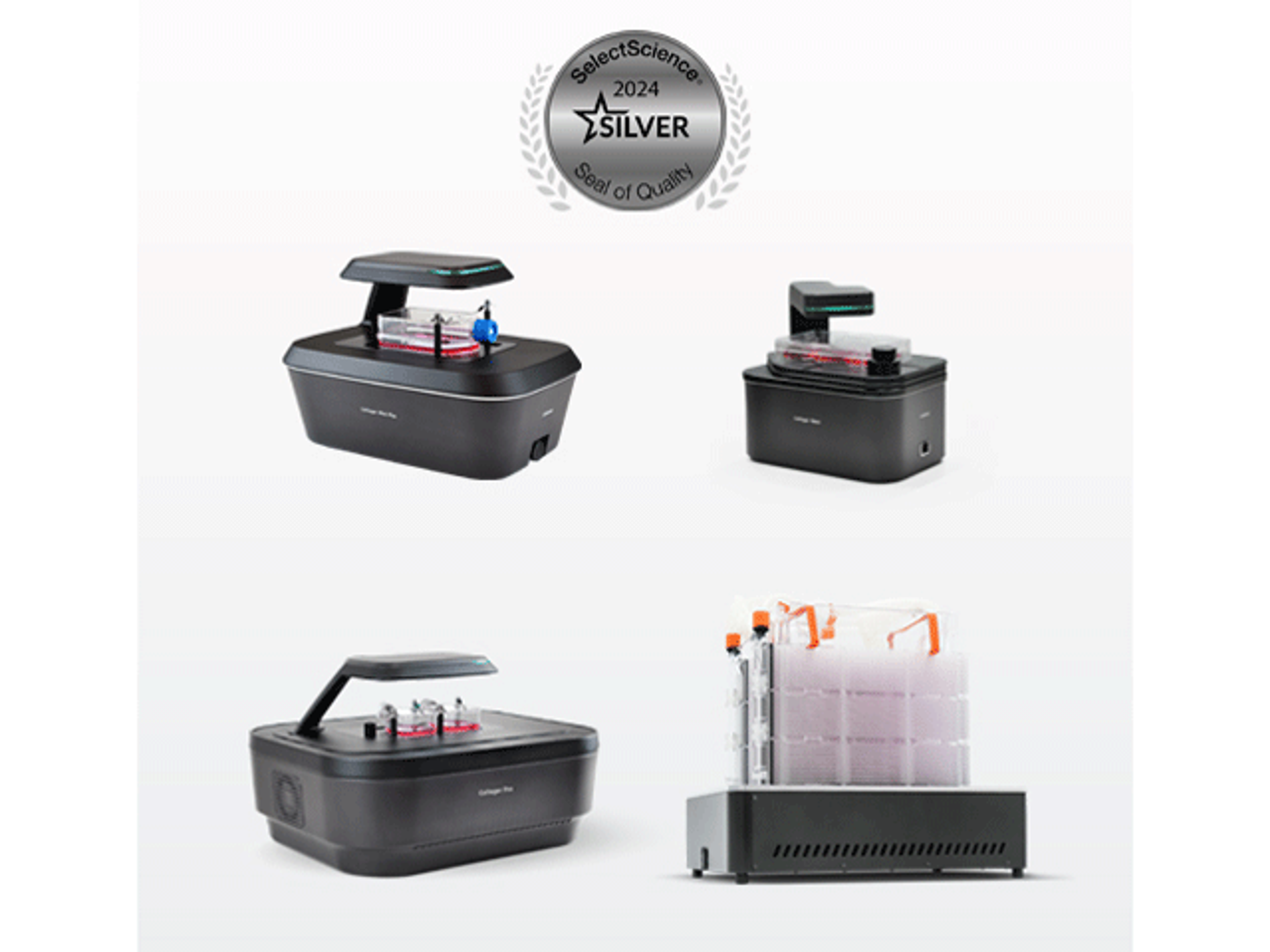
Introducing Curiosis’ automated live-cell imaging systems: The Celloger Series