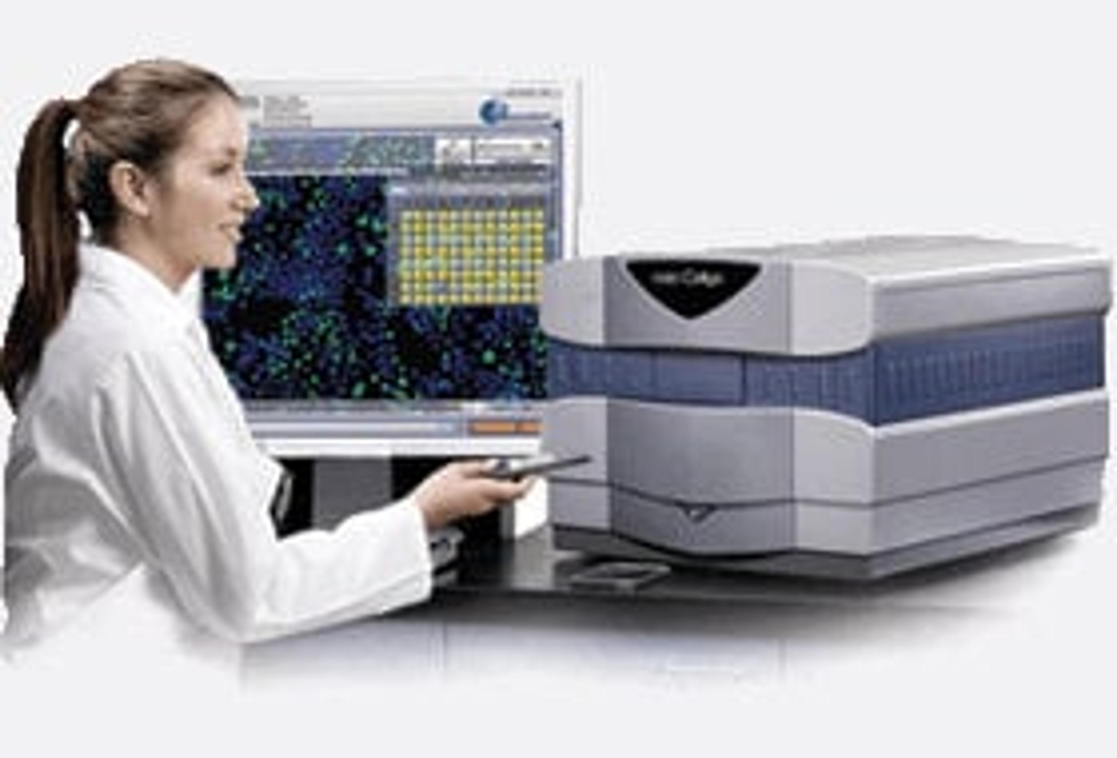
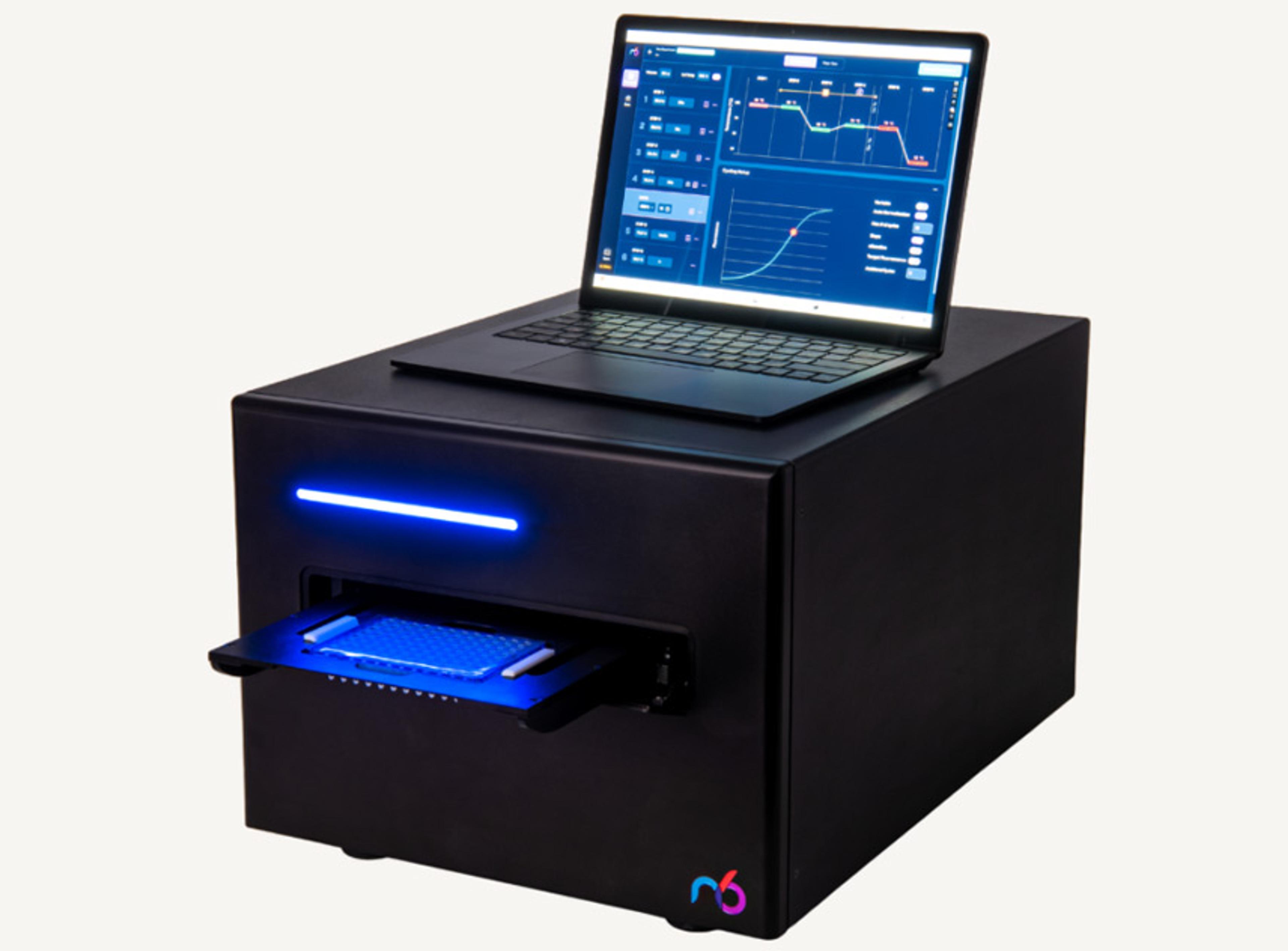
Celigo S Bright Field Image Cytometer
A micro-well, plate-based bright field imaging cytometer for 2D and 3D culture using both adherent and suspension cells.

Best-in-class bright field imaging

The Celigo S Bright Field Image Cytometer offers best-in-class bright field imaging capabilities. It provides high-speed, fully automated imaging and quantification of suspension, adherent, tumor spheroids, iPSC and cancer stem cell colonies. The system images every cell in every well, without any well edge effect. A 384-well plate will take approximately 2 minutes to image. System is compatible with automation platforms for full integration.

Rapid, Label-Free Counting and Characterization of Live Embryoid Bodies
This application note explores the benefits of a fast, automated method for embryoid body (EB) analysis. The Celigo™ Colony Counting: Embryoid Body Application by Nexcelom Bioscience quickly and accurately analyzes EB populations to assess the number, size, and shape of live EBs within multi-well plates.
Automated Cell Growth Tracking for Cytotoxicity and Proliferation
The Growth Tracking application automatically integrates label-free cell counts of the same well/flask from different time points to provide direct measurement of growth rates and doubling times, which is also a good overall assessment of cell health.
Cell Line Development - Single Cell Detection, Clonal Validation, Transfection
The process of developing a cell line to produce a specific protein or antibody involves multiple stages, all of which can be greatly aided by Celigo imaging cytometer.