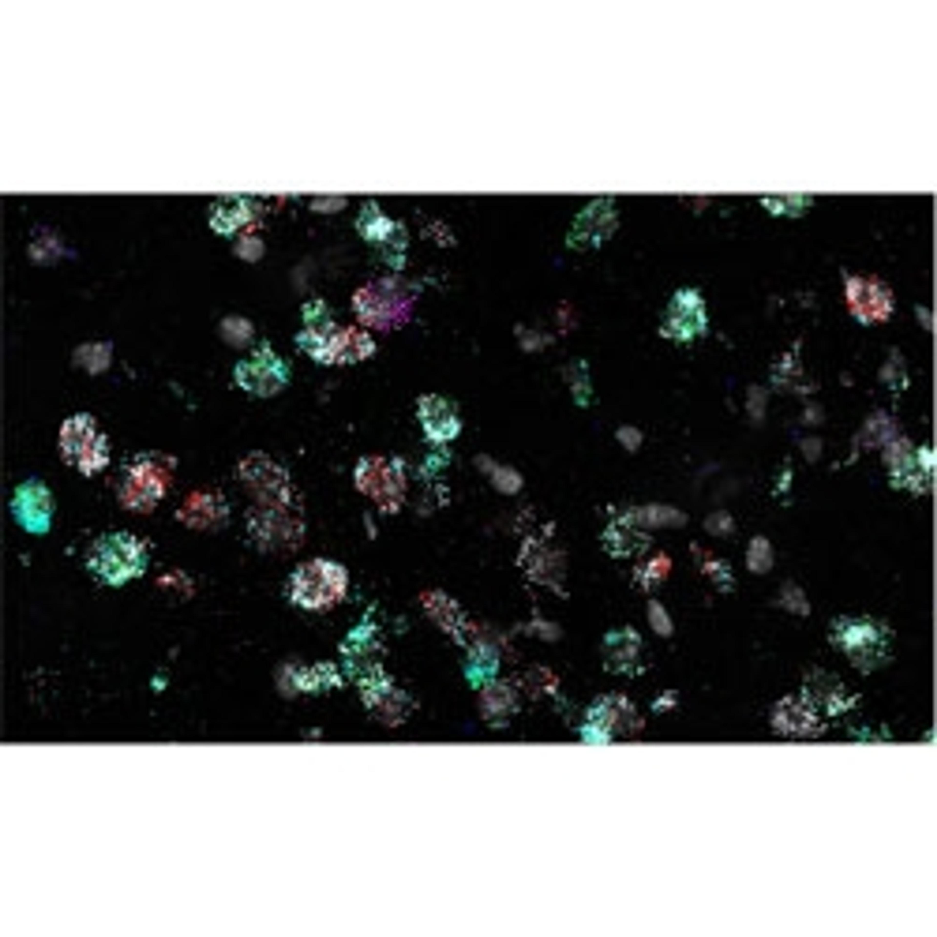
RNAscope™ HiPlex Assays
Introducing RNAscope HiPlex assay to expand your in situ hybridization potential.

The supplier does not provide quotations for this product through SelectScience. You can search for similar products in our Product Directory.
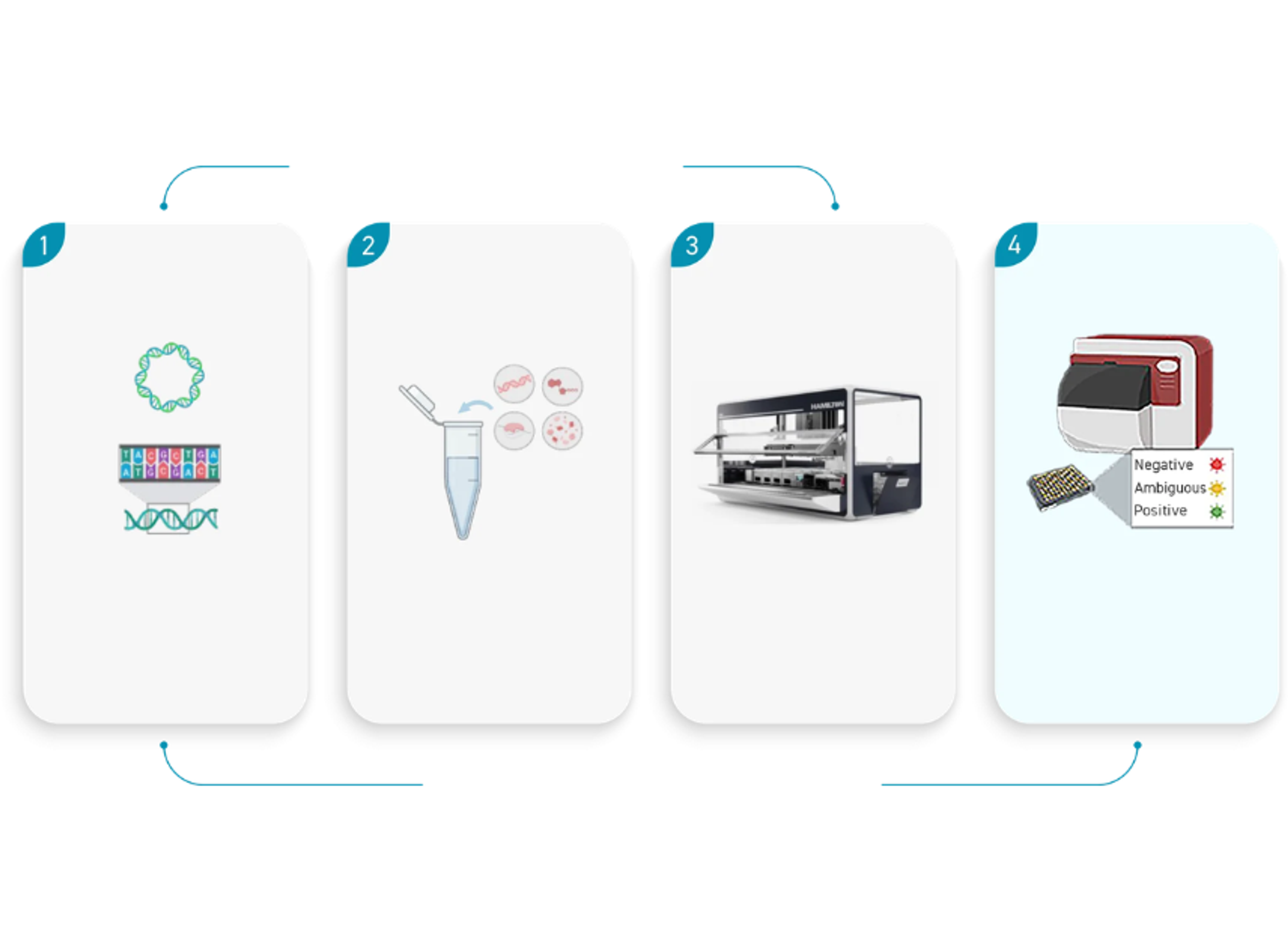
Introducing RNAscope HiPlex assay to expand your in situ hybridization potential. Now allowing simultaneously detection of up to 12 different RNA targets per slide mounted sample. Higher plexing of up to 12 different RNA targets is enabled by employing cleavable fluorophores and iterative detection with the RNAscope HiPlex8 Detection kit, and the RNAscope HiPlex12 Ancillary Kit.
One major application for the RNAscope HiPlex Assay is to validate gene signatures with spatial context at the single cell level.
Taking a multi-omic multiplexing approach to spatial biology
Multiomics has begun to take the center stage in biomedical research as it combines two or more individual omics studies such as genomics, epigenomics, transcriptomics, proteomics, and metagenomics, allowing scientists to interpret and visualize the mechanisms of biological processes with spatial context.
In this free guide, we present a series of case studies illustrating how a multi-omic multiplexing approach can be leveraged to gain a comprehensive view of spatial biology, including a multiplexed in situ transcriptomic method for the spatial mapping of target genes in highly complex and heterogenous FFPE tumor tissues, and much more.

Quantitative RNAscope image analysis guide
In this application note, Advanced Cell Diagnostics (ACD) provides examples of exemplary assays, discusses experimental considerations, addresses common challenges, introduces HALO® image analysis solutions, and answers common quantitative image analysis questions. Plus, learn how RNAscopeTM technology is designed to enable RNA target expression analysis within intact cells and tissues with high sensitivity and specificity.
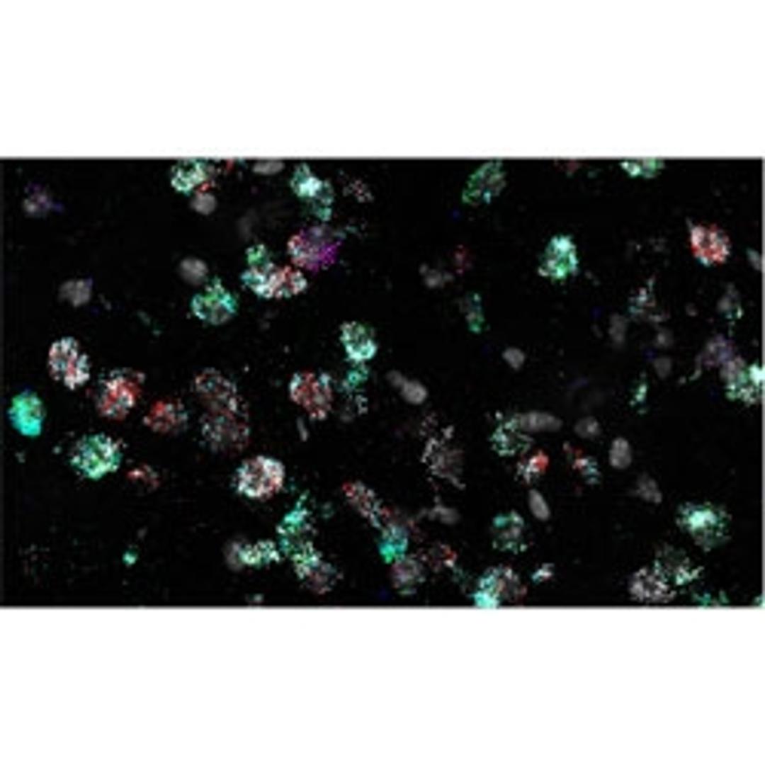
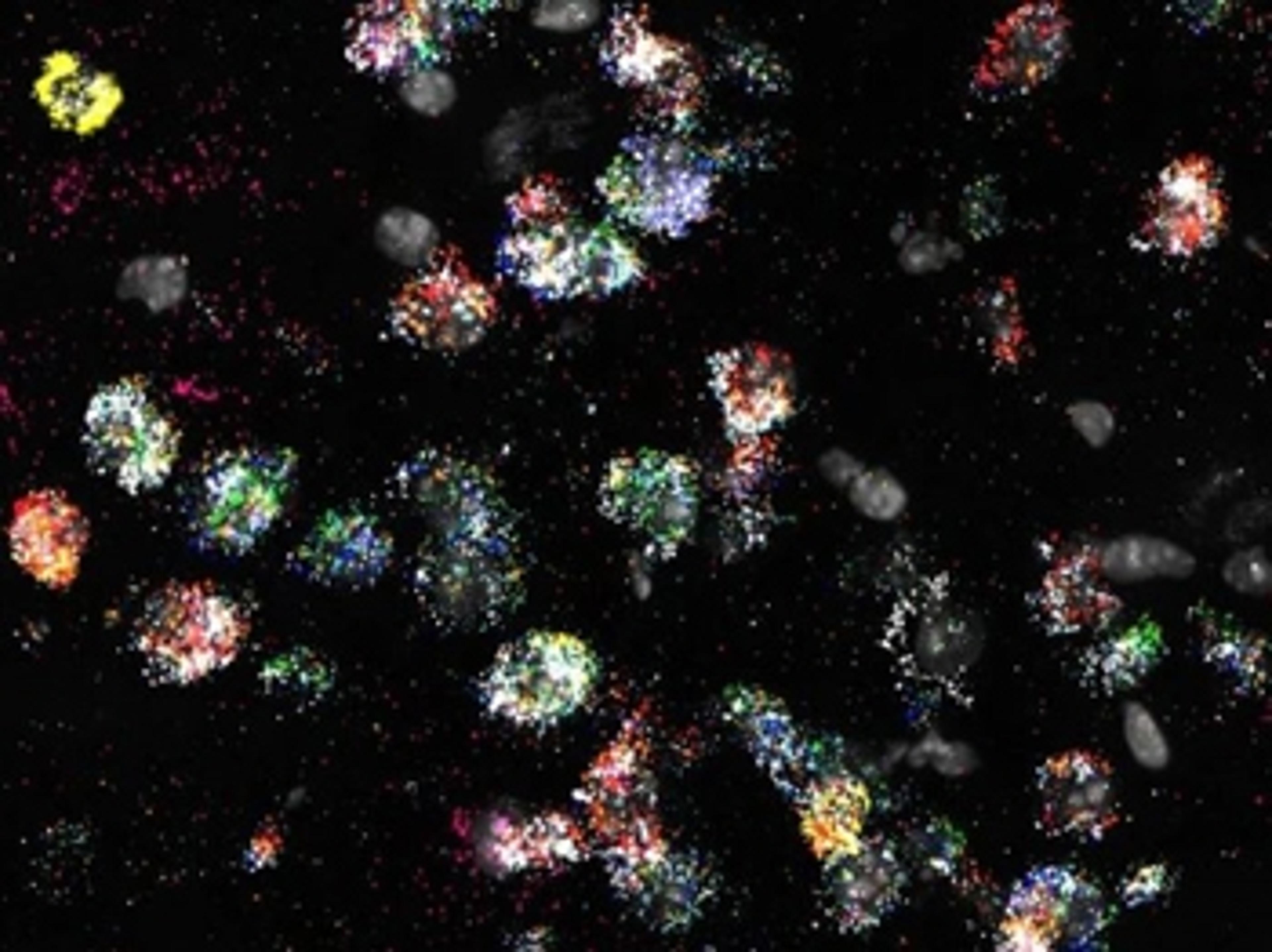
Characterization of immune cell phenotypes through quantification of the 12-plex spatial RNAscope HiPlex v2 assay using the HALO image analysis platform
In this application note, Advanced Cell Diagnostics demonstrates how the HALO® image analysis platform can be employed with HiPlex v2 to quantitatively assess differential expression of 12 RNA targets and quantify six distinct cell phenotypes based on gene expression. Plus, spatial relationships between cell phenotypes within the tumor microenvironment (TME) are analyzed.
RNAscope technology enables the incorporation of spatial mapping and confirmation of gene signatures into single cell RNA sequencing workflows
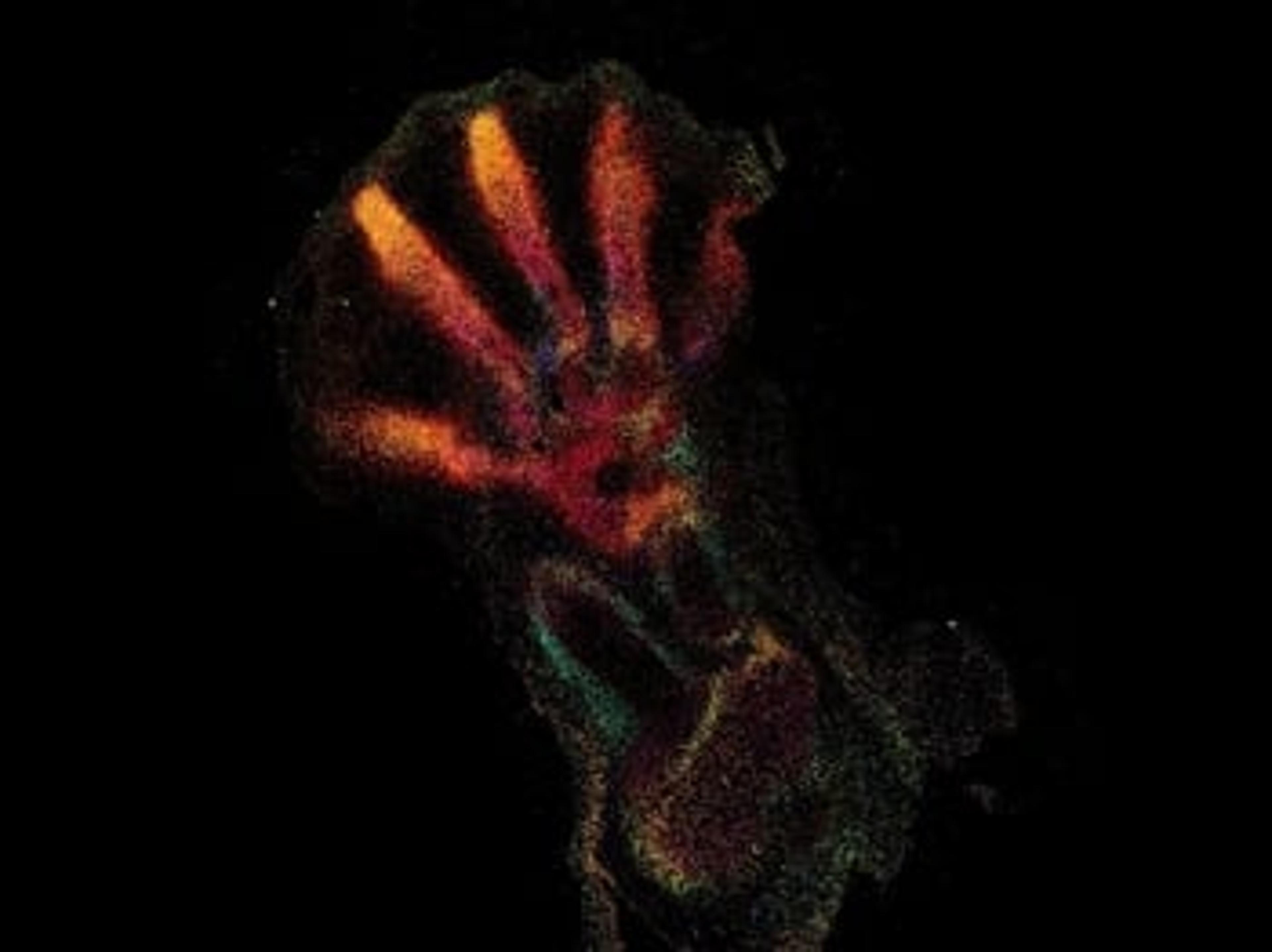
In this application note, the diverse cell types in the mouse striatum that have been previously identified by scRNA-seq were confirmed and spatially mapped using the RNAscopeTM Multiplex Fluorescent assay and the RNAscopeTM HiPlex assay. The major and minor gene signatures identified by scRNA-seq, including discrete D1 and D2 medium spiny neuron (MSN) subtypes, were confirmed.
Further cellular heterogeneity within the MSN subpopulations was marked by a transcriptional gradient, which was spatially resolved with the RNAscopeTM technology.
Spatial profiling of immune cell gene signatures in the tumor microenvironment
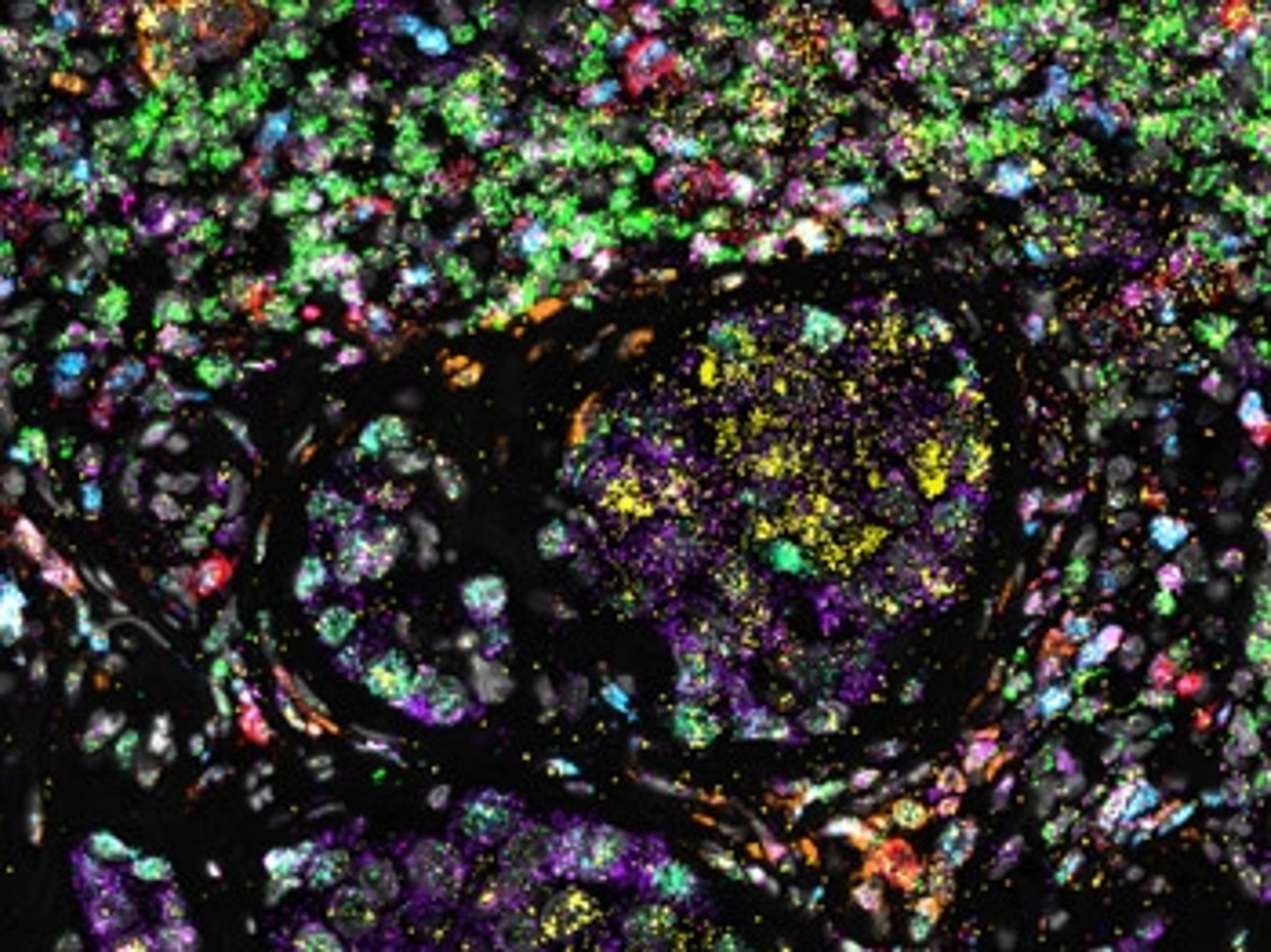
Complex tissues with high cellular heterogeneity require single-cell technologies both at the transcriptomic and spatial level to fully interrogate the cell types within them. This application note demonstrates the capabilities of a multiplexed in situ transcriptomic approach for the spatial mapping of target genes in highly complex and heterogenous FFPE tumor tissues using the RNAscope HiPlex v2 assay.
Upgrade your single-cell data with higher-plexing
RNA plays an important role in the regulation of cellular processes, making it an ideal biomarker for dynamic gene expression changes in both healthy cells and disease phenotypes. In situ hybridization (ISH) technology provides a powerful method to detect this gene expression, at single-cell resolution, within the spatial and morphological context of an intact tissue. Advanced Cell Diagnostics offer a comprehensive portfolio of ISH assays, with various levels of plexing, that can be applied to a wide range of applications, from neuroscience to immuno-oncology to cell therapy.
In this application eBook, we present:
- A series of case studies illustrating the research capabilities of high-plexing assays
- An outline of the benefits of the RNAscope™ technology
- Top tips on how to achieve accurate, reliable results for virtually any target species, tissue, or gene
‘Google Maps’ for the human body – How spatial genomics is revolutionizing our understanding of disease
Discover how the Wellcome Sanger Institute is helping to develop a physical atlas of all human organs

The integral role of human retinal ganglion cells in eye disease
Discover how researchers are better understanding human eye disease by characterizing the subtype diversity and development of RGCs

Demystifying the complex role of GABAergic neurons in sleep deprivation
Discover how cutting-edge single-cell RNA analysis technology is helping make strides in homeostatic sleep pressure research

New insights into reproductive dysfunction with advanced spatial genomics
Dr. Lique Coolen and Dr. Aleisha Moore, Kent State University, present their latest research into the mechanisms underlying spinal cord injury and neuroendocrine dysfunction
New RNA in situ hybridization application guide for single-cell analysis
Check out the new eBook guide that covers a wide range of higher-plexing applications, from neuroscience to immuno-oncology to cell therapy
8 top trends in multiplexing: Could this technology transform your lab?
Discover the latest research applications using multiplex technology to maximize sample use, while saving time and money