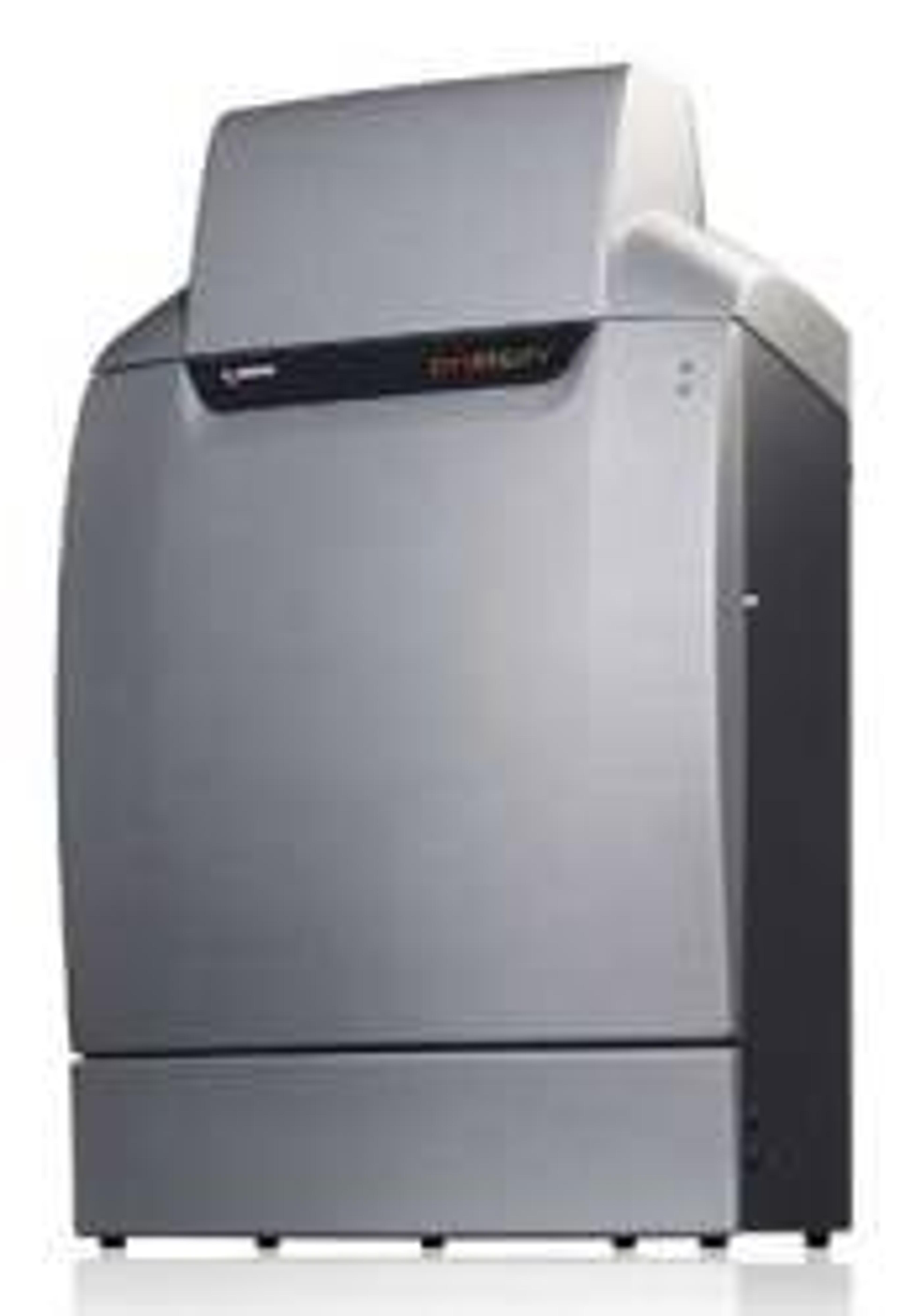
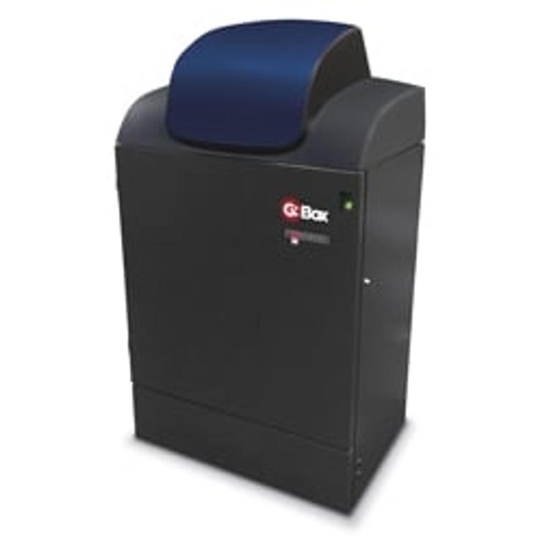
G:BOX Chemi XX9
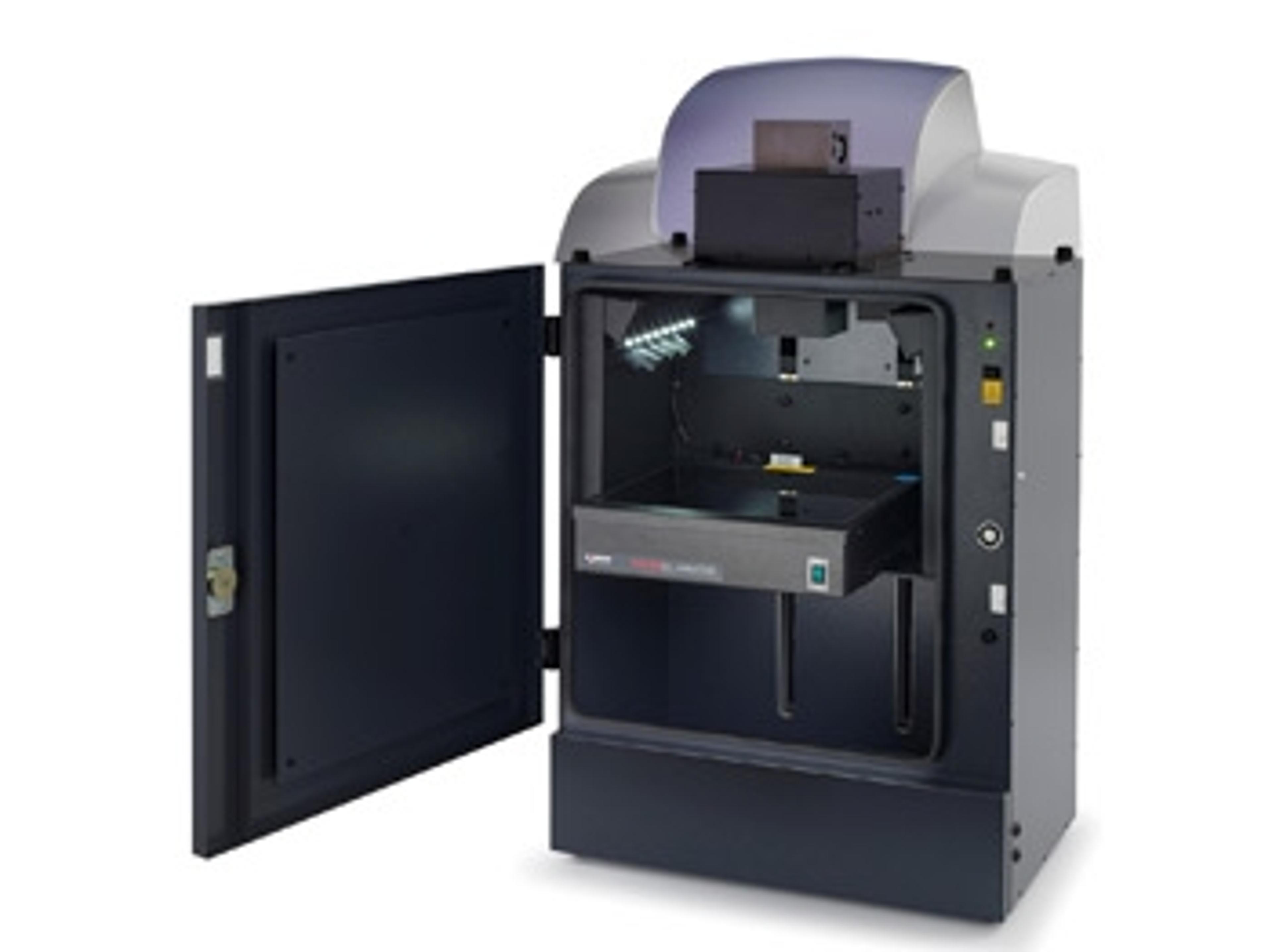
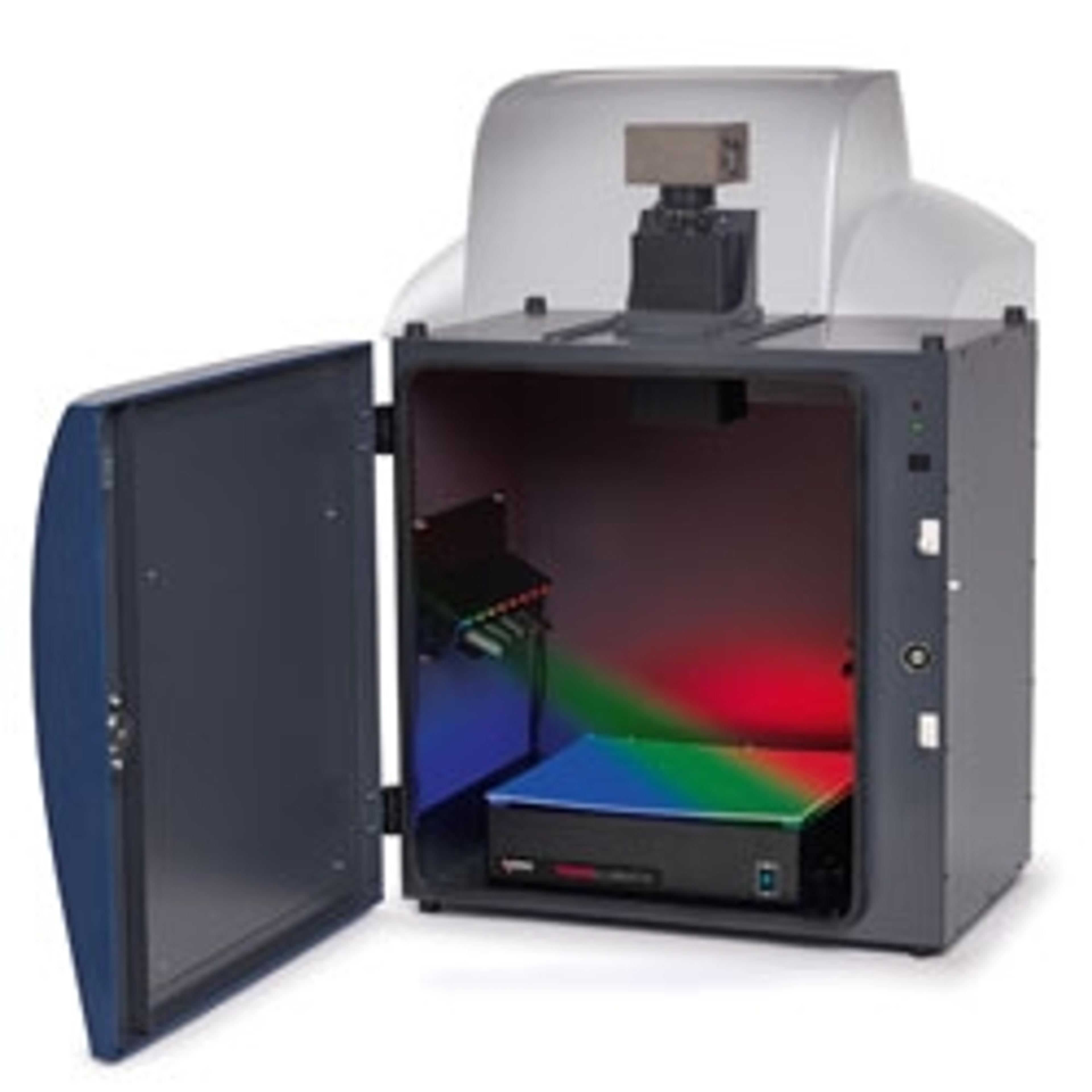
G:BOX Chemi XX9 gel imaging system has a high resolution camera for imaging multiple sample types and sizes, from fluorescence 1D to 2D gels to chemiluminescent blots. Your lab’s imaging system shouldn’t control how you detect proteins on Western blots. Chemiluminescence is great if you want sensitive detection of picogram or femtogram amounts, while fluorescence lets you quantify and detect multiple different proteins on one…

The supplier does not provide quotations for this product through SelectScience. You can search for similar products in our Product Directory.
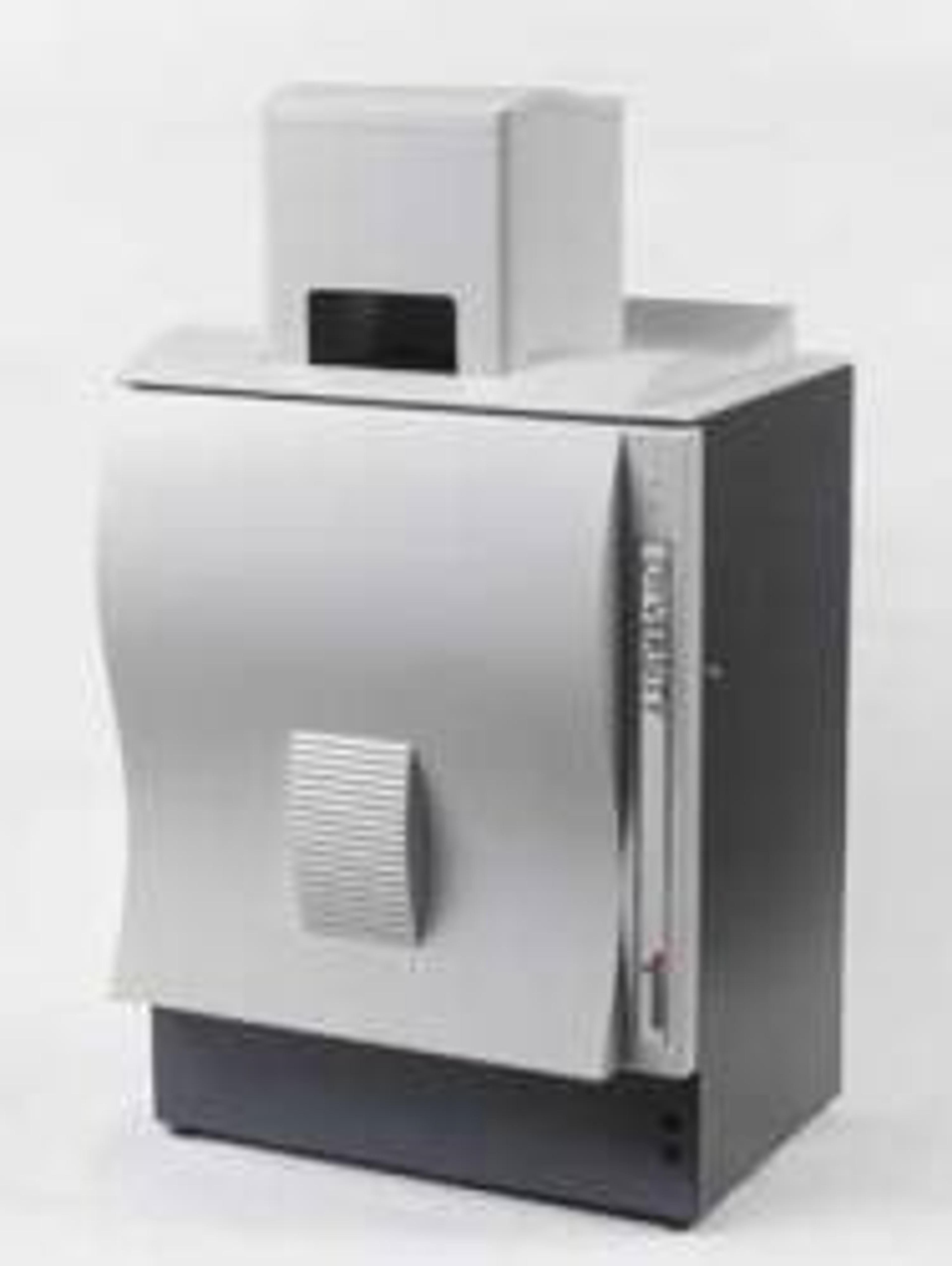
Versatile imaging systems for gels and blots.
Gel imaging and documentation system
Versatile imaging system for fluorescent / visible gel. We regularly use this machine for DNA gels (EtBr), western blots, and PAGE. The software is intuitively designed and has preset modules for different applications like DNA gels with different dyes SYBR Safe, gel Red, western blots with different substrates like ECL, fluorescent probes. The software also allows editing of the captured images, quantification, and annotation. Personally, I feel the software could be made a bit more user-friendly.
Review Date: 18 Jul 2021 | Syngene
G:BOX Chemi XX9 gel imaging system has a high resolution camera for imaging multiple sample types and sizes, from fluorescence 1D to 2D gels to chemiluminescent blots. Your lab’s imaging system shouldn’t control how you detect proteins on Western blots. Chemiluminescence is great if you want sensitive detection of picogram or femtogram amounts, while fluorescence lets you quantify and detect multiple different proteins on one blot. With a G:BOX Chemi XX6/XX9 you can have it all. Powered by Syngene’s revolutionary GeneSys software featuring hundreds of imaging protocols, you’re free to choose chemiluminescence and fluorescence reagents from any manufacturer, putting you in charge of how you detect your proteins.
Featuring an extended darkroom, you can choose between a 6 or 9 mega pixel, cooled, high quantum efficiency camera for unrivaled levels of sensitivity with minimal background interference.
HI-LED lighting options cover the full spectrum of high intensity blue, green, red and infra-red resulting in faster exposure times and publication quality images. An edge lighting option can also be used for 2D gel capture including the use of DIGE gels.
The system is controlled by GeneSys application driven image capture software and comes complete with unlimited copies of GeneTools analysis software.
Features:
- High quantum efficiency (QE) camera
- For unrivalled levels of sensitivity
- Fast chemiluminescence detection
- Walk-away image capture without film
- Motor driven lens and filter wheel
- Easy to use, fully automated set up
- IR and RGB HI-LED lighting options
- Multiplex fluorescence and protein quantification
- Stain-free imaging capability
- Capture images of TGX Stain-Free™ FastCast™ acrylamide gels and many more
- Edge lighting option
- 2D and multiplex in-gel fluorescence DIGE image capture
- White, UV and blue lighting options
- Capture all types of gel and blot images with one system
- Extended darkroom with motor driven stage
- Easily image large gels and blots
- GeneSys application driven image capture software
- Contains extensive database of dyes and imaging protocols. All you need to know is the type of gel you’re using and GeneSys automatically selects the optimal lighting and filters to produce the perfect image
- GeneTools analysis software (unlimited copies)
- Analyse data at your own computer
Brochures

G:BOX Chemi Multi Fluorescence and Chemiluminescence Imaging Systems
Great research comes from accurate Western blot and gel data. With so many ways to image chemiluminescence, fluorescent and visible dyes, you need to know which imaging systems truly capture real results.
Multiplexing LI-COR IRDye 680LT and IRDye 800CW
This application note outlines a method for fluorescence-based detection of proteins on Western blots. Using fluorescence-based fluorophores increases sample throughput and can make multiplexing several fluorophores less time consuming.
SafeView Dye: An Overview
This application note provides a brief overview of SafeView dye from NBS Biologicals, a nucleic acid stain used in gel electrophoresis for the detection of double-strandedA (dsDNA), single-stranded DNA (ssDNA) or RNA.
Overview of Ethidium Bromide Dyes
This application note provides an overview of ethidium bromide dyes, commonly used in agarose gel electrophoresis to detect double-stranded DNA (dsDNA) and single-stranded DNA (ss) or RNA.
An Overview of Alexa Fluor 647 Dyes
This application note provides an overview of Alexa Fluor dyes, a series of fluorescent dyes produced by Molecular Probes that span the visible spectrum.
How to Visualize SYBR Dyes Using a Syngene Image Capture System
Learn how Syngene image capture system,s combined with GeneSnap software are the perfect combination for imaging SYBR dyes.
Chemiluminescent Western Blot Imaging Using ChemiFast Substrate
The commonly used western blotting substrates are luminol-based and produce a chemiluminescent signal. Substrates such as ChemiFast are highly sensitive, enhanced chemiluminescent substrates. In this application note, learn how the ChemiFast substrate’s extremely intense signal output enables detection of HRP using cooled charge-coupled device (CCD) camera imaging methods.
Dynamic Fielding for Syngene Image Capture Systems
Some manufacturers use a Flat Field Correction method in order to address uneven illumination of light sources. This involves subtracting the image information from an empty field of view or ‘perfectly flat’ fluorescent reference sample from that of the same field of view with the gel added. As this process involves the subtraction of one image from another, the integrity of the raw data of the initial captured image is compromised. Such manipulation is incompatible with Good Laboratory Practice (GLP). Syngene uses the Dynamic Fielding Correction method to address uneven light illumination whilst maintaining GLP compliance.
Quantitative Imaging of Chemiluminescent Western Blots: Comparison of Digital Imaging and X-ray Film
Chemiluminescent western blotting is the most frequently used method for detecting proteins. Recently there has been a trend towards chemifluorescence which provides the user with many advantages including shorter exposure times and also means the signal is stable for weeks. This application note will compare the performance of x-ray film with that of Syngene’s G:BOX Chemi IR6 imaging system for the visualisation and quantification of chemiluminescent and chemifluorescence signals on western blots.
New GeneSys software makes imaging chemi blots a breeze
Capturing chemi blot images is tricky, which is why Syngene have redesigned their GeneSys software for G:Box Chemi systems

Syngene Introduces Next Generation G:BOX Imaging Systems
New G:BOX systems feature super-sensitive lens for quick, accurate imaging of all gels and blots