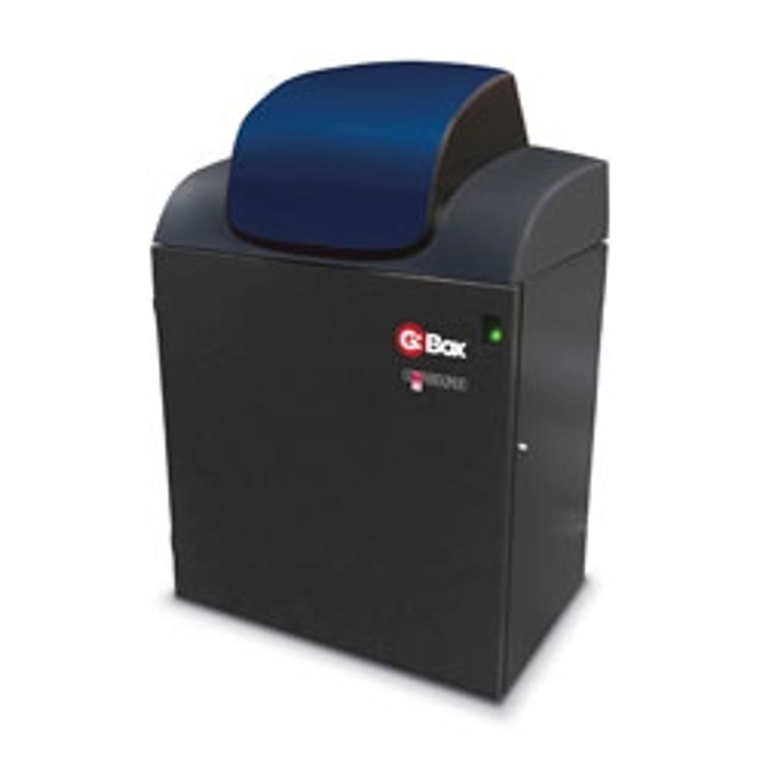
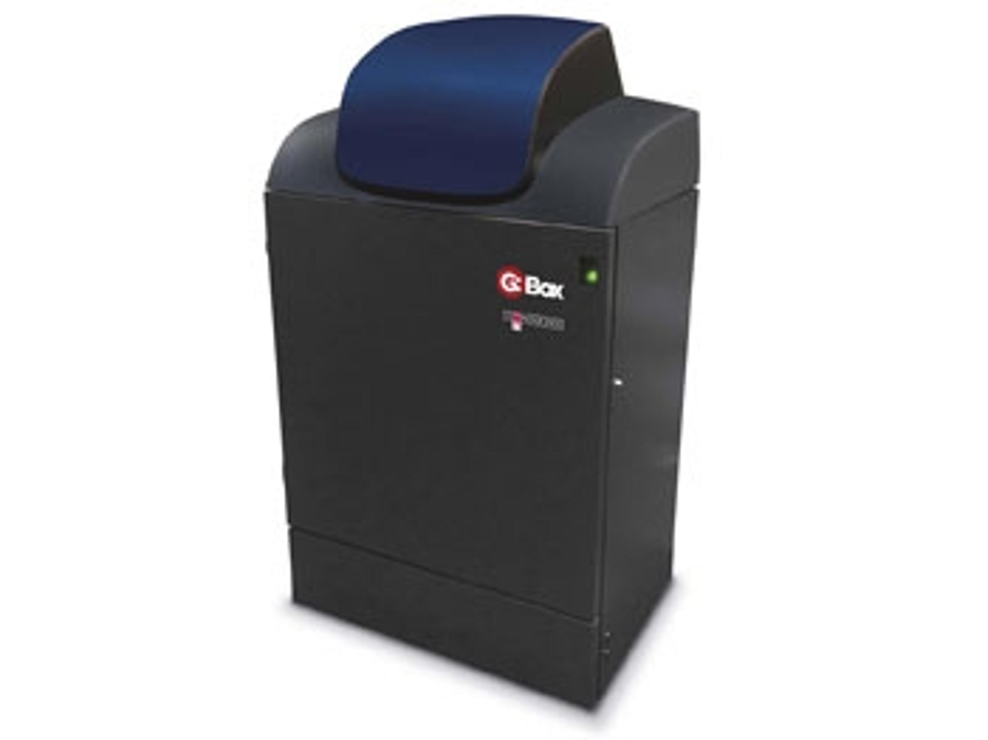
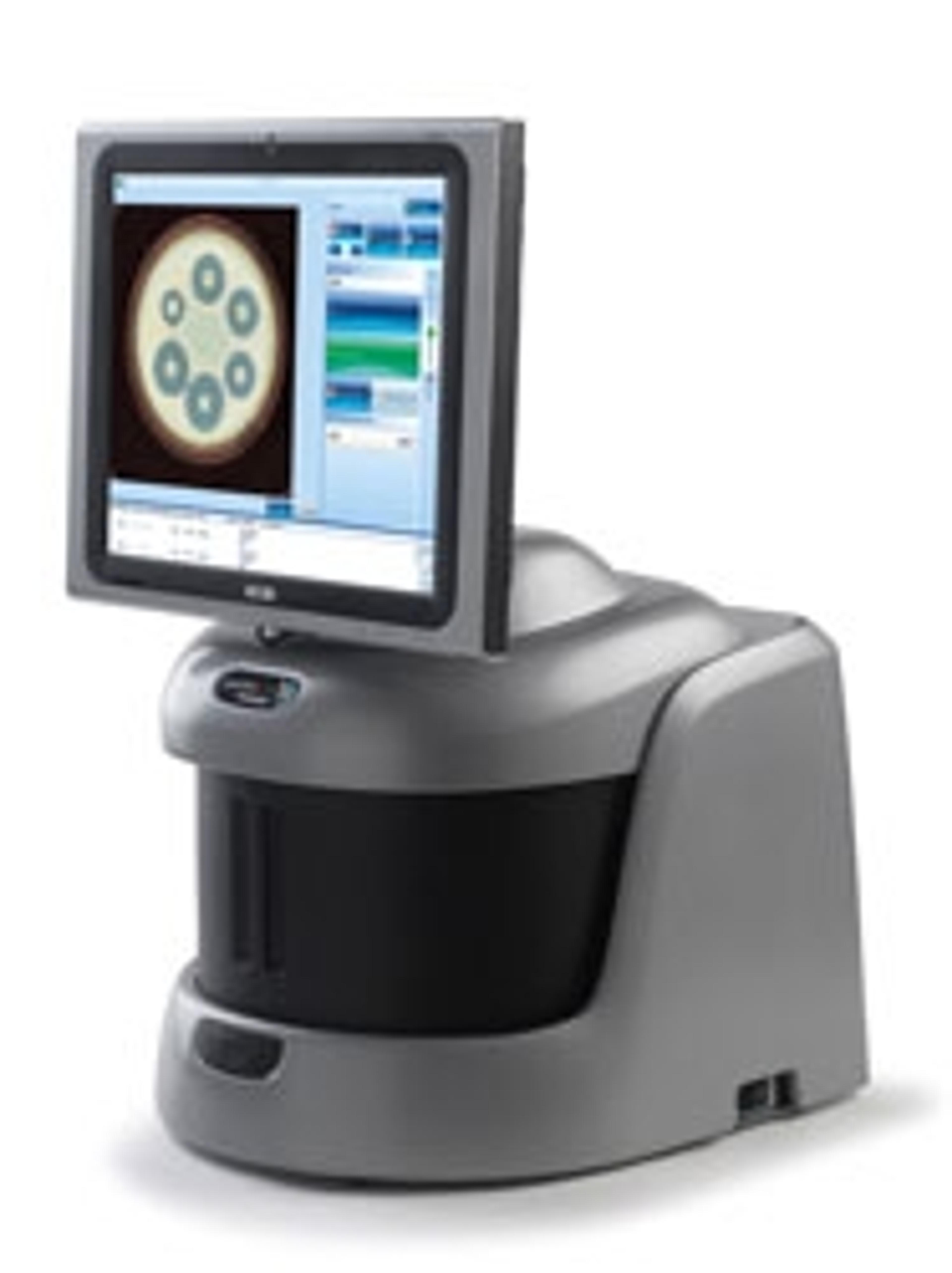
G:BOX Chemi XRQ
G:BOX Chemi XRQ is a cost-efficient chemiluminescence imaging and gel documentation system. For a laboratory that needs hassle-free chemiluminescent detection, as well as routine gel documentation, using the G:BOX Chemi XRQ’s powerful GeneSys software to switch between applications is simplicity itself.

The supplier does not provide quotations for this product through SelectScience. You can search for similar products in our Product Directory.
Easy operation, high quality images, reproducibility guaranteed! Highly recommended!
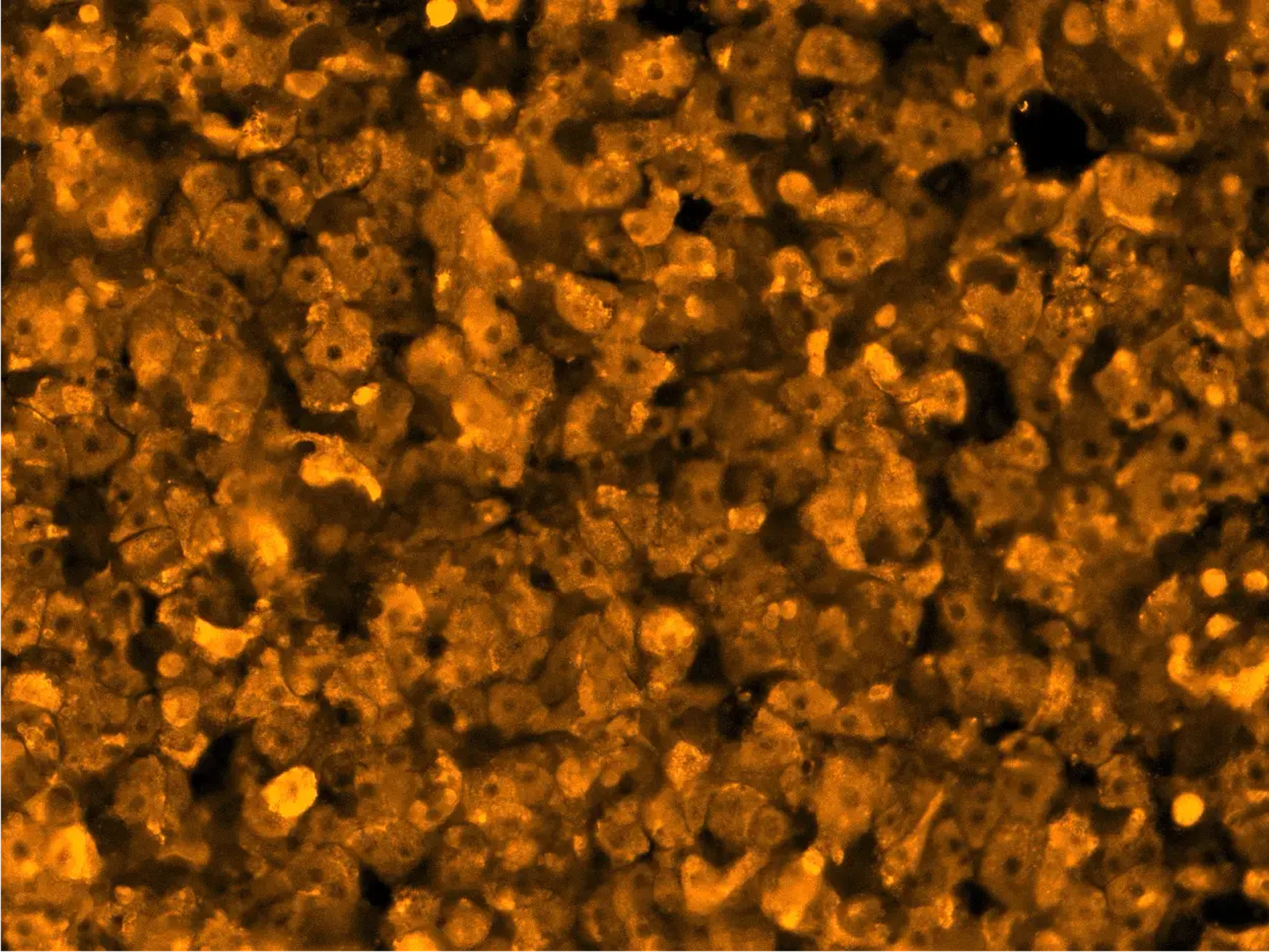
DNA, RNA and protein gel imaging and analysis, Chemi luminescent western blotting imaging
Ease of Operation: Ease of gel doc system operation and very user friendly software. Auto-focusing and captures high quality images in just few clicks. Never required to manually adjust the IRIS/focus and other related settings. High quality images for DNA / protein gels and chemiluminecent western blots with reproducibility. Covers entire range fluorescent dyes which can be selected based on our desired application. For chemiluminescent imaging also, no need to do manual settings, auto-setting always give high quality images and are reproducible. Automatic overlaying of visible marker over chemi-western blot is very convenient. After Sales Care: Highly competent application scientist and service engineers available. Promptly attempted and resolved all queries. For emergency, even immediate support provided through online software access whenever needed. Value for Money: Definitely value for money. Compared to my previous experience with other gel docs, it has much more advanced features, high quality image and very user friendly software at equivalent or lower price than other top players. Highly recommended!
Review Date: 10 Dec 2020 | Syngene
Great images, wonderful customer care and the price was right!
Imaging westernblot membranes
We recently switched over to this digital imaging system from the traditional film and chemical method. We have no regrets and love the images that we are able to get from this system. We were provided with unlimited access to the application specialist who came in to help us with the training. He offered to come in and help as much as we needed until we were all comfortable with the software. Their customer care surpassed our expectation. The best part was the price!
Review Date: 22 Dec 2017 | Syngene
Great results very quickly.
Viewing DNA gels and western blots
The G:Box is extremely user friendly. The equipment is heavily used by multiple labs and we have had no problems. Anytime someone has a question the sales rep, is accessible with assistance and further follow up. After sales care is the most important issue after any equipment is purchased. It is excellent for the G:Box. For all the use it gets the value for Money is excellent.
Review Date: 14 Dec 2017 | Syngene
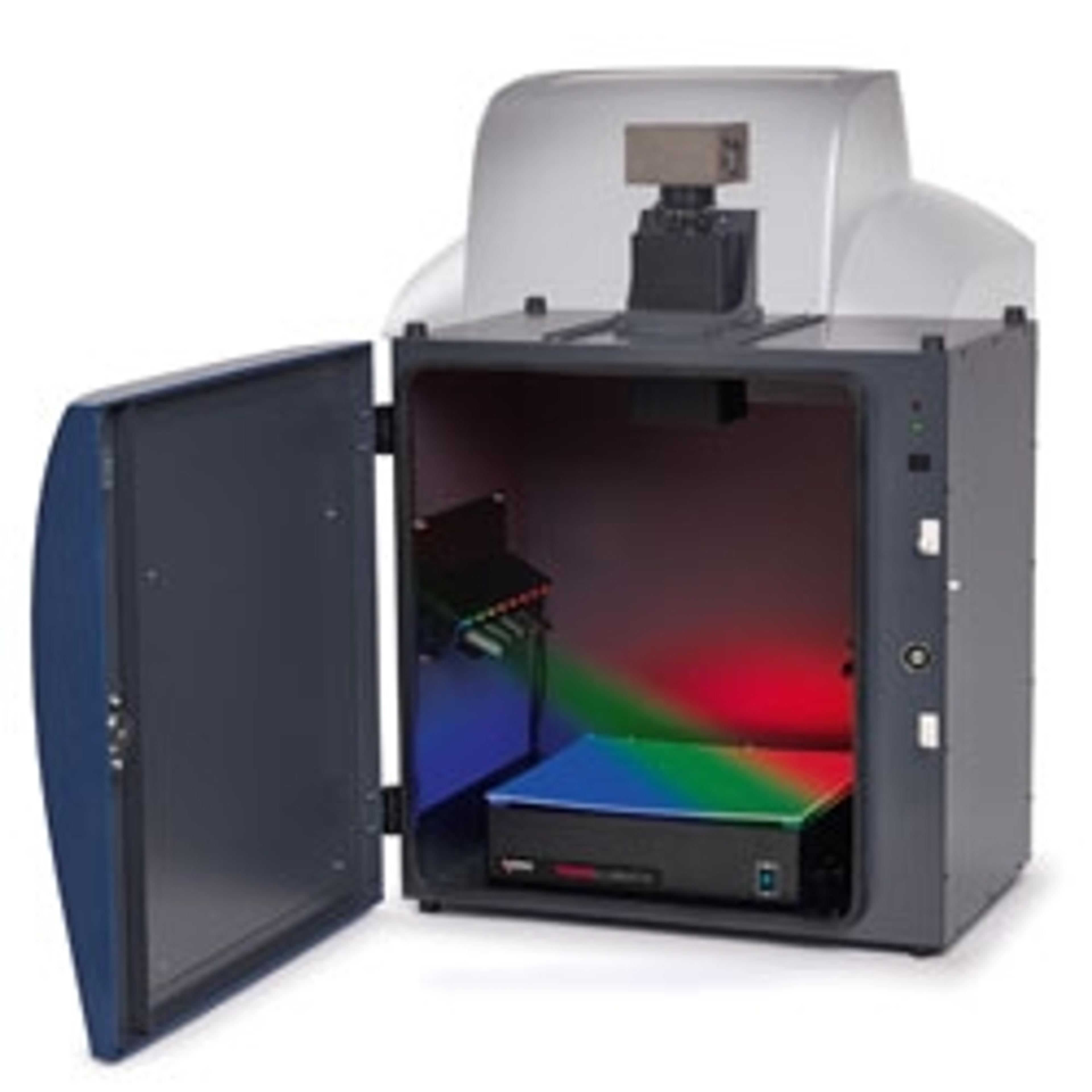
G:BOX Chemi XRQ is a cost-efficient chemiluminescence imaging and gel documentation system. For a laboratory that needs hassle-free chemiluminescent detection, as well as routine gel documentation, using the G:BOX Chemi XRQ’s powerful GeneSys software to switch between applications is simplicity itself. Place your chemi blot in, and the system’s cooled camera captures images with a sensitivity equal to film or slide in your gels to generate quality pictures of your DNA and proteins anytime you want.
Featuring a standard darkroom and a 4 million pixel image resolution, the G:BOX Chemi XRQ camera is cooled to provide a low signal to noise ratio with perfect background.
HI-LED lighting options cover the full spectrum of high intensity blue, green, red and infra-red resulting in faster exposure times and publication quality images.
The G:BOX Chemi XRQ is a cost-effective alternative to laser based technology offering a faster workflow for imaging complex multiplex fluorescent gels and blots.
The system is controlled by GeneSys application driven image capture software and comes complete with unlimited copies of GeneTools analysis software.
Features:
- High quantum efficiency (QE) camera
- Picogram detection
- Multiplex imaging of up to 5 different channels with UV, blue and RGB HI-LED lighting options
- Easily switch between blot or gel imaging
- Stain-free imaging capability
- Capture images of TGX Stain-Free™ FastCast™ acrylamide gels and many more
- Auto-exposure
- Perfectly exposed chemi images without film
- Excellent signal to noise ratio
- No annoying backgrounds on your blots
- Protocol driven image capture
- No camera expertise needed for great results
- Motor driven lens and filter wheel
- Simple, automated set up
- Reproducible and quantifiable data
- Accurate results time after time
- CFR21 Part 11 compliant
- Fully traceable results suitable for regulatory audits
- GeneSys application driven image capture software
- Contains extensive database of dyes and imaging protocols. All you need to know is the type of gel you’re using and GeneSys automatically selects the optimal lighting and filters to produce the perfect image
- GeneTools analysis software (unlimited copies)
- Analyse data at your own computer
Brochures

G:BOX Chemi Multi Fluorescence and Chemiluminescence Imaging Systems
Great research comes from accurate Western blot and gel data. With so many ways to image chemiluminescence, fluorescent and visible dyes, you need to know which imaging systems truly capture real results.
Multiplexing LI-COR IRDye 680LT and IRDye 800CW
This application note outlines a method for fluorescence-based detection of proteins on Western blots. Using fluorescence-based fluorophores increases sample throughput and can make multiplexing several fluorophores less time consuming.
SafeView Dye: An Overview
This application note provides a brief overview of SafeView dye from NBS Biologicals, a nucleic acid stain used in gel electrophoresis for the detection of double-strandedA (dsDNA), single-stranded DNA (ssDNA) or RNA.
G:BOX iChemi XR & XT forImaging Ribonuclease Protection Assays
Gene Expression and protein activity within a cell are of great interest to researchers. Changes in cellular mRNA levels are of particular interest as these changes directly correlate in their corresponding protein levels, there are however exceptions to the rule. Many scientists study gene expression by monitoring the response of mRNA molecules to genetic manipulations this is often accomplished by using a number of different techniques including Northern blotting, RTPCR and nuclease protection assays.
Overview of Ethidium Bromide Dyes
This application note provides an overview of ethidium bromide dyes, commonly used in agarose gel electrophoresis to detect double-stranded DNA (dsDNA) and single-stranded DNA (ss) or RNA.
An Overview of Alexa Fluor 647 Dyes
This application note provides an overview of Alexa Fluor dyes, a series of fluorescent dyes produced by Molecular Probes that span the visible spectrum.
How to Visualize SYBR Dyes Using a Syngene Image Capture System
Learn how Syngene image capture system,s combined with GeneSnap software are the perfect combination for imaging SYBR dyes.
Chemiluminescent Western Blot Imaging Using ChemiFast Substrate
The commonly used western blotting substrates are luminol-based and produce a chemiluminescent signal. Substrates such as ChemiFast are highly sensitive, enhanced chemiluminescent substrates. In this application note, learn how the ChemiFast substrate’s extremely intense signal output enables detection of HRP using cooled charge-coupled device (CCD) camera imaging methods.
Stain-Free Method for Western Blotting
Western blotting is a molecular biology tool commonly used for the detection of relative expression. The reliable assessment of changes in target protein levels by Western blot requires measurements of both the target and loading control proteins in the linear dynamic range. Stain-free technology allows the visualization and quantification of proteins in gels and blots.
Dynamic Fielding for Syngene Image Capture Systems
Some manufacturers use a Flat Field Correction method in order to address uneven illumination of light sources. This involves subtracting the image information from an empty field of view or ‘perfectly flat’ fluorescent reference sample from that of the same field of view with the gel added. As this process involves the subtraction of one image from another, the integrity of the raw data of the initial captured image is compromised. Such manipulation is incompatible with Good Laboratory Practice (GLP). Syngene uses the Dynamic Fielding Correction method to address uneven light illumination whilst maintaining GLP compliance.
Pixel Binning for Improved Sensitivity
Pixel binning is a process that further enhances sensitivity of a CCD sensor in terms of the speed of image acquisition. This process involves taking square groups of pixels and combining them into one ‘super’ pixel, capable of holding much more light. This has the effect of reducing required exposure times. It is however important to realize that the drawback of pixel binning is a reduction in image resolution. This application note explains the different types of pixel binning available, to maximize speed of image collection whilst maintaining the quality of your images.
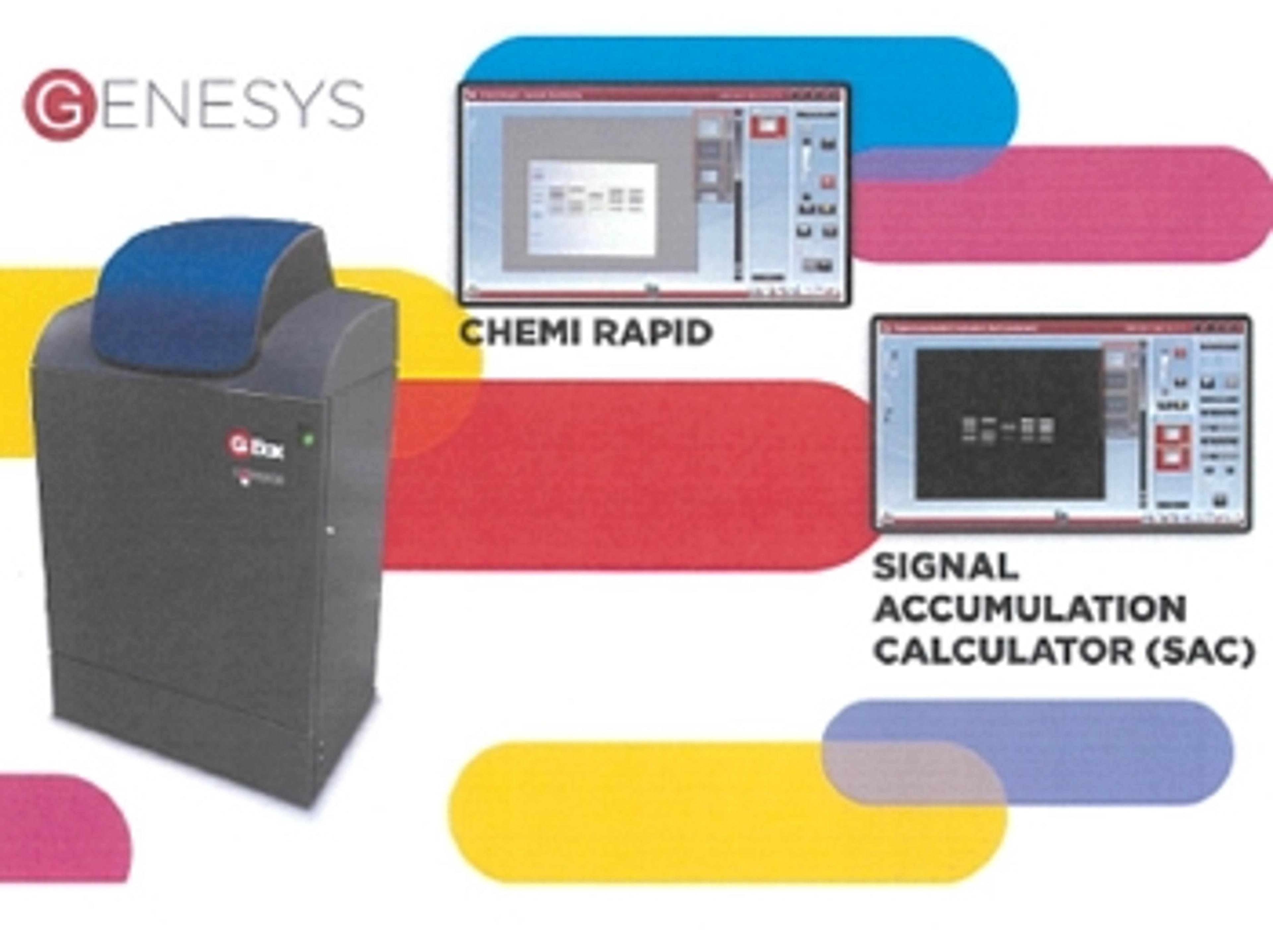
New GeneSys software makes imaging chemi blots a breeze
Capturing chemi blot images is tricky, which is why Syngene have redesigned their GeneSys software for G:Box Chemi systems


Syngene G:BOX Chemi XRQ System Successfully Used to Image Proteins Associated with Fibrosis, Angiogenesis and Wound Healing
G:BOX Chemi XRQ used in extracellular imaging in the School of Biochemistry at the University of Bristol
G:BOX Chemi XRQ used to Study Mechanisms of Cardiac Myocyte Function
Research could assist in developing stem cell therapy for heart repair
Syngene and Synbiosis Present Quality Systems to Accelerate Molecular Biology and Microbiology Research at ARAB LAB 2015
Live on the Syngene/Synbiosis Stand 134