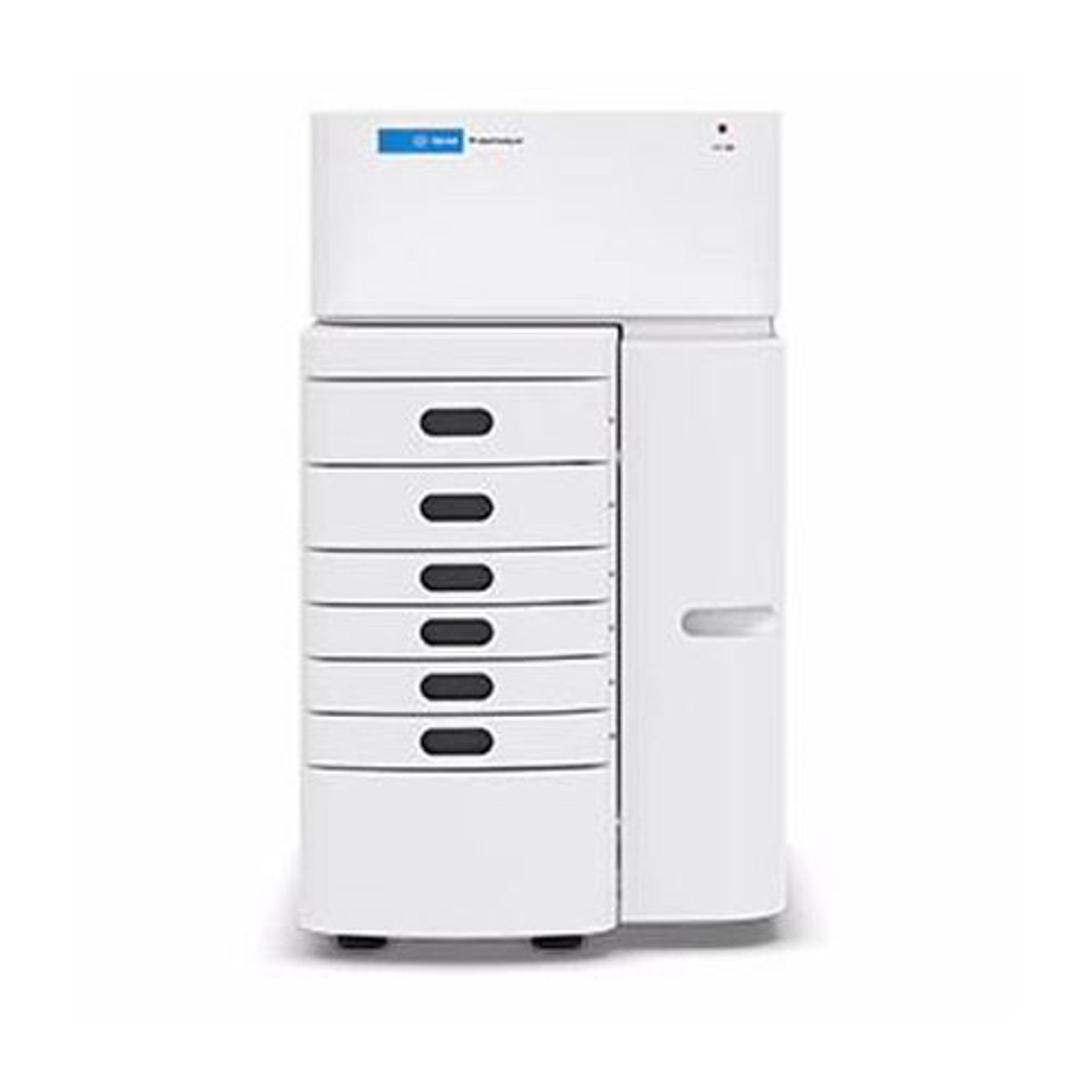
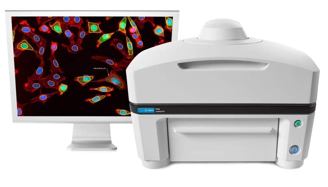
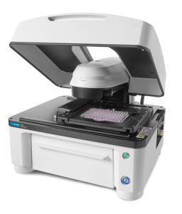
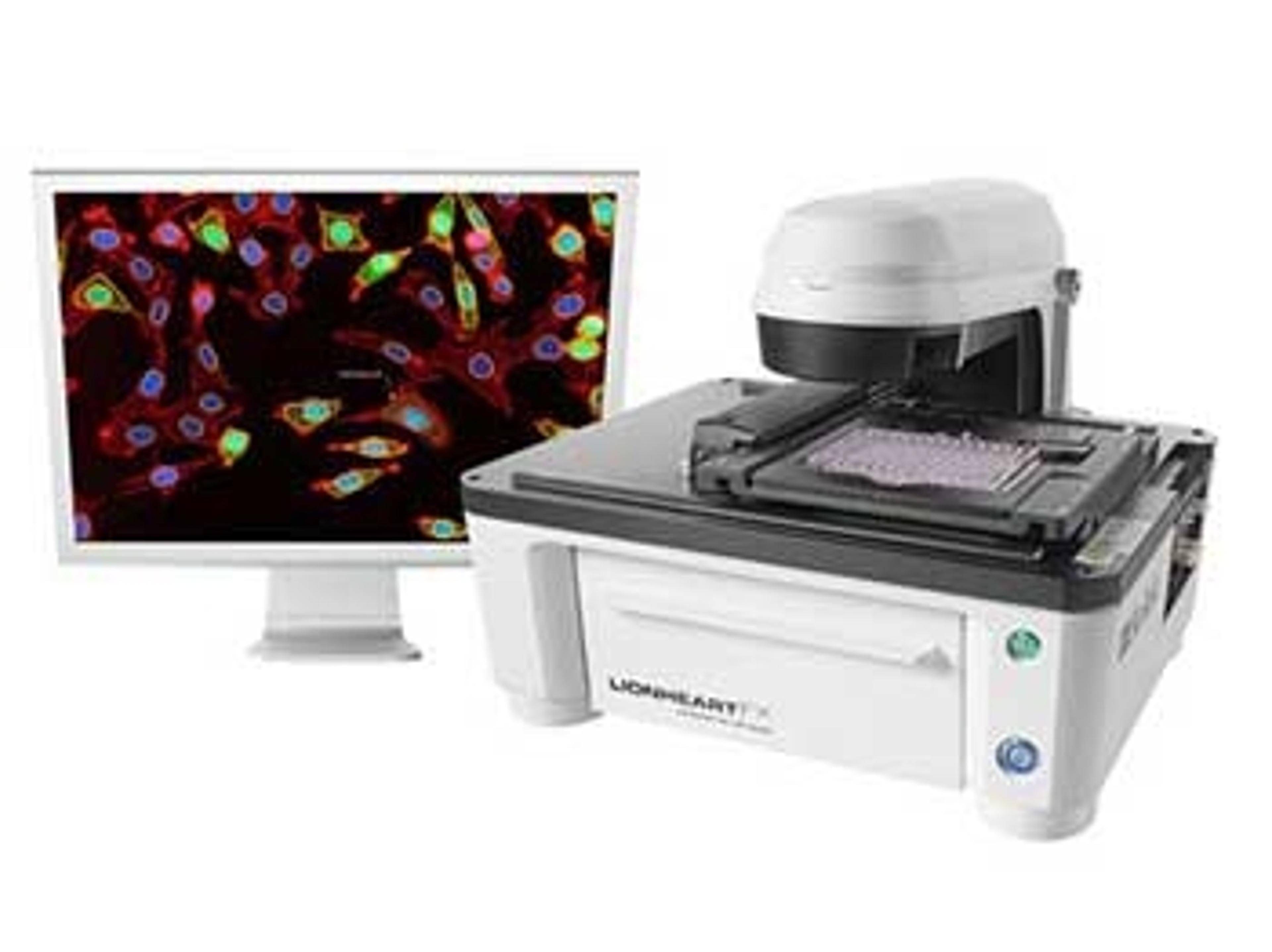
Agilent BioTek Lionheart FX Automated Microscope
Agilent BioTek Lionheart FX Automated Microscope is an inclusive microscopy system for fixed and live-cell applications.

Lid half open
Lid closed

No lid


Receive your quote directly from the manufacturer.
Great outcomes
3D cell phenotyping
This serves at the centre of our dry lab and wet lab pipelines providing high quality imaging information for all our 3D cell models
Review Date: 7 Dec 2023 | Agilent Technologies
We cannot image our lab without it.
Using it to analyze abnormal cell for cancer research.
The product is very effective it has produce perfect visual data for results. It was very cost effective, because of the excellent data it provides publication have been written along with grants, so the equipment have paid for itself.
Review Date: 9 Dec 2021 | Agilent Technologies
Love this equipment, very easy to use.
Lab
Great results produced and easy to use and read.
Review Date: 9 Dec 2021 | Agilent Technologies
Biotek Lionheart™ FX Automated Microscope is perfect for live-cell imaging
Live cell imaging
We have been using Lionheart™ FX Automated Microscope for several years. It is an extremely robust system which can handle different tasks. Excellent system for small-medium scale screening. Integrated incubator allows live-cell imaging for prolonged periods. Sensitivity is also excellent.
Review Date: 20 Oct 2021 | Agilent Technologies
Good results; good instrument
Live-cell assay support with optional CO2/O2, humidity chamber, and temperature control to 45 °C
Great tool for live-cell imaging, simultaneous multi-well plate analysis, capability with the CO2 and temperature module for time lapse experiments.
Review Date: 26 Feb 2020 | Agilent Technologies
Versatile platform and easy to use.
Plate Imaging
The technology is good for open-ended plate housing + for people who want to develop new technology. It has nice imaging modality, versatile platform and easy to use control.
Review Date: 14 Feb 2019 | Agilent Technologies
Great tool that just needs a few updates to make it a home run!
Live cell imaging
Great in theory, and a useful tool in our lab. Just a few minor updates would go a long way in making this a home run, e.g.: 1. The ability to add beacons anywhere on the plate. Current system requires user to create a plate definition, and add "wells" that are quite restrictive. 2. Adding objectives could be easier. It takes us a long time to install and calibrate a new objective. Sales and support have been great from beginning to end. Just some technical issues to iron out.
Review Date: 25 Oct 2018 | Agilent Technologies
We obtain high quality kinetic data.
HIV and cancer
We obtain high quality kinetic data. The kinetics data acquisition is made possible through time-lapse imaging.
Review Date: 3 May 2018 | Agilent Technologies
Great tool for automation and high throughput imaging
Automated high throughput fluorescence microscopy
The LionHeart FX saved us significant time and removed the subjectivity of the analysis. The after sales technical support is great and helped in developing specific protocols and analysis algorithms.
Review Date: 2 May 2018 | Agilent Technologies
Excellent!
live cell imaging, automated microscopy
The Lionheart is amazing. We have been able to get high quality live and fixed cell images with ease. The support has been outstanding. We are looking forward to many years with our Lionheart.
Review Date: 30 Apr 2018 | Agilent Technologies
The Agilent BioTek Lionheart FX automated microscope is a compact system for a broad range of imaging workflows. It offers up to 60x air; 60x and 100x oil immersion magnification, with fluorescence, brightfield, color brightfield, and phase contrast channels for maximum application reach.
An optional environment control cover provides incubation to 40°C and effective containment for CO2/O2 control. A humidity chamber optimizes conditions for long-term live-cell imaging applications, and an available dual reagent injector facilitates rapid kinetic assays.

Microbial monitoring: Ultimate guide to success
The ability to manipulate and monitor microbial organisms, such as bacteria and yeasts, is critical to exploiting their many applications, from use in medical products and biomedical research to environmental remediation and food production. Meeting the increasing demands of microbial monitoring is therefore of vital importance, requiring laboratories to optimize their workflows with the most accurate, reliable and up-to-date instrumentation.
Download this application guide to learn how to take your bacterial monitoring to the next level and achieve real-time results, increased productivity and greater efficiency with the latest monitoring technologies, as we cover:
- The principles of light scattering techniques
- New ways to enhance the performance of brewing yeasts
- The advantages of brightfield microscopy for monitoring in real time
- How to achieve optimum productivity, without multiple instruments, whilst reducing costs

Calculate cell count/volume using automated label-free image analysis
With BioTek’s Lionheart FX Automated Microscope and Gen5 Image Analysis Software, total cells/volume can be calculated for almost any type of cell, using digitally captured and analyzed images, independent of probes or stains. This process is demonstrated in this free infographic that shows how to quantify pre- and post-fermented yeast cells in corn mash.
Nuclear translocation of RelA in stimulated macrophages
As critical components of the innate immune system, macrophages respond to microbes by recognizing molecules, such as the gram-negative bacteria product lipopolysaccharide (LPS), via Toll-like receptors. Receptor activation stimulates a complex signaling network that involves, among others, the NF-κB pathway. Here we quantitate the nuclear translocation of the protein RelA, a component of the NF-κB transcription factor complex, in macrophages using image-based analysis.
Automated image screening of zebrafish embryos exposed to developmental toxins
In this application note, BioTek demonstrates high-throughput screening and analysis of zebrafish embryos treated with several developmental toxins. Here, BioTek exposes embryos to ethanol, retinoid acid, and cyclopamine, then capture brightfield images of the embryos in multi-well plates with Lionheart™ Automated Microscope. The images are analyzed for size and shape using Gen5™ software. The combination of high-throughput imaging and analysis using a vertebrate model makes this an attractive method for toxicology screening.
Autophagy Analysis Using Object Spot Counting
Autophagy is critical for the maintenance of cellular homeostasis. However, dysregulated autophagy can lead to death of healthy cells and survival of cancerous cells. Here we describe the use of CYTO-ID® Autophagy Detection Kit in combination with automatedobject-based spot counting to quantitatively assess the effects of starvation and rapamycin on cellular autophagy by determining the size and number of autophagosomes per cell.
Automated Imaging Assay for Characterizing Ca2+ Flux with R-GECO Biosensor
GPCR activation was kinetically monitored using a Ca2+ biosensor that was transfected along with the human muscarinic M1 receptor into HEK 293 cells. Rapid Ca2+ flux was evident upon carbachol stimulation that peaked 1 second after injection, followed by a decay back to baseline fluorescence over 90 seconds. Imaging also allowed for the monitoring of single cells within the cell population in the field of view where differences in kinetics can be assessed. Suitable assay performance was achieved using a % Responders readout (% of cells responding to carbachol stimulation) that provided z’ values above 0.5 and consistent pharmacology.
Automated Imaging-Based Technique for Monitoring of Ca2+ and DAG Biosensors Enables Detailed Characterization of GPCR Kinetics
This poster describes an automated imaging-based approach to characterize the IP3 /DAG signaling pathway using multiplexed R-GECO Ca2+ and DAG fluorescent biosensors from Montana Molecular. This method produces detailed kinetic profiles of Ca2+ flux and DAG levels following activation of Gq-coupled receptors within the same population of cells. Background subtraction and image analysis tools enable detection of changes in Ca2+ and DAG levels that are more sensitive than techniques relying on bulk fluorescence measurements. Additionally, this method enables single-cell analysis for detailed characterization of response dynamics and subcellular effects.
Characterizing Calcium Mobilization using Kinetic Live Cell Imaging
Monitoring calcium flux is an important method for characterizing GPCR (G-protein coupled receptor) activation. This application note describes a live cell imaging based approach to quantify Ca2+ flux kinetics using the Lionheart™ FX and Fluo-4 Ca2+ indicator dye that delivers sub-second resolution and a large assay window. This method enabled continuous monitoring of intracellular calcium, provided very good sensitivity and generated detailed kinetic profiles for GPCRs.
Monitoring Saccharomyces cerevisiea Growth with Brightfield Microscopy in Real Time
In this application note describes the use of the Lionheart™ FX Automated Live Cell Imager and the ONIX2 Microfluidic Platform to monitor yeast cell growth through the analysis of kinetic brightfield images of yeast cells under a variety of conditions in real time. The yeast under inspection is the yeast Saccharomyces cerevisiae. Strains of the yeast Saccharomyces cerevisiea serve critical roles in the production of many different products including food staples, medicines, and biofuels.
Oridonin Perfusion Causes Cytotoxicity in U-2 OS Cells
This application note describes using the ONIX2 System in conjunction with the Lionheart™ FX Imager to rapidly image and analyze perfused tissue culture cells in multiple fluorescent colors and brightfield.
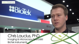
From Media Exchange to Image Acquisition: Optimize Your Entire In Vitro Workflow
Chris Laucius, Ph.D., Imaging and Microscopy Specialist at BioTek Instruments, explains how the plate washers and imagers from BioTek Instruments can help with every step of your workflow: from gentle media exchange, sample prep, image acquisition all the way to data analysis.
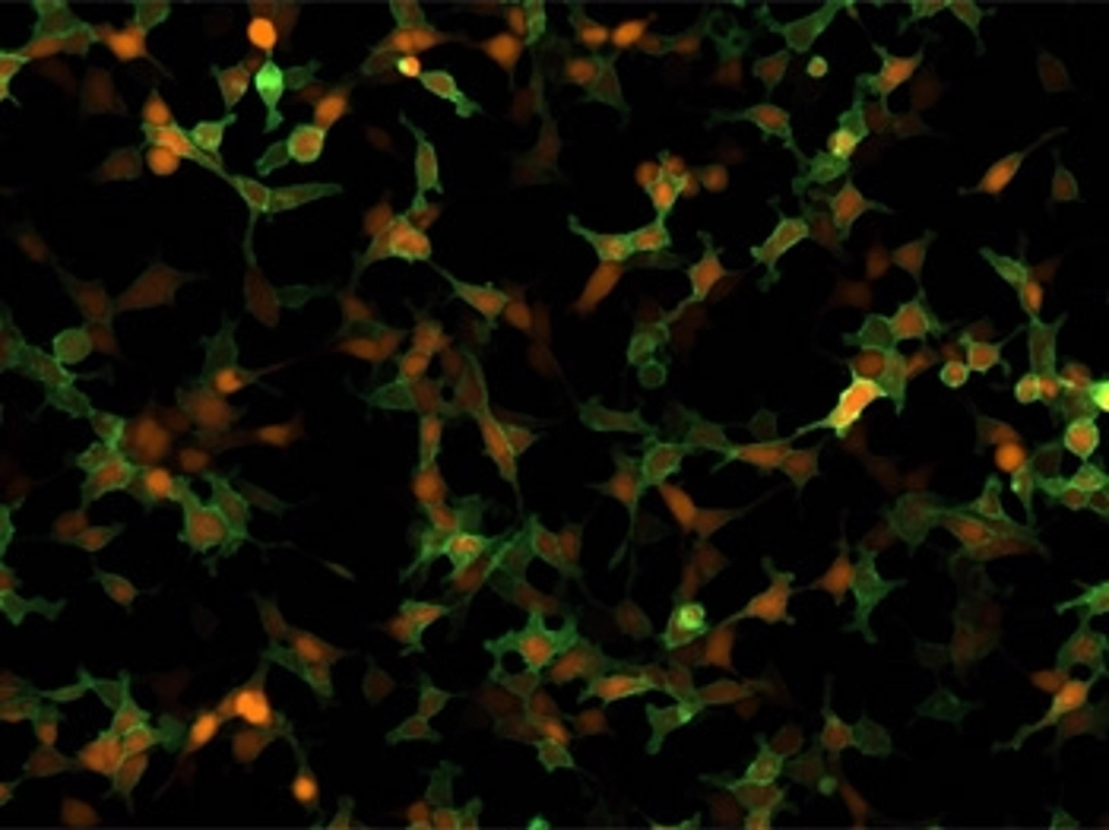
Understanding Novel Oncogenic Regulators in Breast Cancer with the Lionheart FX
Scientists at Stony Brook University, New York, have discovered that the previously known antiviral protein BST2, also known as tetherin or CD371, functions as an oncogenic driver in breast cancer-promoting cell adhesion, growth, migration and invasion of tumor cells. In this video, discover how the automated imaging system of BioTek's LionheartTM FX platform has enabled the group to unearth many of the structure-function relationships of BST2 in cancer, whilst eliminating investigator bias and enabling highly accurate acquisition of kinetic data. Read our article for more information on research in the Okeoma lab.
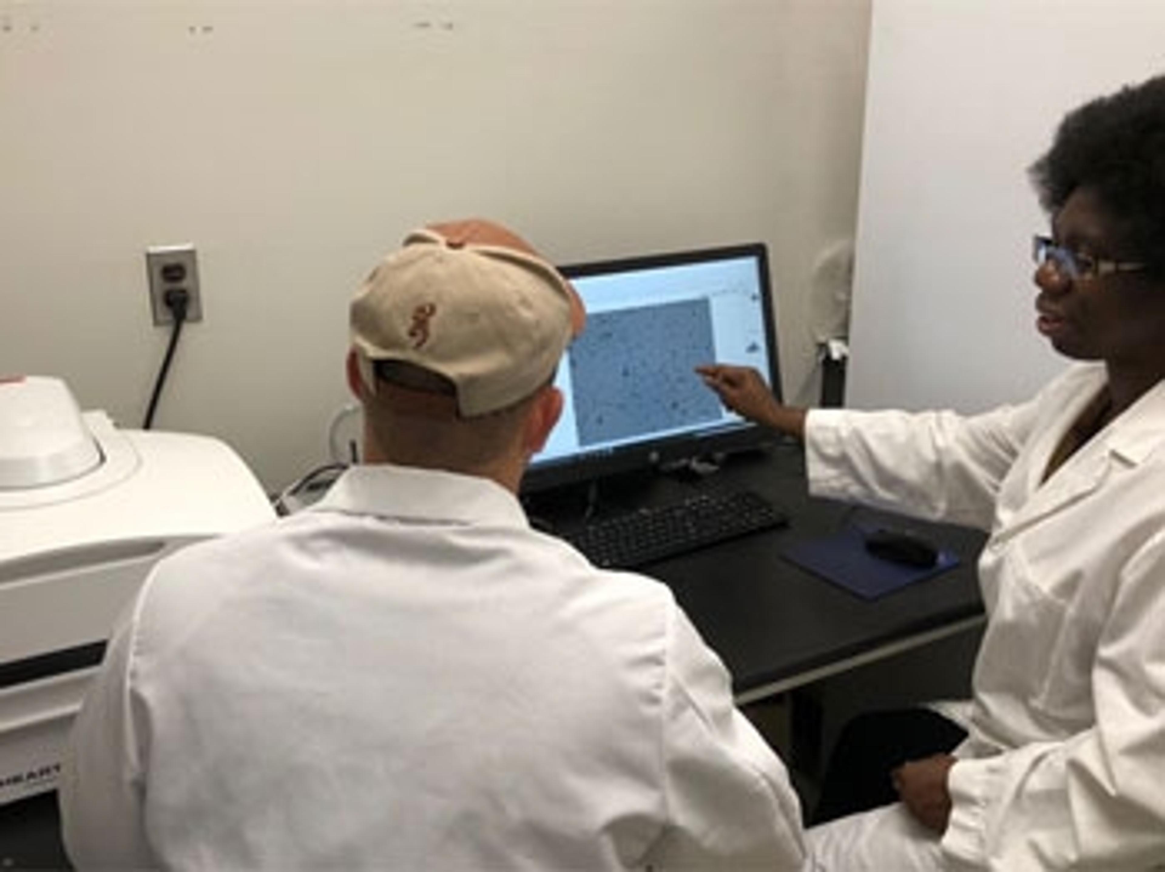
Improve Your Cell Imaging with a Fully Automated, Versatile Inverted Microscope
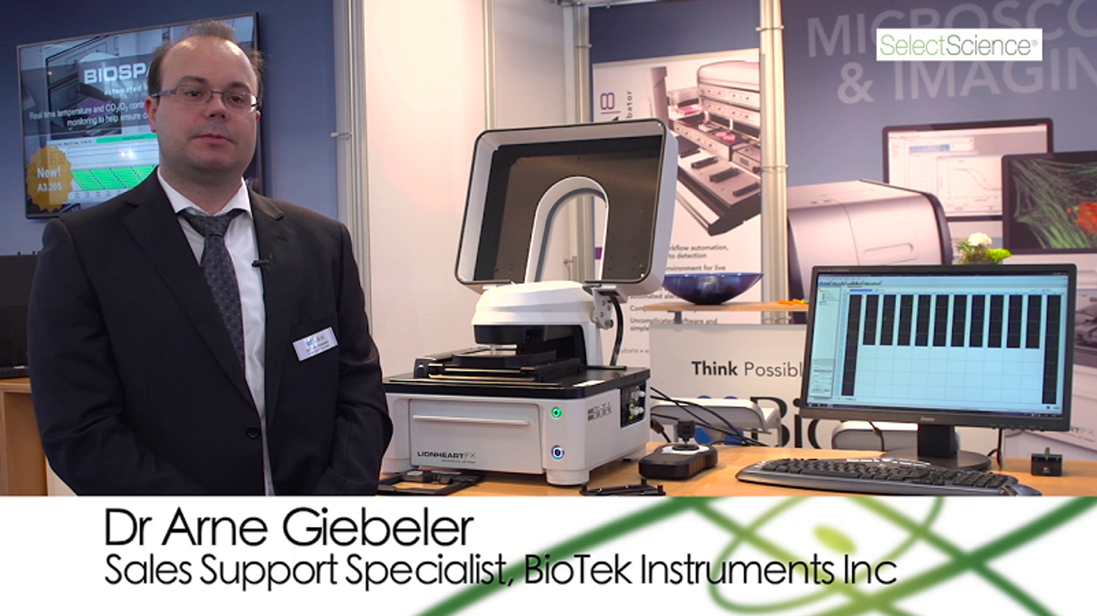
Dr Arne Giebeler, Sales Support Specialist at BioTek Instruments Inc., describes how the compact Lionheart™ FX has enhanced environmental and focusing controls to facilitate multiple cell imaging applications.


Your Neuroscience Playlist: 9 Essential Video Interviews
Watch video highlights from the Neuroscience 2018 annual meeting in San Diego, from the latest research presented, to the new technologies innovating neurobiology research
Illuminate Intracellular Calcium Kinetics in Real Time with Live-Cell Imaging
How advanced imaging technology is progressing drug discovery through understanding of GPCR-dependent second-messenger systems

Understanding Breast Cancer – Using Advanced Imaging to Unravel its Molecular Secrets
A laboratory in the US is using advanced imaging to decipher the role of a ‘double-agent’ protein in breast cancer, and to help develop drugs targeted at disrupting its function
Leading Scientists Rate BioTek’s Lionheart FX Automated Microscope
Find out how experts have been using this new imaging technology and read their verdicts on its performance
Beating Cystic Fibrosis – Perfecting an Intestinal Assay in the Hunt for New Gene Modulators
Learn how a lab in the US has perfected a high-throughput assay in mouse organoids for testing potential new cystic fibrosis drugs