Differential staining in biological tissue
19 Aug 2019Energy dispersive spectrometry (EDS) can be used to analyse differential staining protocols, allowing access to new information about biological samples. Differential staining is an important step in generating contrast when preparing cells and tissues for electron microscopy. EDS produces qualitative data about the distribution of elements associated with the stain and comparative quantitative data about relative concentrations of elements.
Related Products
Request Quote for All Products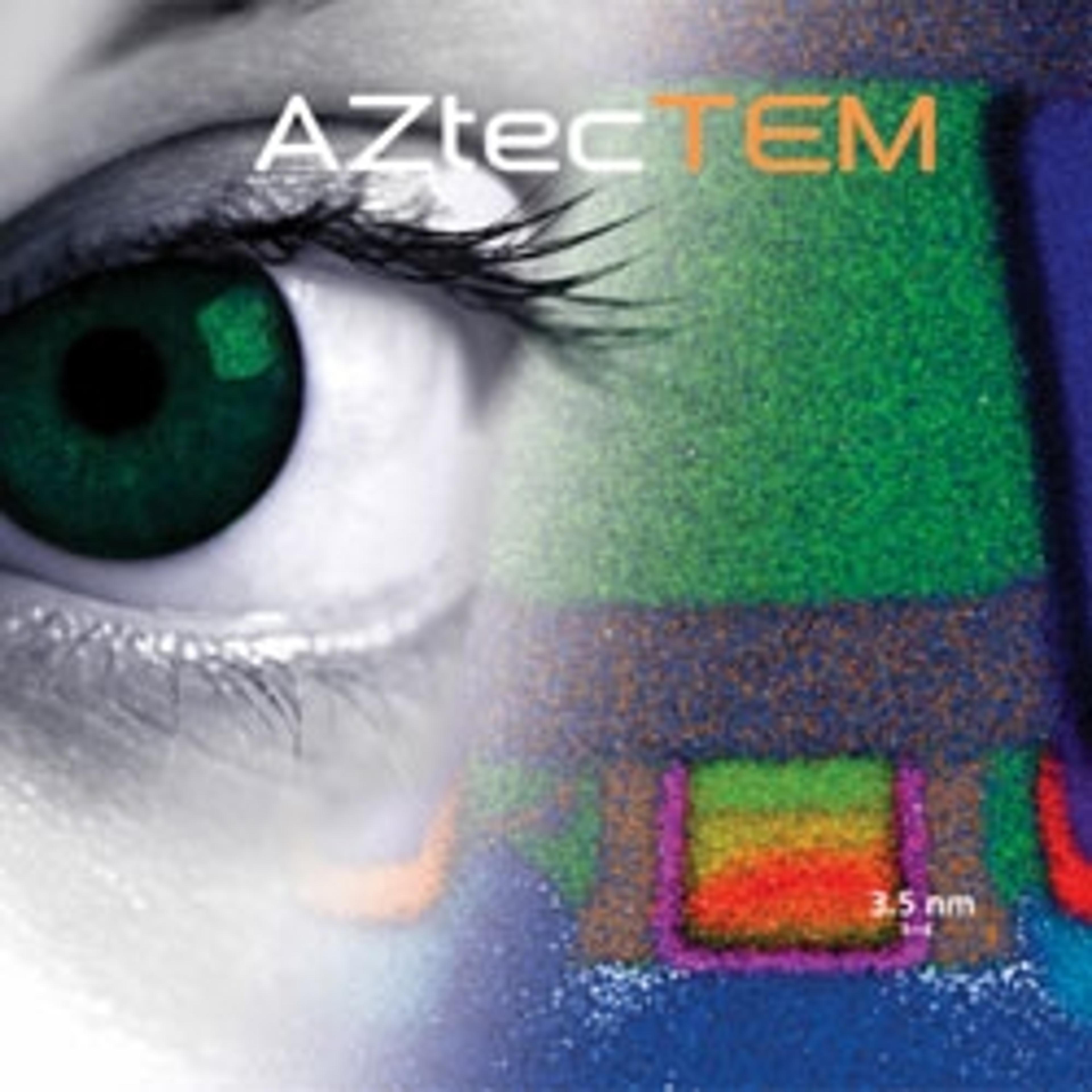
Ultim® Max and AZtecTEM
Oxford InstrumentsAZtecTEM, powered by Ultim™ Max (Silicon Drift Detector), provides exceptional elemental characterisation abilities in the TEM.
Ultim® Extreme
Oxford InstrumentsUltim Extreme Silicon Drift Detector is a breakthrough solution for ultra high resolution FEG-SEM applications and delivers solutions beyond conventional micro- and nano-analysis.