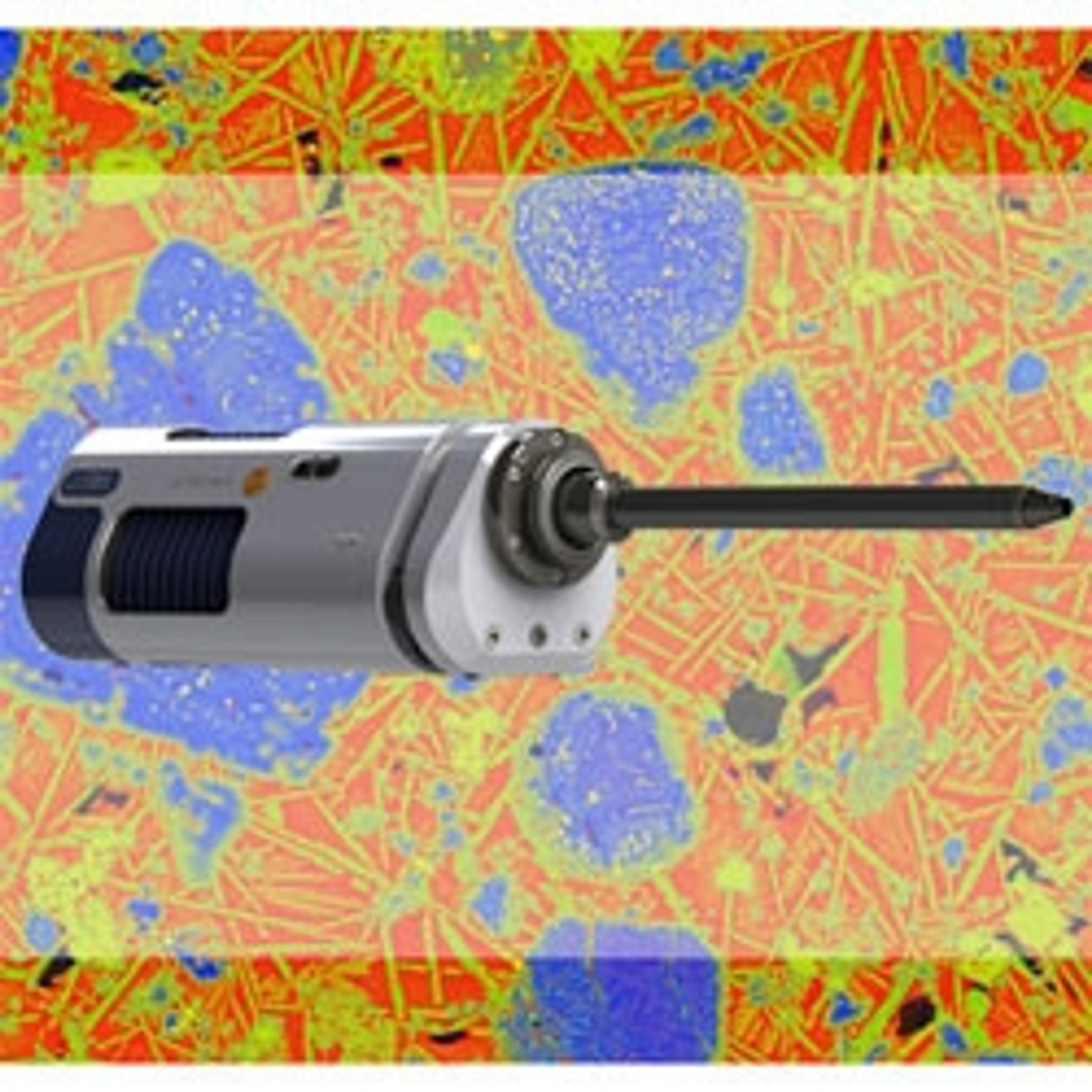
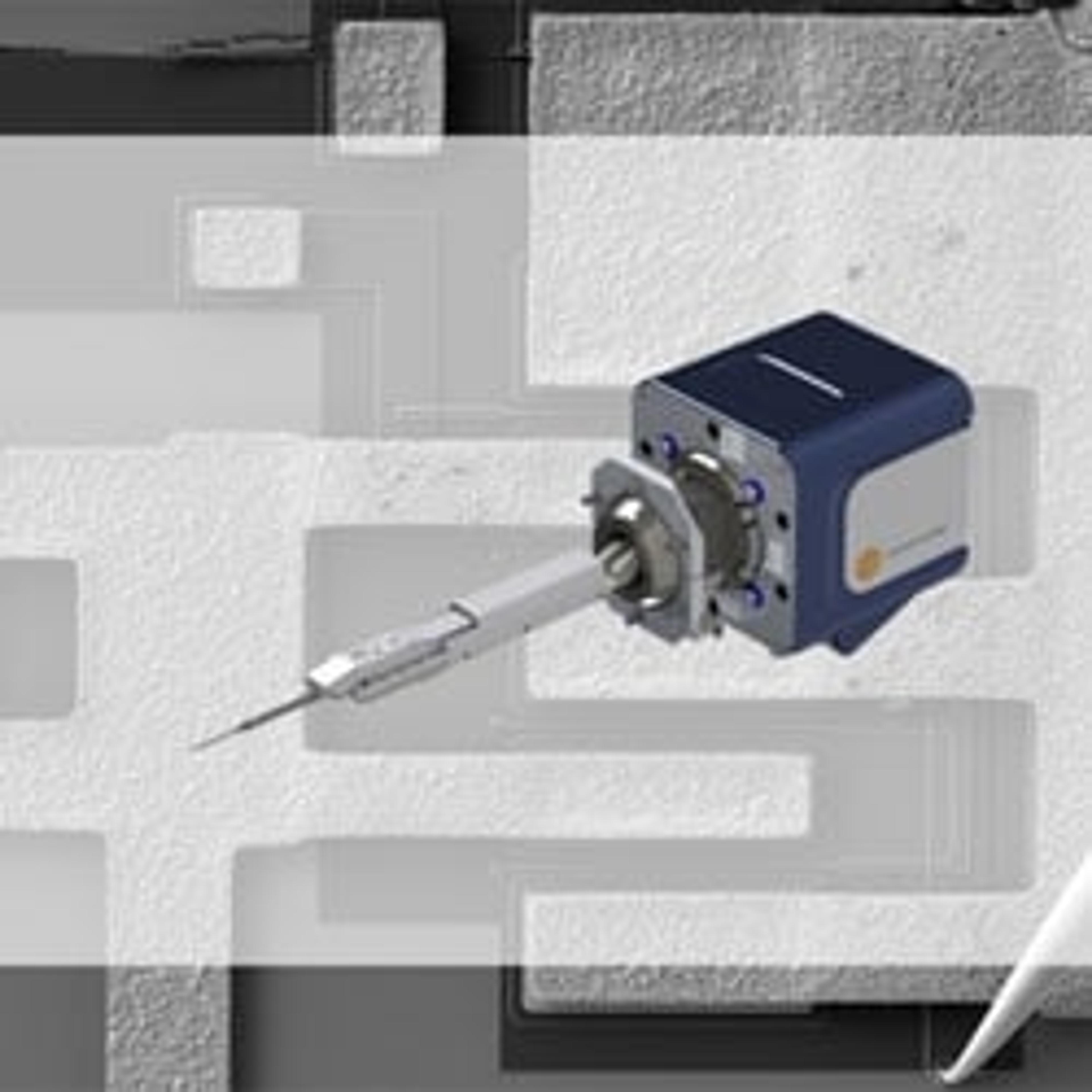
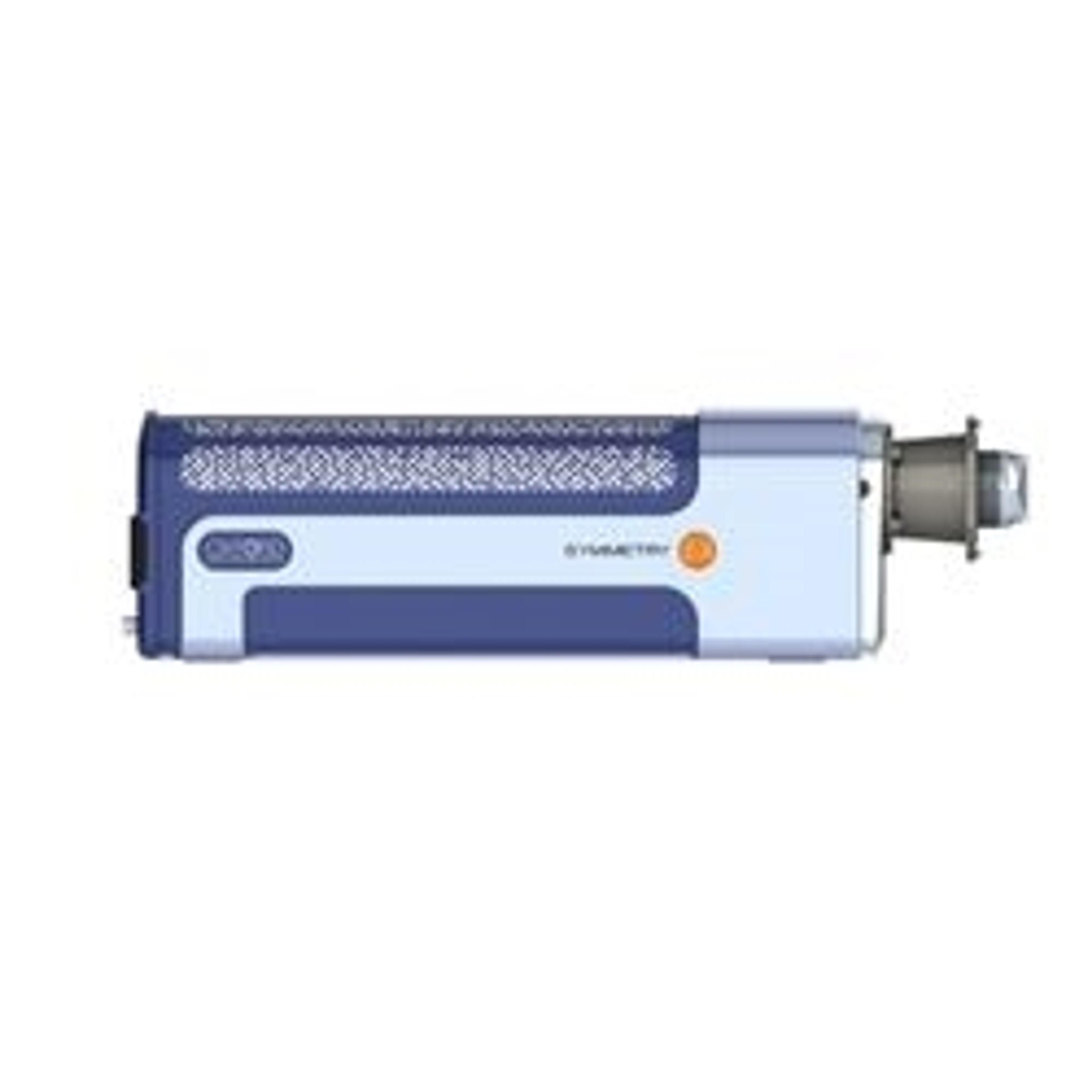
Ultim® Extreme
Ultim Extreme Silicon Drift Detector is a breakthrough solution for ultra high resolution FEG-SEM applications and delivers solutions beyond conventional micro- and nano-analysis.
Super Sensitive at low kV
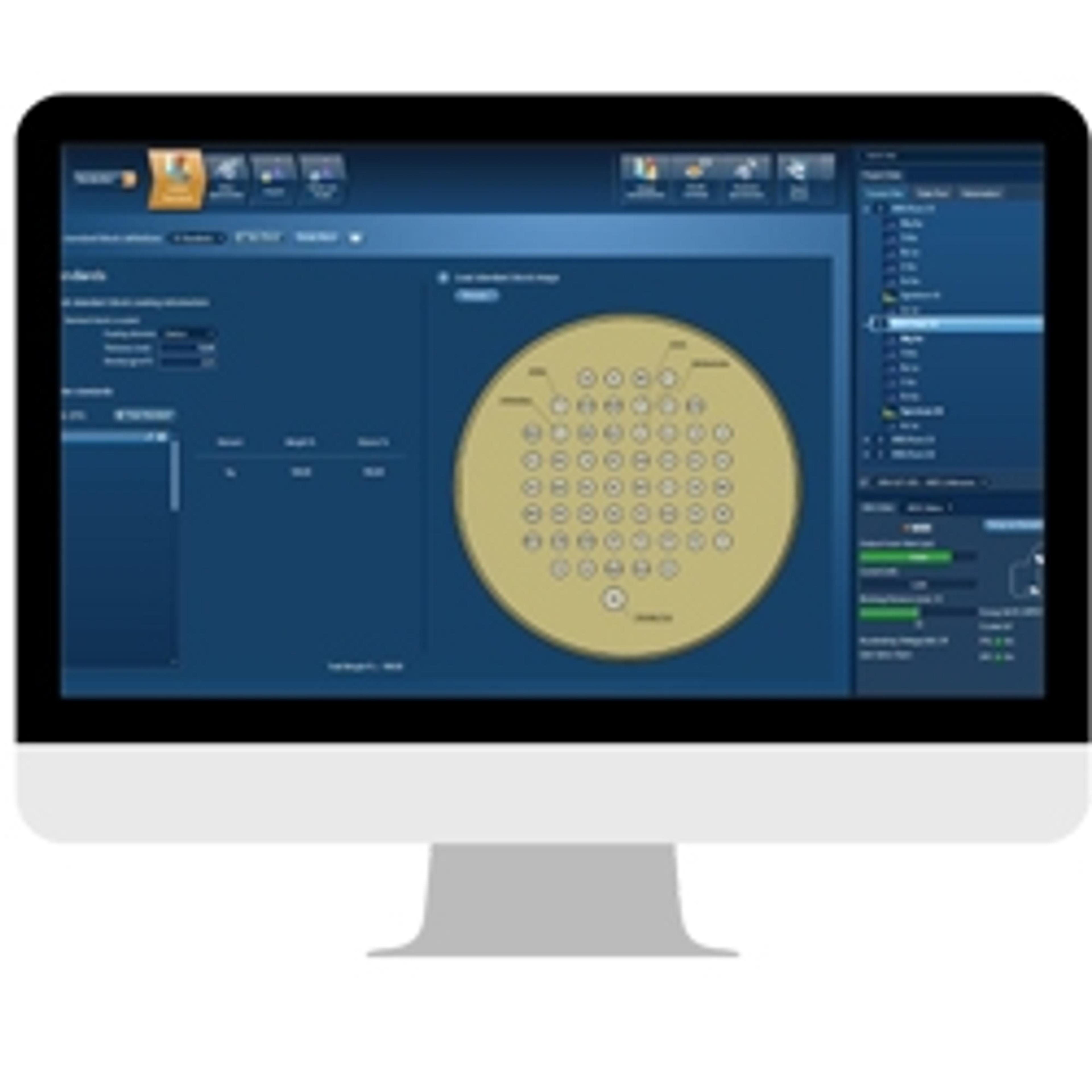
Energy dispersive spectroscopy
Easy to use. Maps in seconds. Excellent training and follow up webinars.
Review Date: 19 Nov 2021 | Oxford Instruments
Superb instrument, very innovative.
Chemical composition
The Ultim Extreme brings a whole new level to EDS in the FE-SEM. The service is extraordinary. The ability to work at a smaller working distance is a perfect marriage of the Ultim Extreme and FE-SEM.
Review Date: 5 Nov 2019 | Oxford Instruments
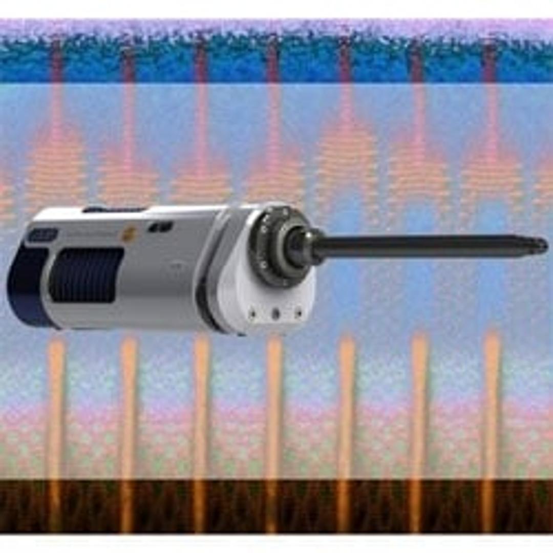
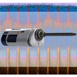
A windowless 100 mm2 version of Ultim, this detector was designed to optimise sensitivity and spatial resolution. Utilising radical geometry allows the highest standard of both imaging and EDS performance even at short working distances and at low kV.
Features:
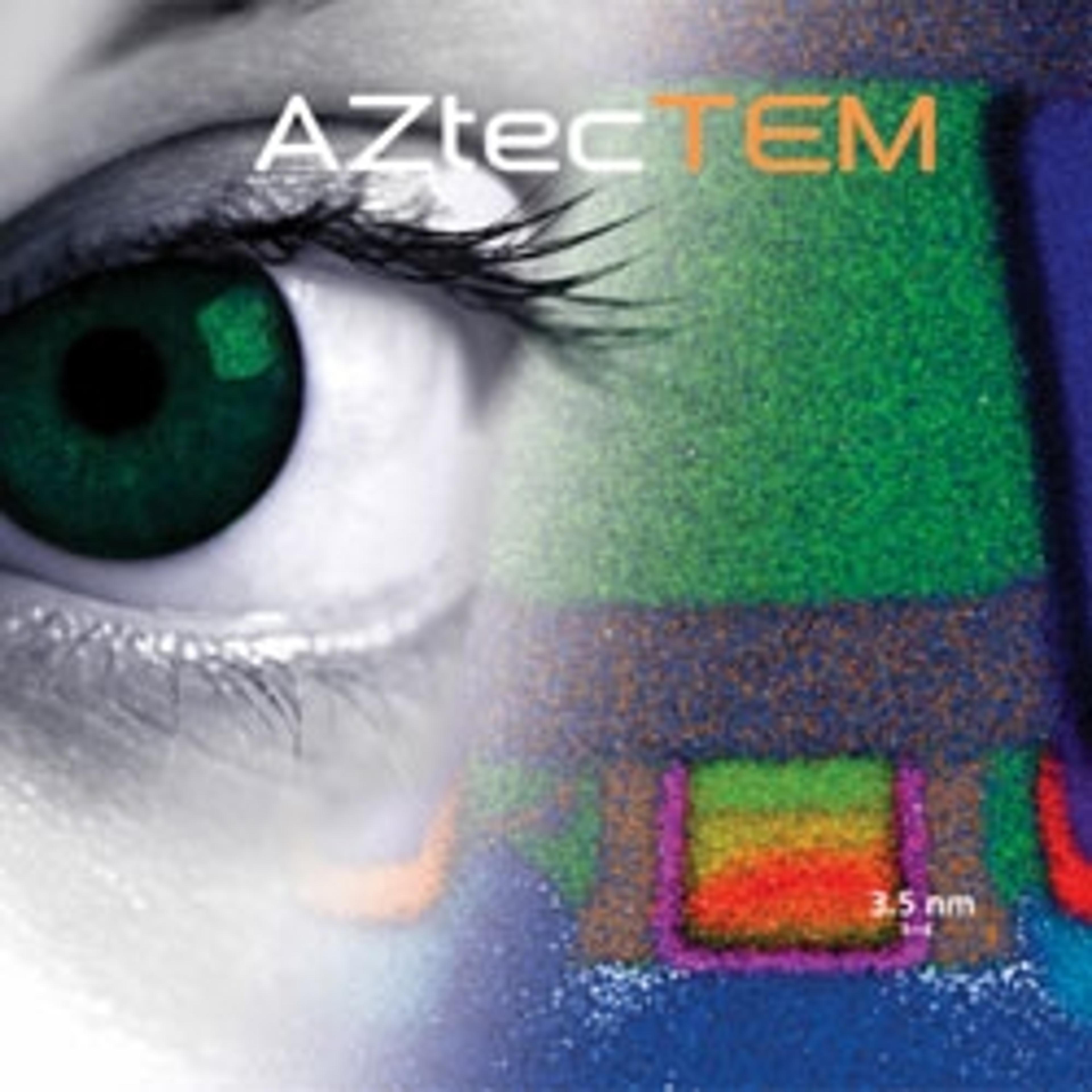
- Sub 10 nm element characterisation in the FEG-SEM
- Sensitivity for surface science
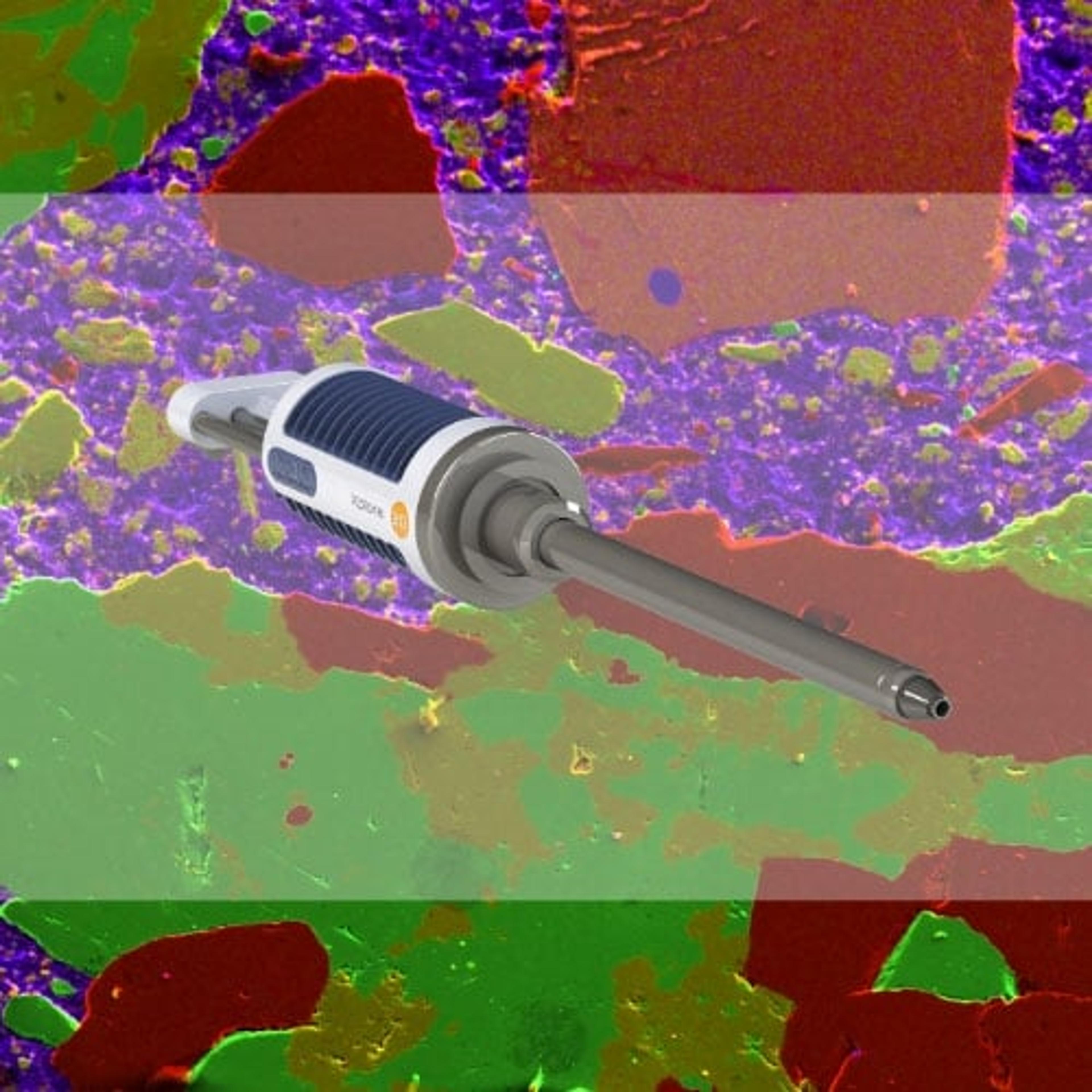
- Material characterisation down to the lowest kV
- Real-time nano-characterisation
- Sensitivity for light elements such as lithium, nitrogen and oxygen
Your guide to EDS analysis in electron microscopy
Harness energy-dispersive X-ray spectrometry (EDS) to transform your scanning electron microscopy (SEM) research with this free guide which covers:
- Questions EDS can answer
- How to prepare biological specimens for EDS
- Benefits of combining EDS with advanced SEM techniques
- The ideal EDS detector
Explore a variety of application resources and find out how to enhance your EM research using EDS. Download to discover how EDS can provide invaluable compositional information to greyscale ultrastructural images via color EM, how to select your detector for low-energy X-ray analysis, and how EDS can unlock a simpler and smarter sample preparation.
Energy dispersive x-ray spectrometry (EDS) for biology
In this white paper, Oxford Instruments presents a brief introduction to energy dispersive X-ray spectrometry (EDS) and discusses the improvements this method provides in terms of sensitivity and stability when compared to older detectors. It also discusses how EDS can be used to analyze differential staining protocols of biological samples.
Scanning electron microscopy-based analysis of lithium ion battery materials
In this application note, Oxford Instruments NanoAnalysis shows the latest progress towards characterizing Li-ion battery materials with energy dispersive spectroscopy (EDS) in scanning electron microscopy (SEM). Here, Oxford Instruments NanoAnalysis explores how lithiation can be studied on graphite anodes with the Extreme detector and on lithium-containing ceramics using a conventional detector.
Differential staining in biological tissue
Energy dispersive spectrometry (EDS) can be used to analyse differential staining protocols, allowing access to new information about biological samples. Differential staining is an important step in generating contrast when preparing cells and tissues for electron microscopy. EDS produces qualitative data about the distribution of elements associated with the stain and comparative quantitative data about relative concentrations of elements.
Advanced techniques in SEM and EDS for biological samples
Watch this on-demand webinar to explore the applications of biological energy-dispersive X-ray spectrometry and how it can be used in combination with advanced scanning electron microscopy


