Products & ReviewLife Sciences

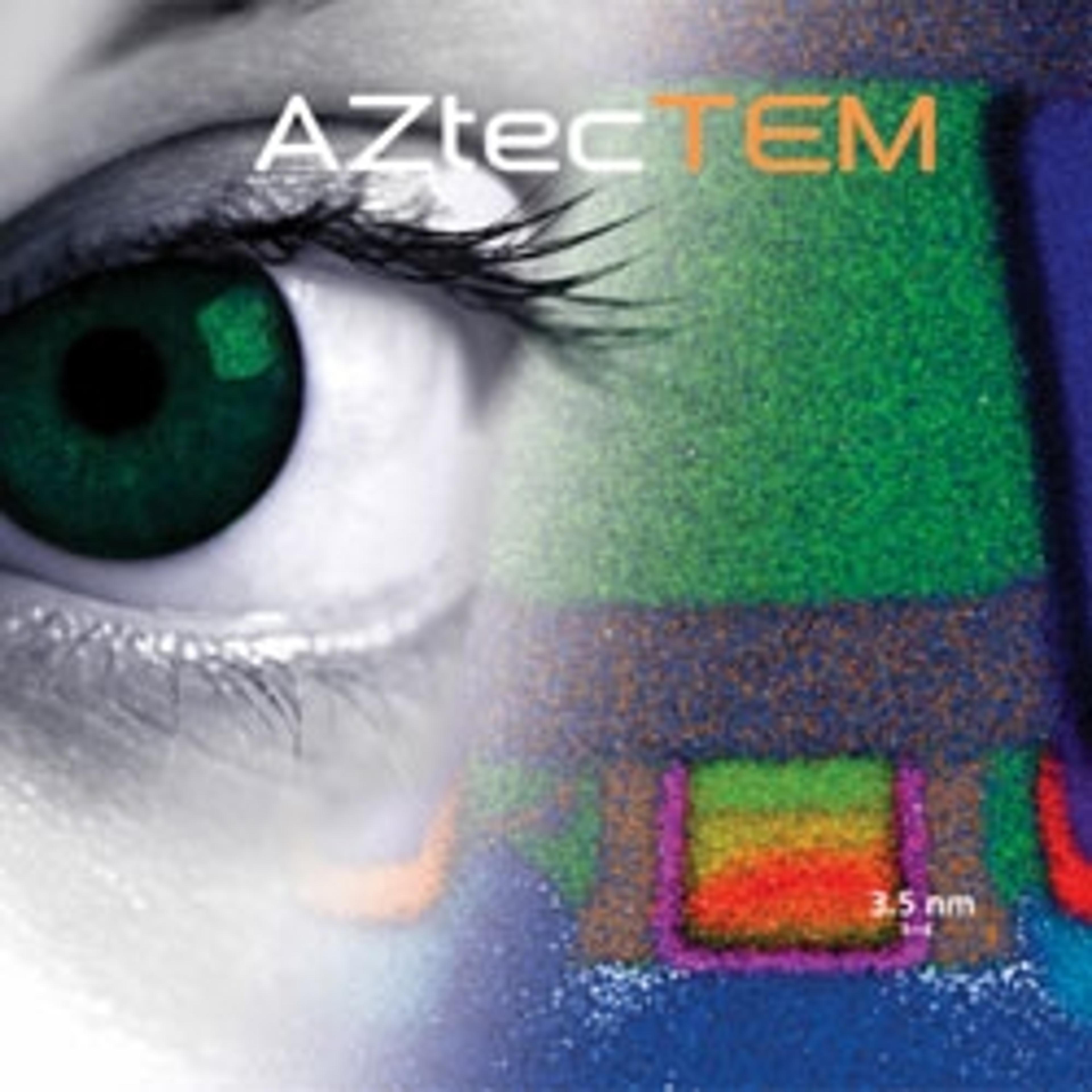
Ultim® Max and AZtecTEM

Oxford InstrumentsAvailable: Worldwide

AZtecTEM, powered by Ultim™ Max (Silicon Drift Detector), provides exceptional elemental characterisation abilities in the TEM.

Request Pricing
Oxford Instruments

Receive your quote directly from the manufacturer.

Ease of Use
After Sales Service
Value for Money
Be the first to leave a review
Description

AZtecTEM is an EDS software, optimised for TEM applications. The software allows you to view real time in situ chemistry changes as they happen.

AZtecTEM is powered by Ultim™ Max, the newest generation of Silicon Drift Detectors for the TEM. Optimised to deliver elemental characterisation at the atomic scale providing maximal count rates at minimal probe sizes.

Features

  • Maximised sensitivity
  • High throughput
  • M2T quant for sample thickness measurements
  • Drift correction
Application NoteSpectroscopy

Analyzing a NAND Flash memory device using low kV EDS

In this application note, Oxford Instruments NanoAnalysis demonstrates that increasing detector sensitivity, either by sensor size, sensor-sample distance, or window removal, it is possible to collect X-ray counts at high resolution imaging condition and achieve elemental maps with sub 20nm spatial resolution even on the lightest of elements. This, combined with the data processing of Aztec TruMap, allows the accurate mapping of elemental distributions in complex specimens with nano-structures such as a NAND flash memory device.


Application NoteLab Automation

Semiconductor mapping in transmission electron microscopy

Elemental analysis of semiconductors is typically difficult due to strong overlaps of X-ray lines between commonly used elements and low concentrations of dopants. Not only are concentrations of dopants small but their X-ray lines often overlap with other materials used in semiconductor processing. In this application note, Oxford Instruments NanoAnalysis shows how AZtecTEM solves these overlaps to achieve an accurate elemental analysis.


Application NoteSpectroscopy

Differential staining in biological tissue

Energy dispersive spectrometry (EDS) can be used to analyse differential staining protocols, allowing access to new information about biological samples. Differential staining is an important step in generating contrast when preparing cells and tissues for electron microscopy. EDS produces qualitative data about the distribution of elements associated with the stain and comparative quantitative data about relative concentrations of elements.

Product Overview

Links

Ultim Max and AZtecLive

Oxford Instruments

AZtecLive and Ultim™ Max (Silicon Drift Detector) for EDS analysis, combine live electron images with live X-ray chemical imaging to allow a revolutionary change in how users investigate their samples in the SEM.

(0)