Doppler Flow Velocity System
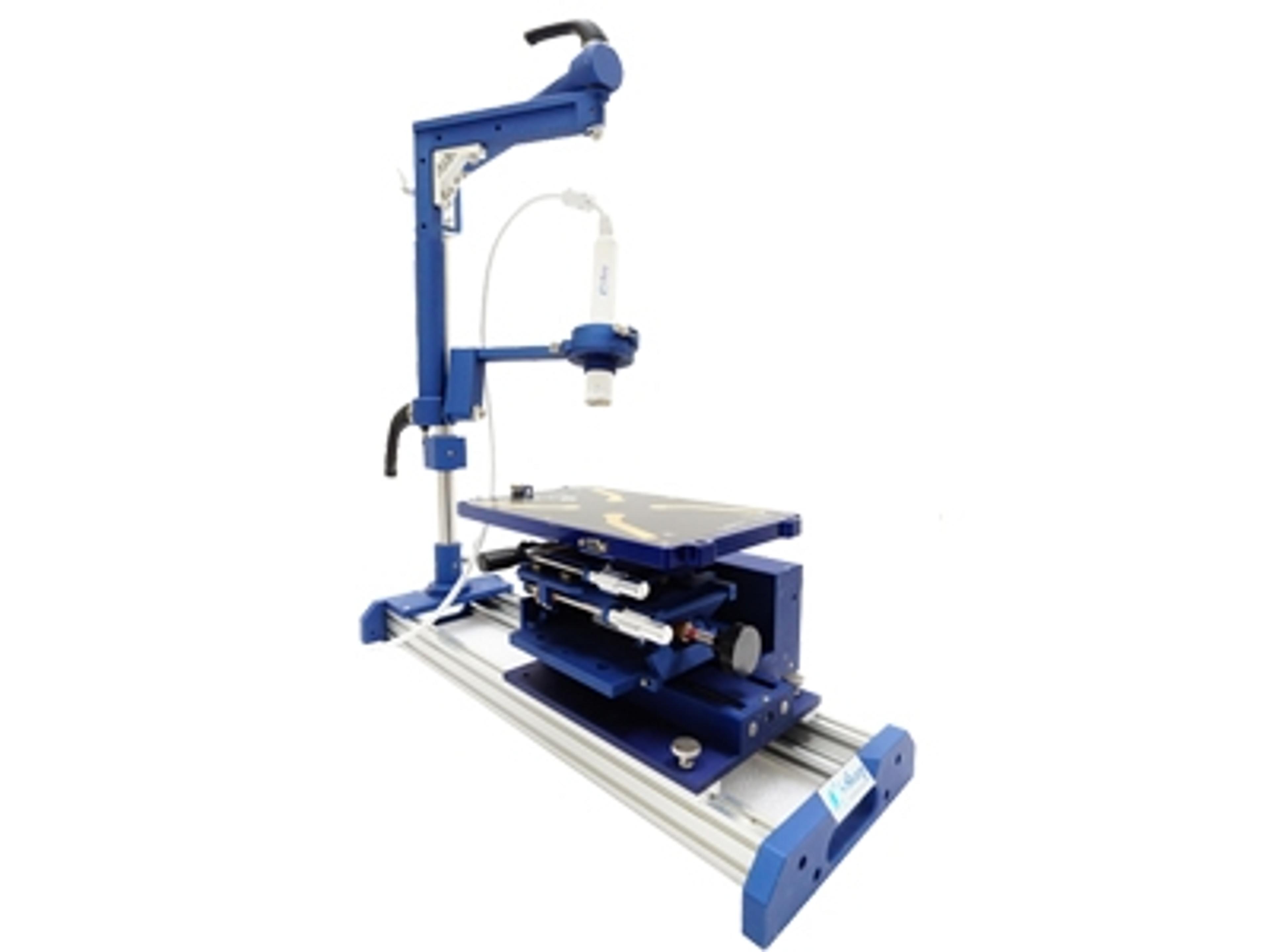
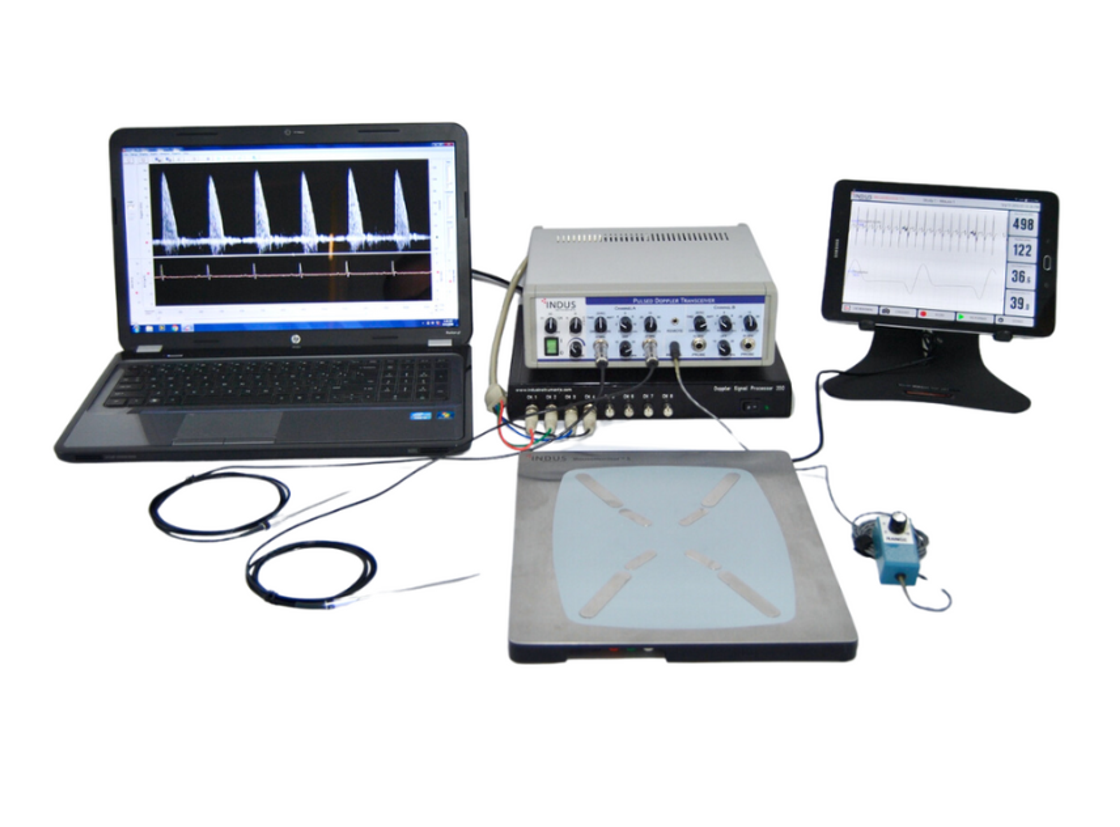
ScinticaThe Indus Doppler Flow Velocity System is a high-frequency, real-time pulsed Doppler measurement device with integrated data analysis software designed for measuring cardiovascular function in small animals.
The Indus Doppler Flow Velocity System is a high-frequency, real-time pulsed Doppler measurement device with integrated data analysis software designed for measuring cardiovascular function in small animals.
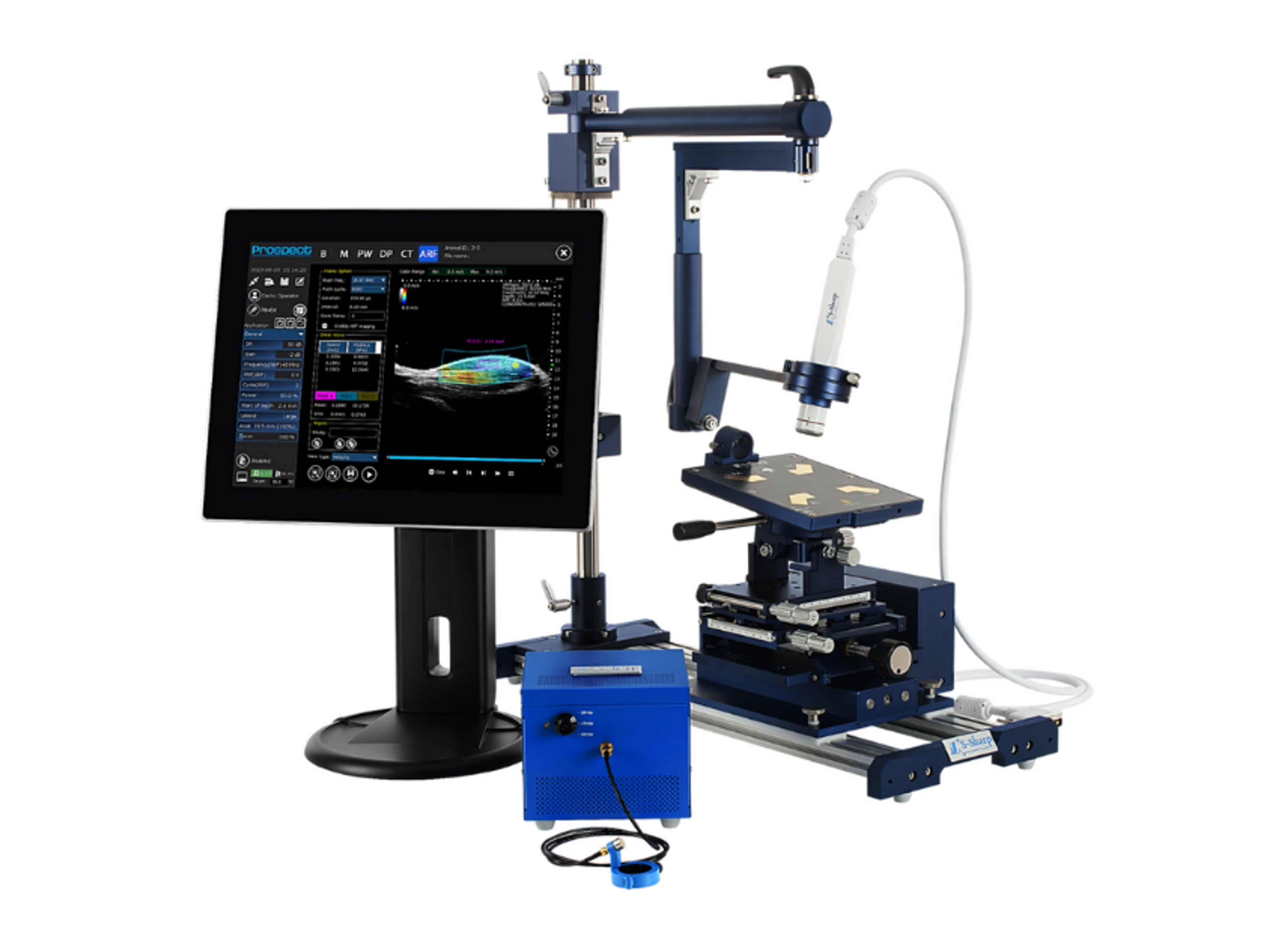
The Prospect T1 is an innovative high-frequency ultrasound system designed specifically for in vivo preclinical imaging in small animals such as mice and rats. This compact and cost-effective tablet-based system provides high-resolution images (up to 30 µm) and advanced capabilities to monitor changes in hemodynamics and observe anatomical structures in real-time.
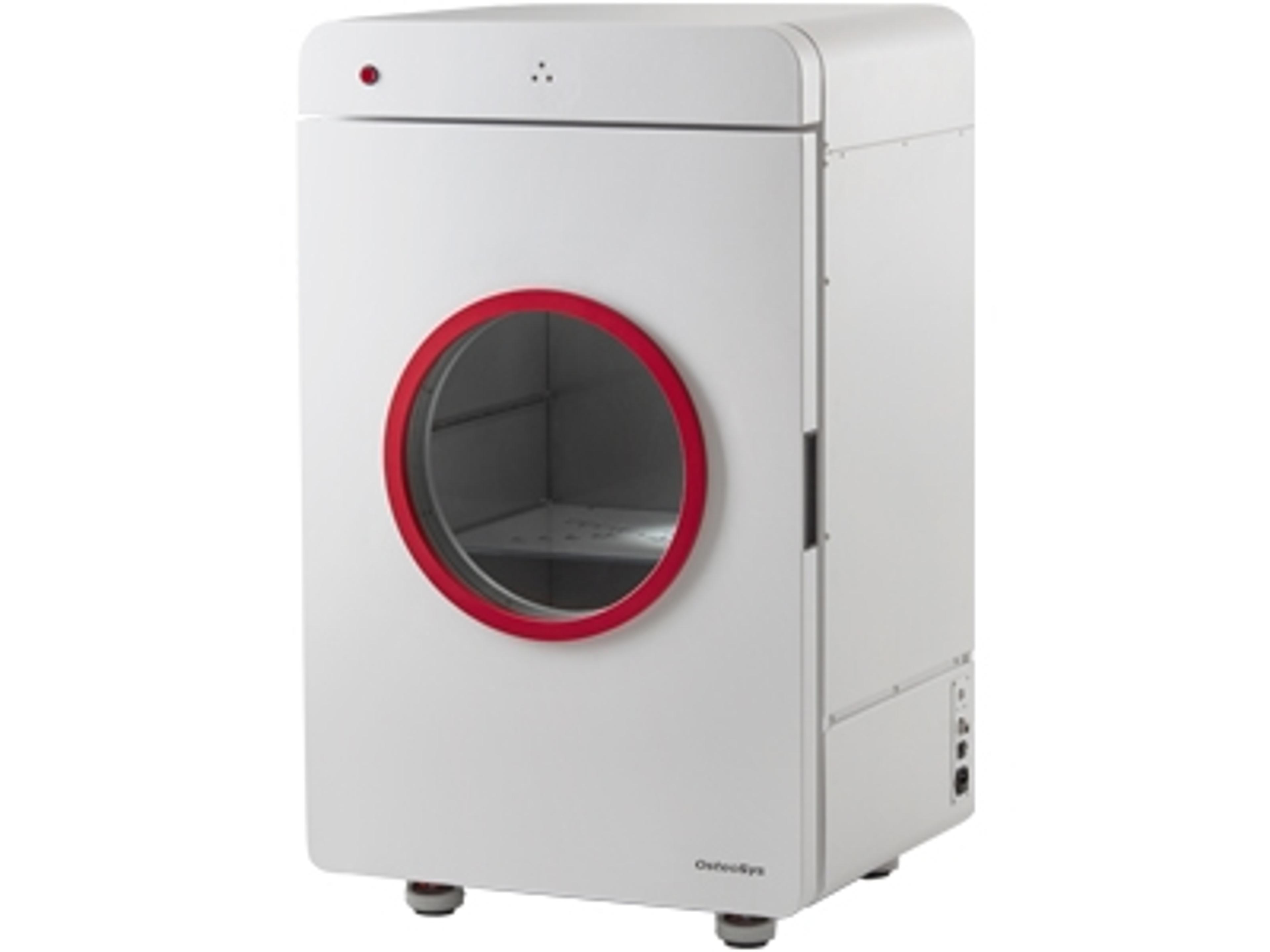
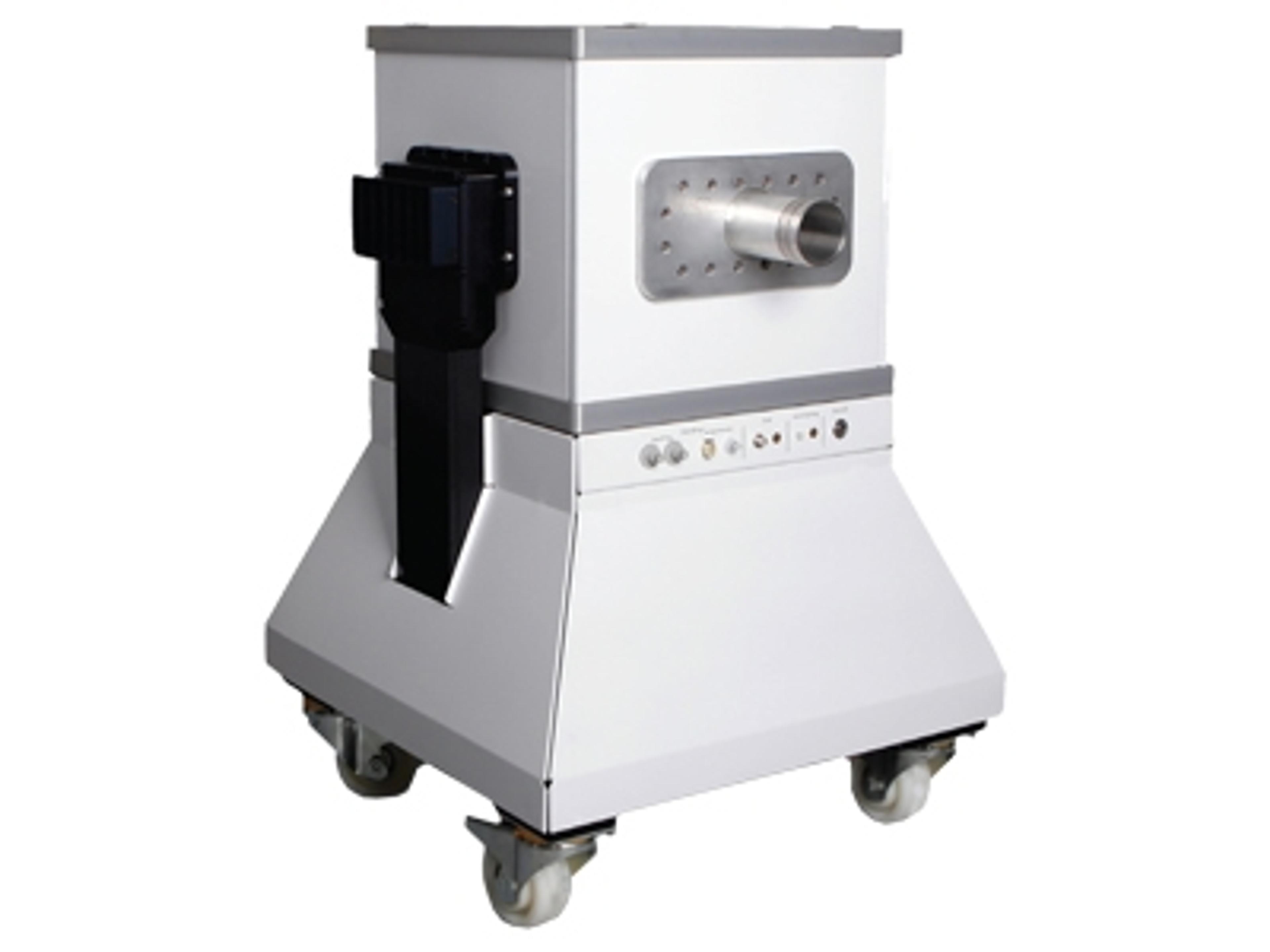
The iNSiGHT DXA system is an in vivo Dual Energy X-Ray Absorptiometry (DXA/DEXA) technology for preclinical research. This fully shielded X-ray cabinet is suitable for small animal DXA applications – mice, rats, and animals of similar size (up to 5kg). Efficient body composition measurements allow for fast scan times of 25 seconds. Combined with its low-dose radiation, it’s ideal for longitudinal studies.
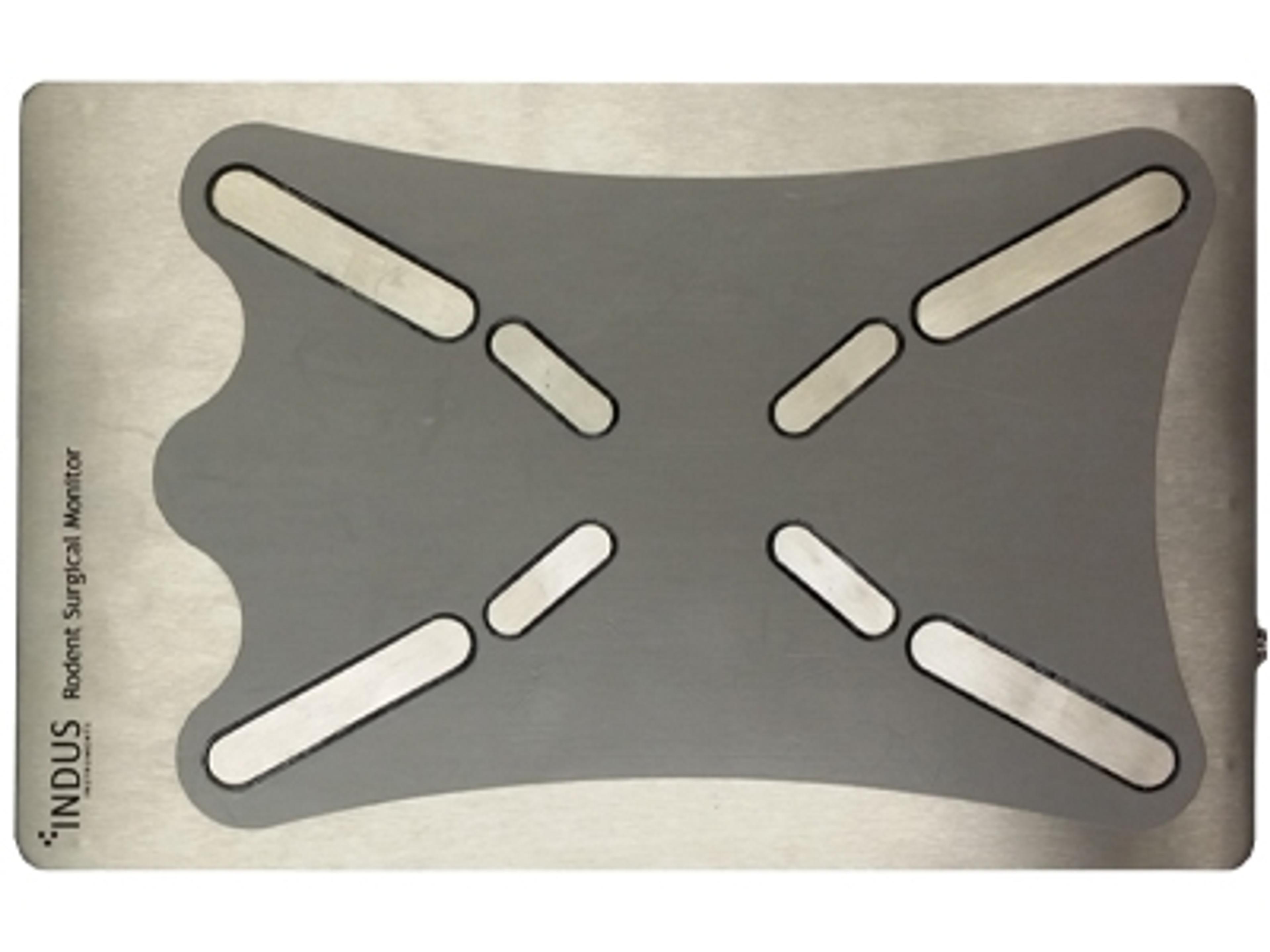
The Indus Rodent Surgical Monitor+ is an advanced, integrated surgical warming and vital signs monitoring solution for preclinical research in mice, rats and other small animals. The system provides detailed information, in real time, regarding subject body temperature, ECG, heart rate, pressure and respiration.
The Prospect T2 is an innovative high-frequency ultrasound system designed specifically for in vivo preclinical imaging in small animals such as mice and rats. This compact and cost-effective tablet-based system provides high-resolution images (up to 30 µm) and advanced capabilities to monitor changes in hemodynamics and observe anatomical structures in real-time.
The M-Series™ systems are cryogen/cooling-free, self-shielded, high-performance MRI systems based on permanent magnet technology. The M-Series systems allow preclinical researchers, with or without in-depth knowledge of MR physics, to utilize the gold standard method in soft tissue imaging without the cost, complexity, and technical burden of superconducting MRI systems.
The NEWTON FT-900 is a deeply cooled CCD imager dedicated to SWIR (NIR-I, NIR-II), fluorescence and bioluminescence multi-spectral imaging as it allows for in vivo applications in the visible, near and short-wave infrared spectrum (VIS/ NIR/ NIR-II). Smart and ultra sensitive, it provides non-ionizing and non-invasive visualization of biological processes in real-time.
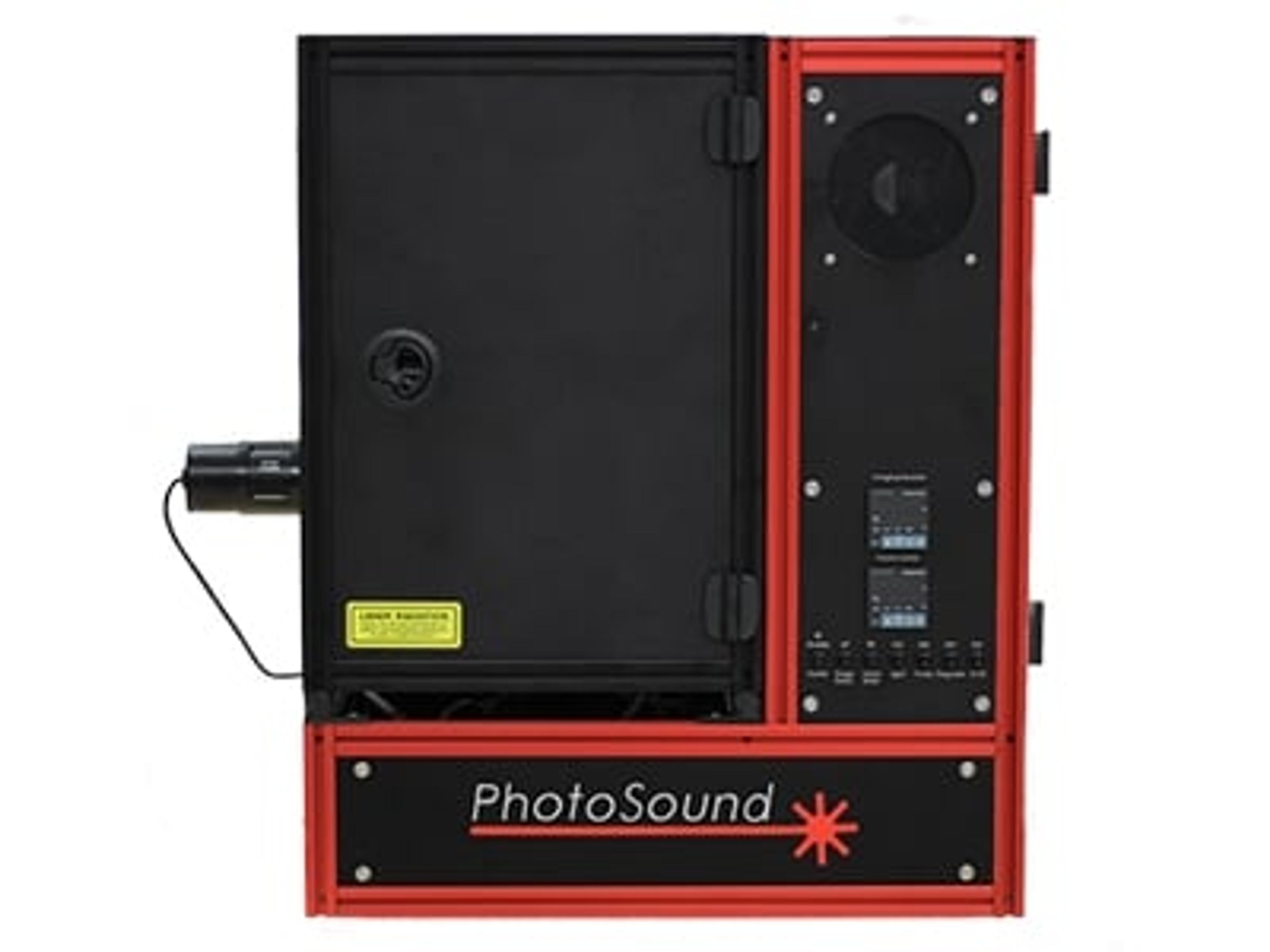
The TriTom imaging platform utilizes photoacoustic and fluorescence tomographies (PAFT) for high-resolution, non-invasive whole-body imaging in small animal models, achieving up to 160 µm resolution. This multi-modality system enables simultaneous acquisition of photoacoustic and fluorescence data in large volumes (> 25 cm3), allowing for spectroscopic molecular analysis.
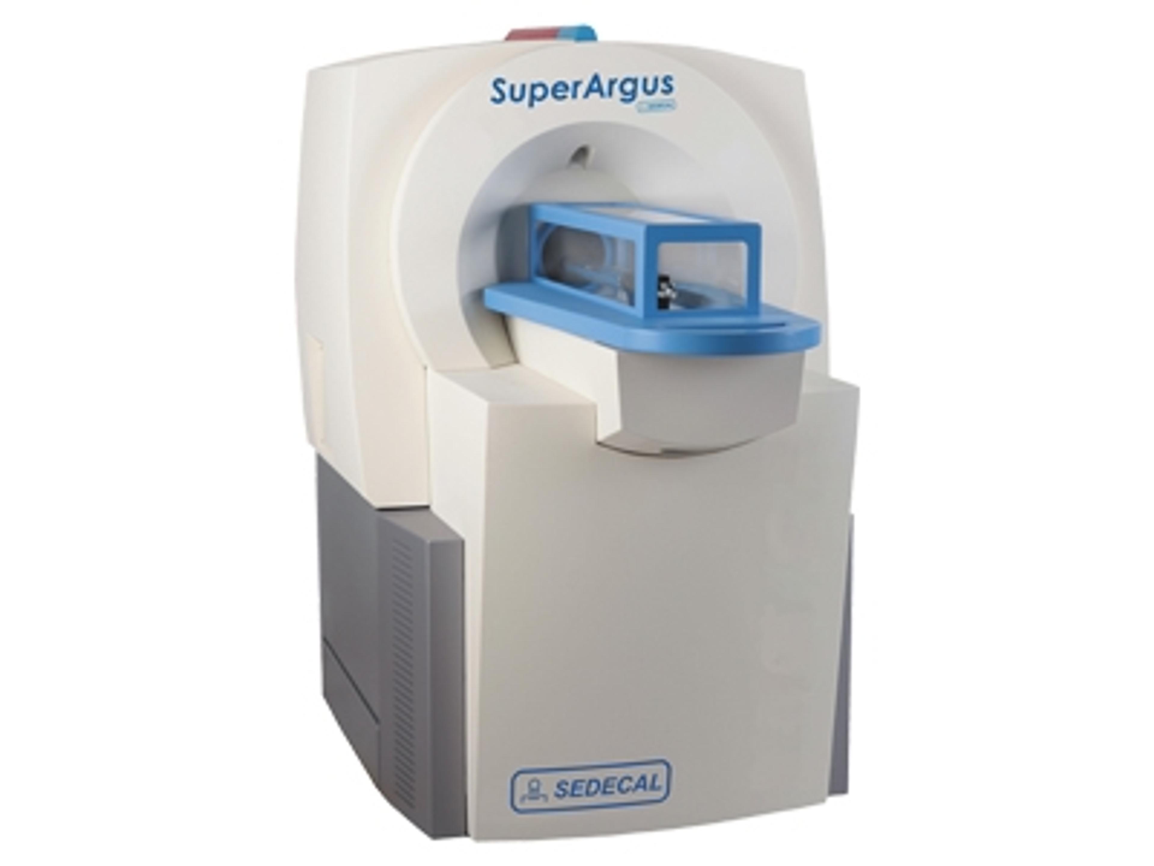
The SuperArgus PET/CT is a high-performance imaging system it can be configured as a combined PET/CT or PET or CT only system, and features state-of-the-art phoswich PET detectors with true depth-of-interaction (tDOI) for resolution uniformity and high sensitivity. The system offers real-time imaging up to 2.5msec frame rate and advanced capabilities like sensorless cardiac gating and conscious/awake imaging.
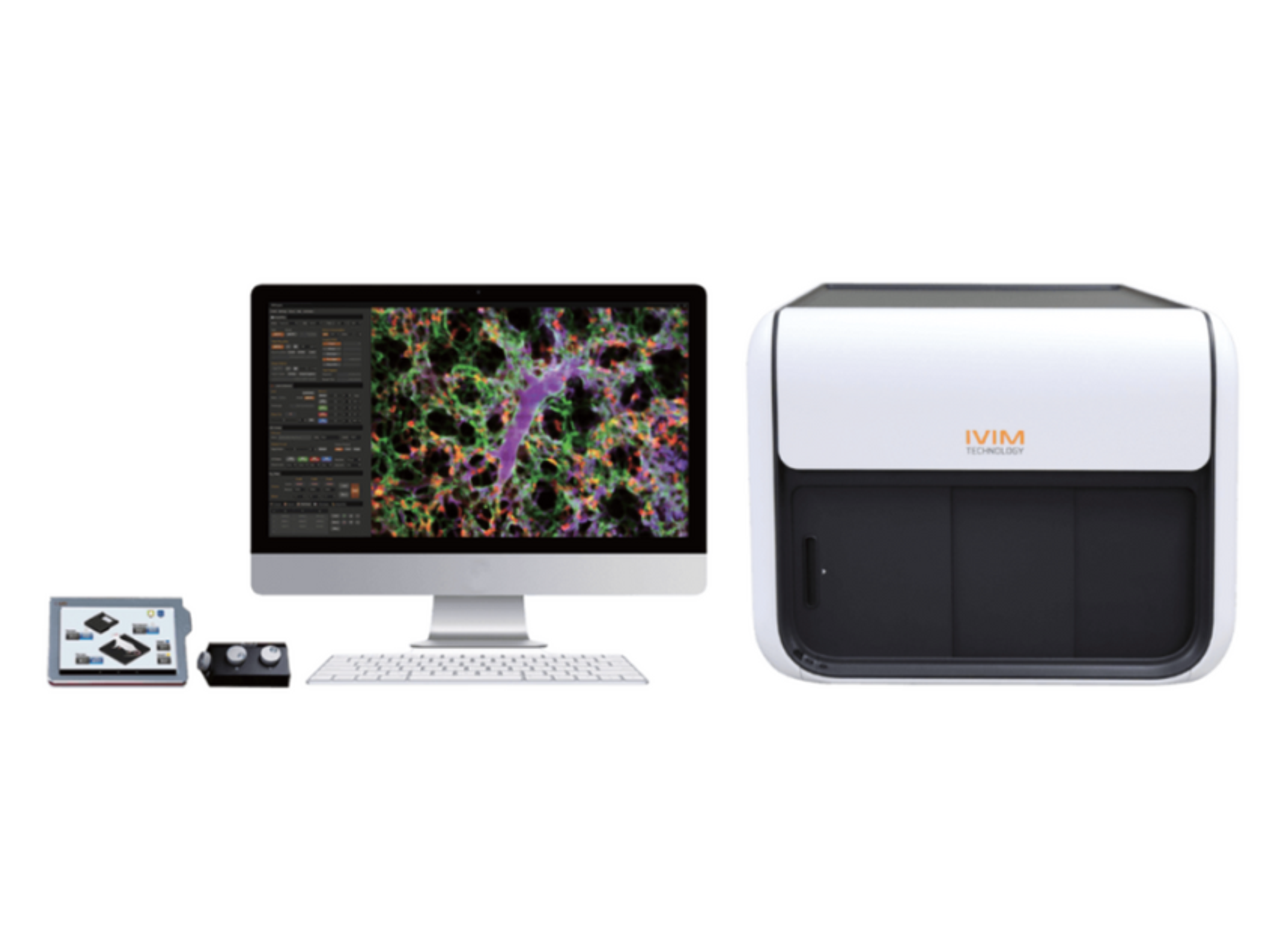
Intravital Microscopy(IVM) is an all-in-one two-photon and/or confocal microscopy system designed and optimized for longitudinal imaging of live animal models in vivo. This equipment has been designed around ease-of-use and augmented throughput as a next-generation core technology for biologists and translational scientists to elucidate the underlaying mechanism of every biological phenomena at tissue and cellular level.
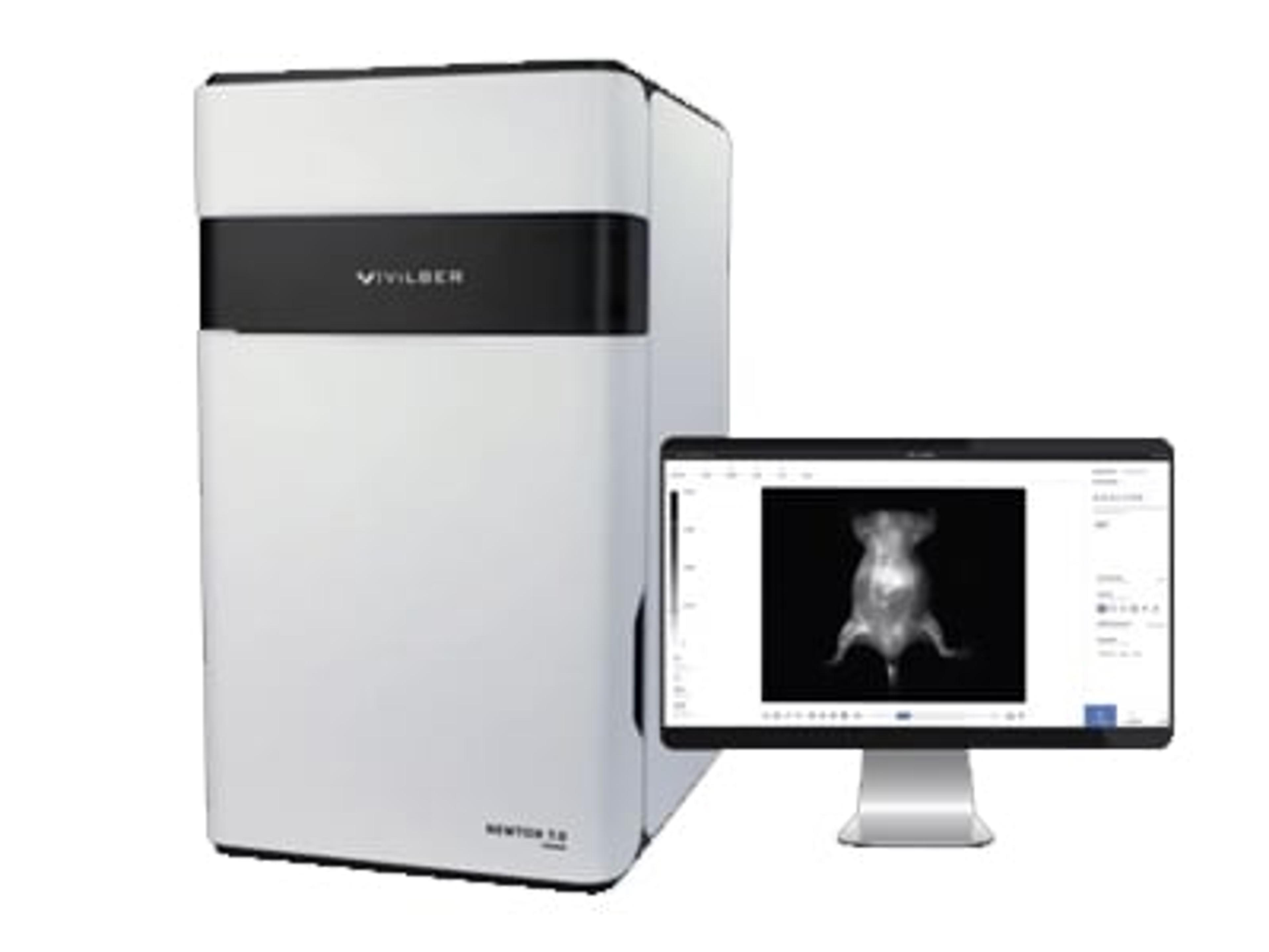
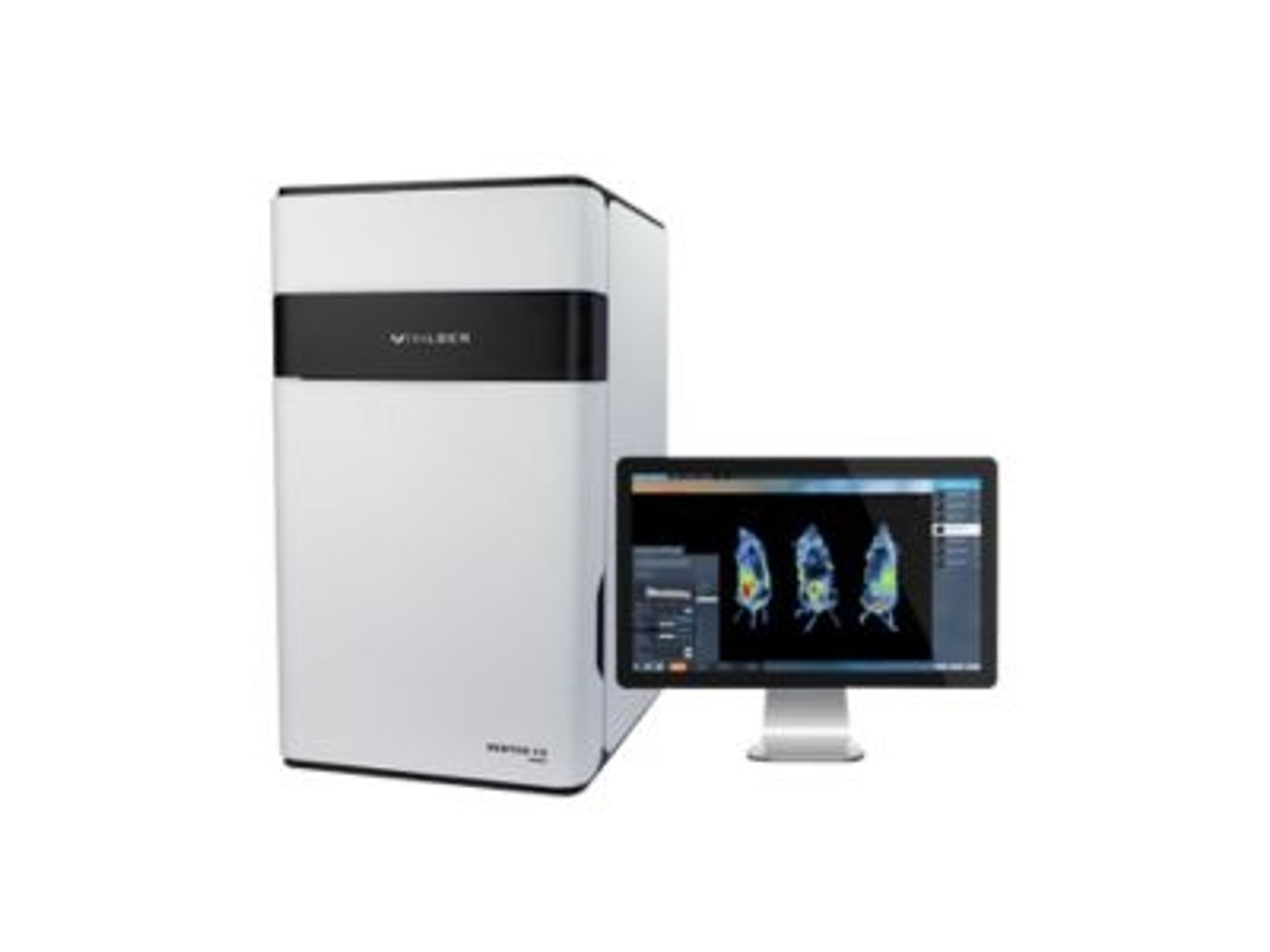
The Newton 7.0 is a cutting-edge optical imaging system that offers the versatility to perform bioluminescence, fluorescence, and 3D tomographic imaging in a single device. Its user-friendly interface and advanced features make it ideal for in vivo, ex vivo, and in vitro imaging applications, as well as simultaneous imaging of multiple specimens.