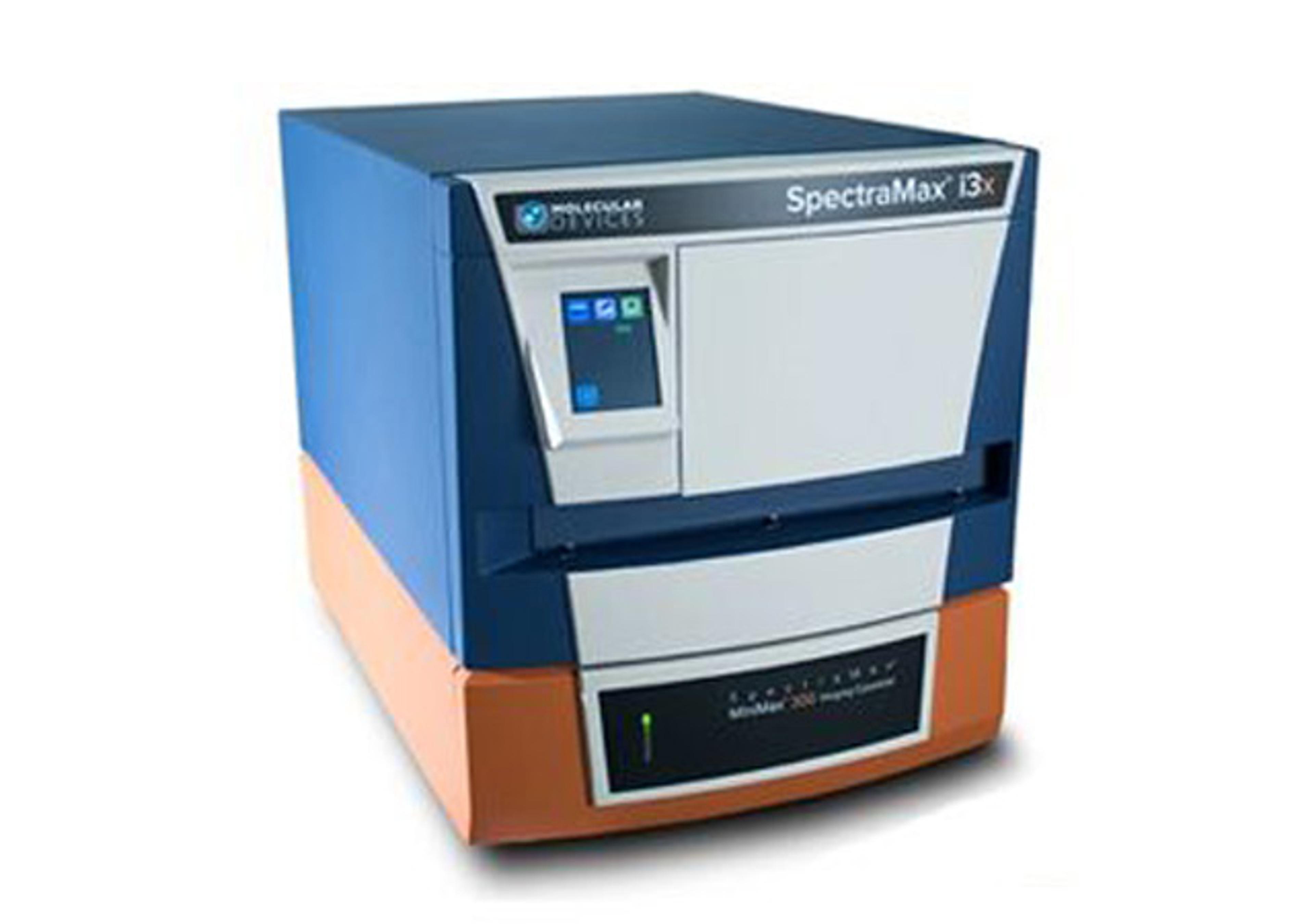
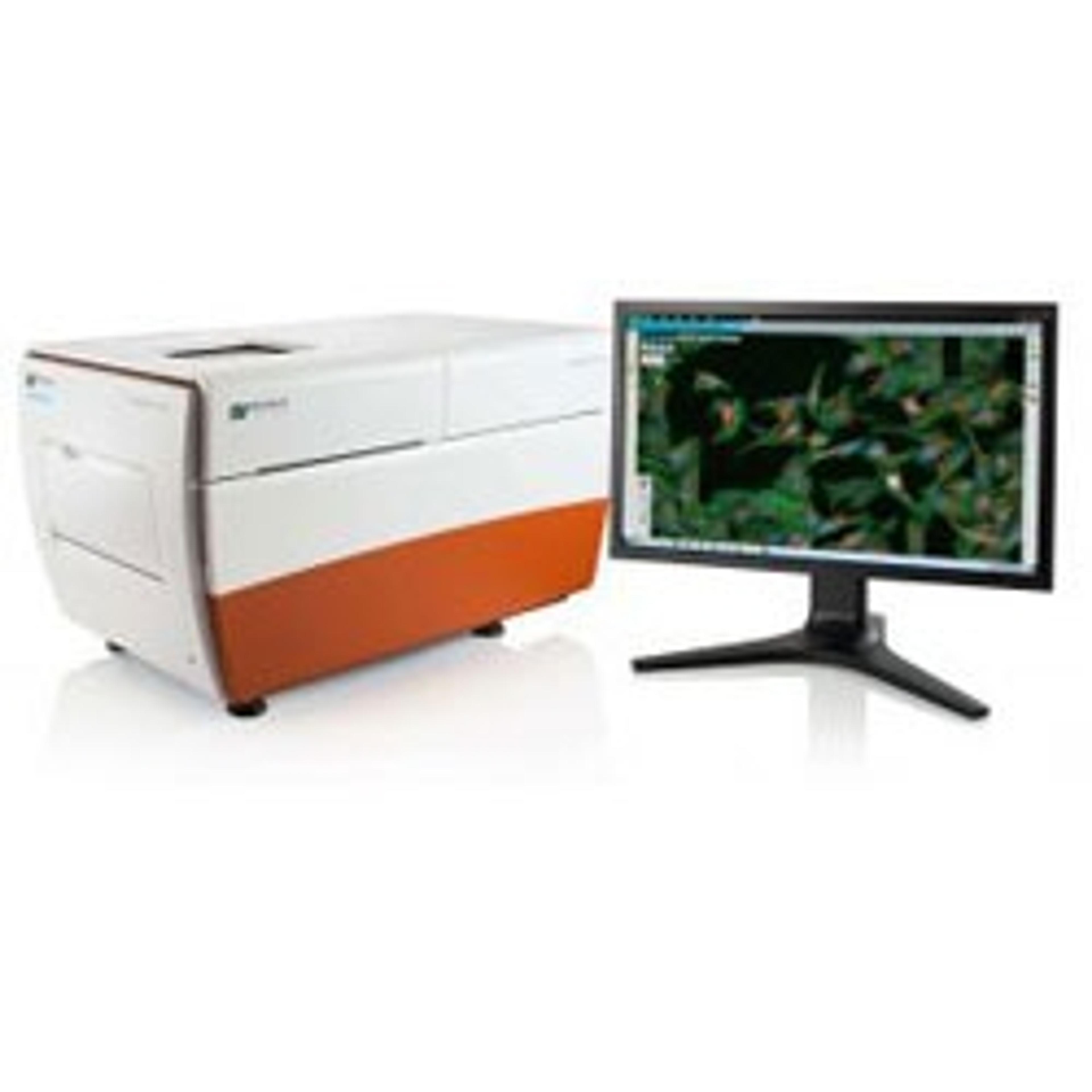
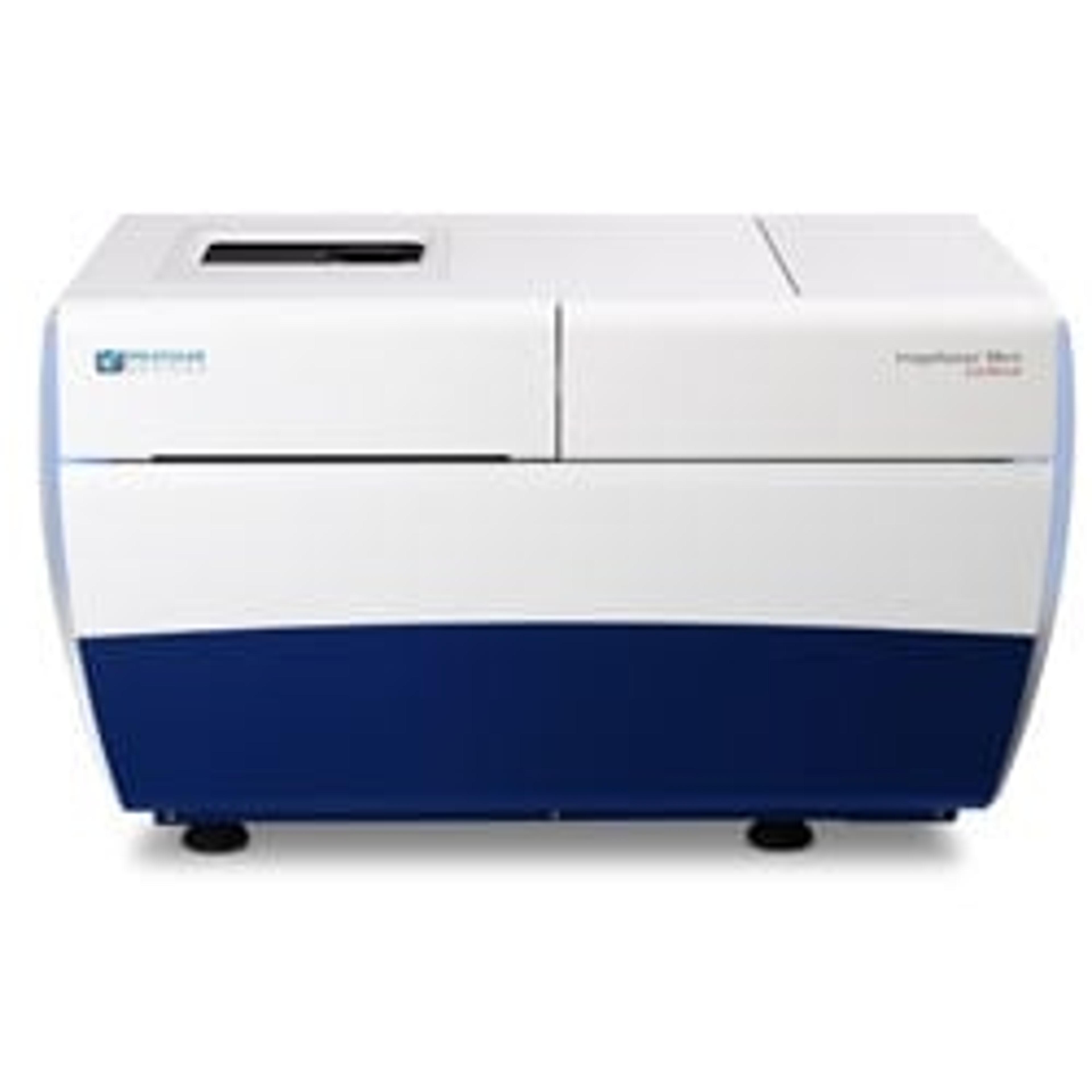
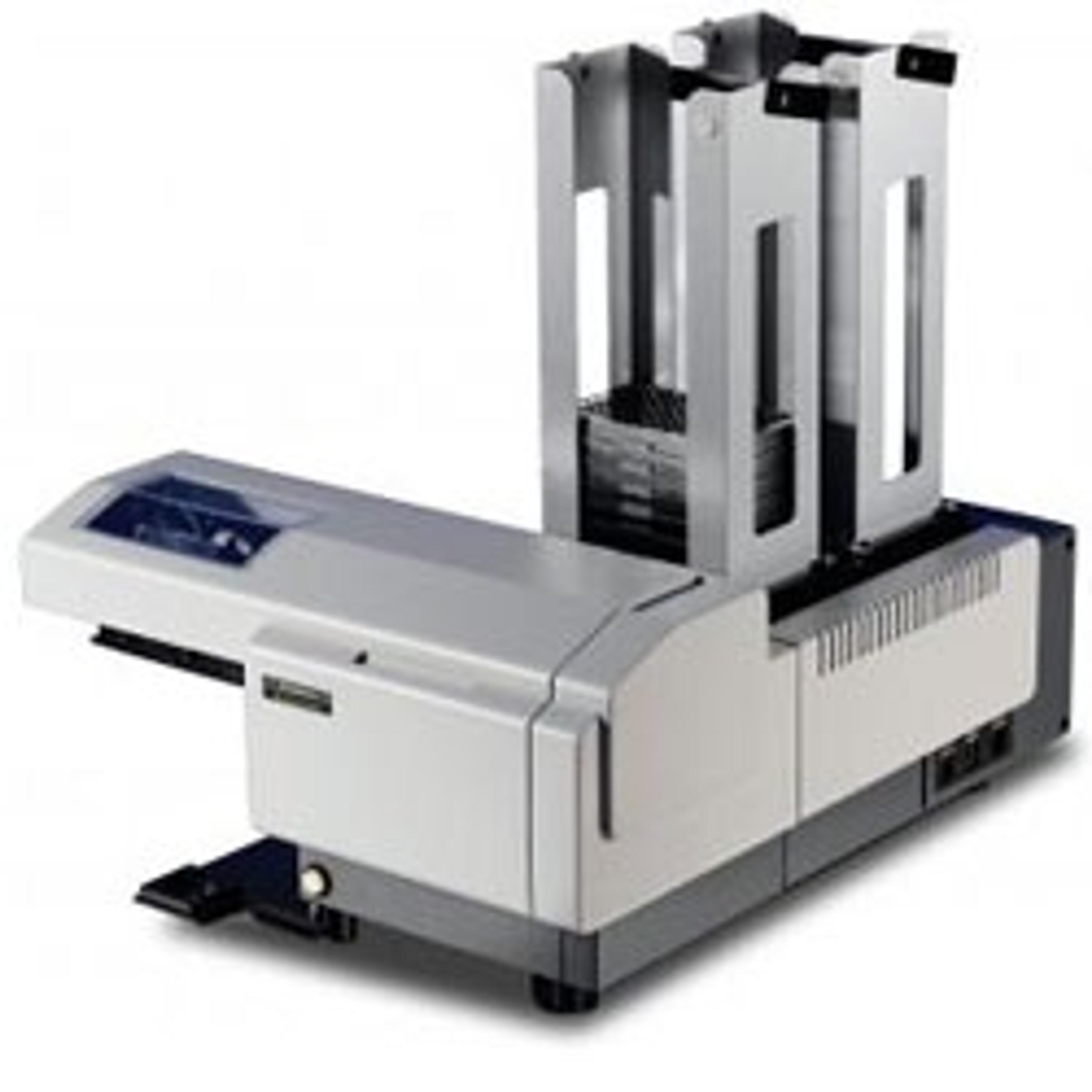
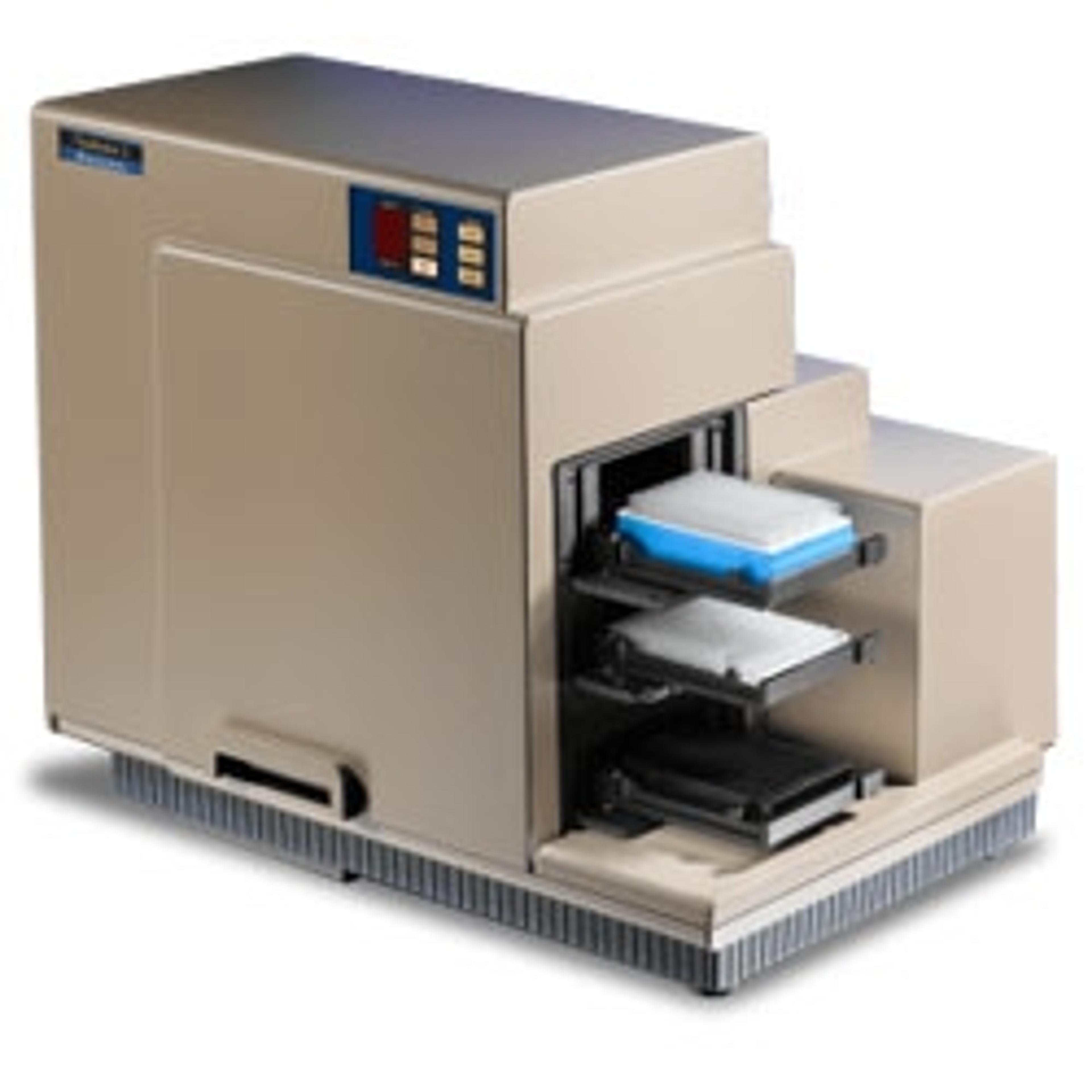
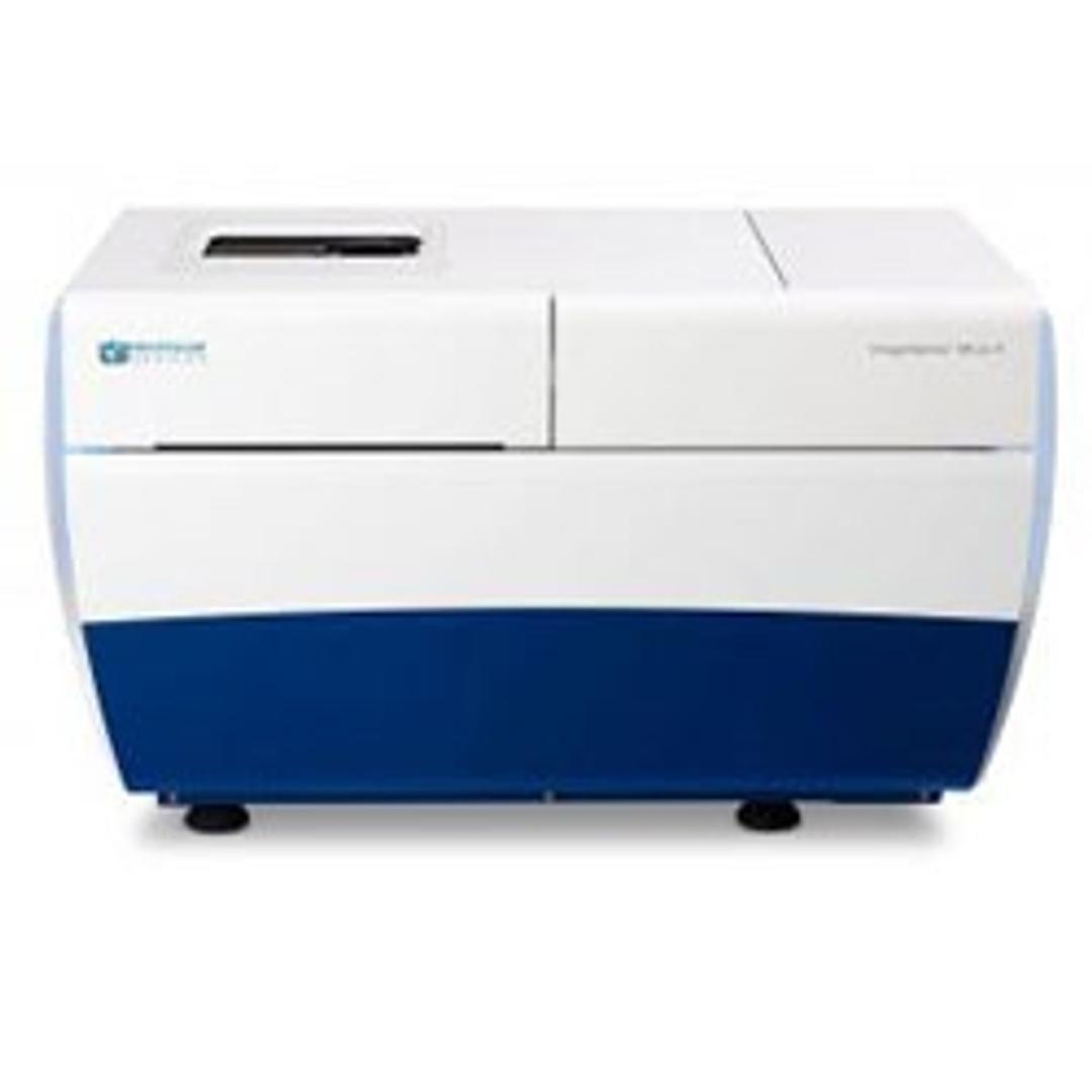
ImageXpress Micro 4 High-Content Imaging System
Position your research – with breakthrough, intelligent imaging solutions.


Receive your quote directly from the manufacturer.
Great company support
Cell counts and IHC analysis
This instrument is not very easy to use, but I believe this is because of it's power and versatility. We inherited an older instrument, and none of us had training. While the learning curve is long, the best part of this is the support we have been getting from Chris Nishioka. He has been fabulous about spending time with us via zoom to help us get up and running. Can't say enough good things about this company and their support.
Review Date: 15 Nov 2021 | Molecular Devices®
Great workhorse instrument!
High-content screening, phenotypic screening, quantitative IF
This product is a robust instrument. It is used for a wide variety of applications and has been a workhorse instrument for our core lab. Users love it and its reliability has been outstanding. Great value for the price you pay, it has paid for itself and more.
Review Date: 18 May 2020 | Molecular Devices®
An excellent addition to our core facility.
Cell biology
This instrument of easy to use and customize. The out of the box analysis modules are amazing. Cell segmentation and analysis algorithms work very well. The tech support is very knowledgable and thorough.
Review Date: 8 Nov 2017 | Molecular Devices®
The ImageXpress® Micro 4 High-Content Imaging System represents the culmination of four generations of imaging expertise. The latest agile design allows you to boost your research with this faster-than-ever system, while providing the option to upgrade to confocal in the future to align with your research needs.
Capture images of whole organism and cellular or intracellular events with the ImageXpress Micro 4 system configured to suit your specific biological needs. We offer unlimited configurability with user exchangeable filter cubes, the widest range of objective lenses and environmental control, transmitted light, confocal imaging, and fluidics options.
Built on over 30 years of cell-based imaging experience, the ImageXpress Micro 4 High-Content Imaging system is engineered for performance to reliably accelerate the pace of your research.
Features:
- FASTER-THAN-EVER, AGILE SYSTEM, WHICH GROWS WITH YOUR NEEDS.
4th Generation Platform with unlimited configurability, developed to address your research needs. - FOCUS ON YOUR BIOLOGY.
The automated 5-position filter cube changer facilitates multiplex assays with up to five filter sets and four objectives for a single experiment. - ACQUIRE STATISTICALLY RELEVANT DATA FASTER WITH LARGE FIELD OF VIEW AND >3 LOG DYNAMIC RANGE.
Minimize tiling effect and reduce your acquisition time with the ability to image a whole 384 well at once. - EXPAND YOUR RESEARCH CAPABILITIES WITH AVAILABLE OPTIONS.
Available options include Environmental Control (temperature, humidity and CO2 control for multi-day live-cell experiments), Transmitted Light True Phase Contrast, Confocal Imaging (field upgradable), and Fluidics (on board disposable pipetting).
Brochures

ImageXpress Micro Confocal: The confocal solution for your complex biology
In this brochure, Molecular Devices provides key features, applications and specifications of the ImageXpress® Micro Confocal system, a high-content solution that can switch between widefield and confocal imaging for improved quantification of live or fixed cell assays.
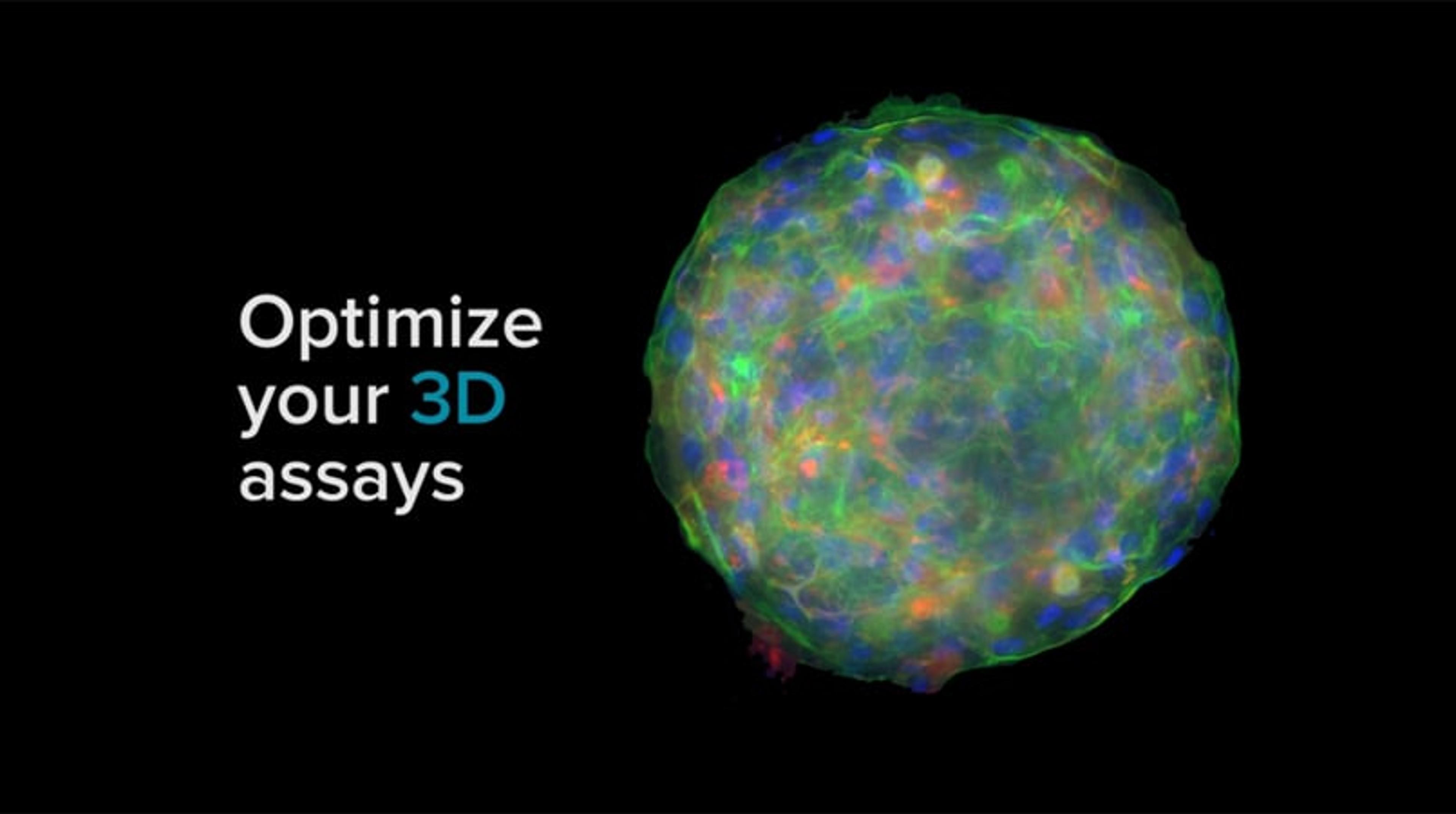
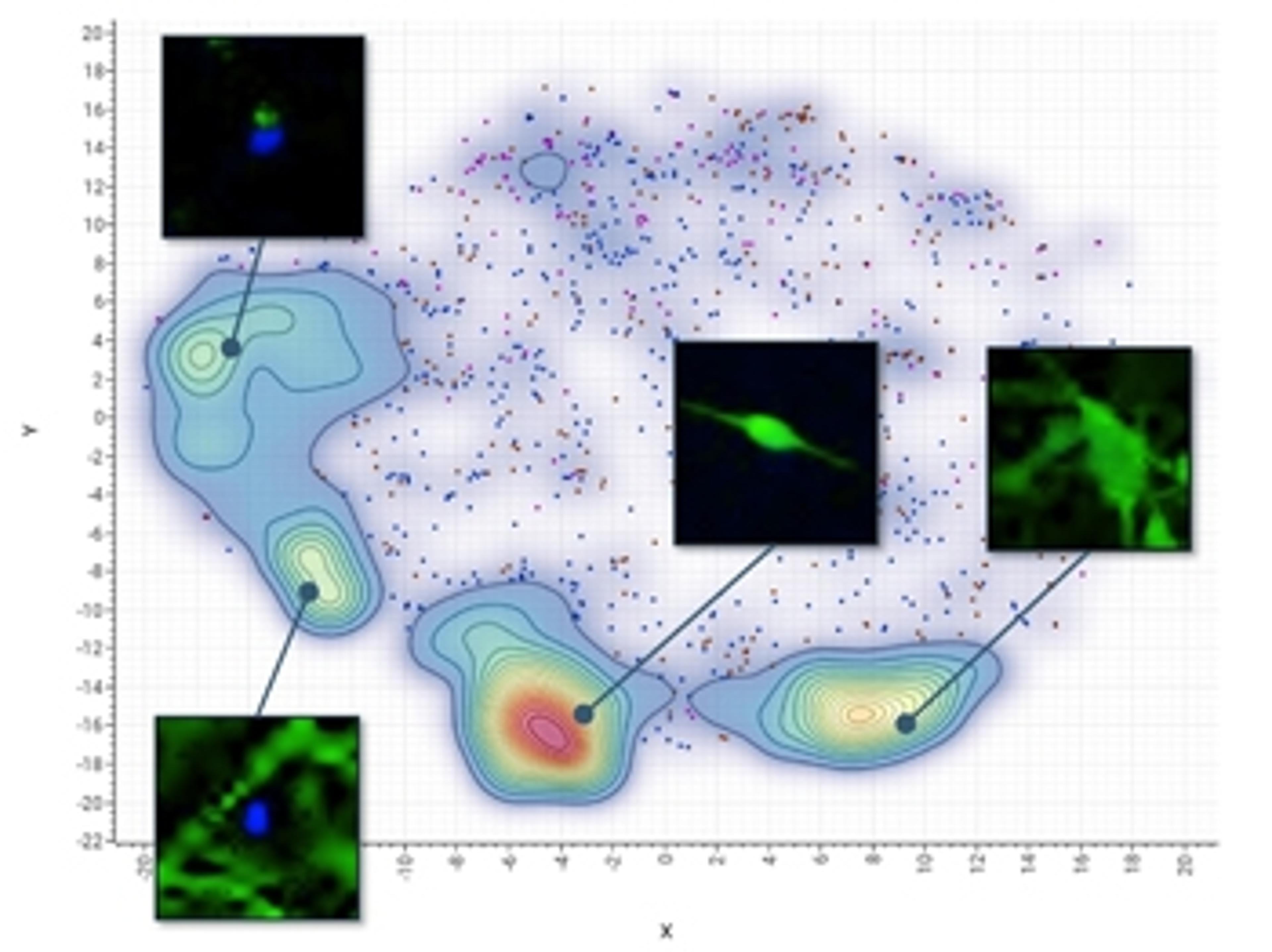
High-content imaging for diverse 3D cell culture models
3D cell models are morphologically diverse with varying characteristics based on the cell type and the underlying research questions. In this application compendium, we bring you helpful case studies to perform high-content imaging on a range of 3D models and resolve common challenges experienced in 3D cell culture assays.
Learn how to capture in-depth and high-quality images of spheroids, stem cells, organs-on-chips and whole organisms and read case studies from the scientists developing 3D cell assays to delve into the complex biology of neurodegenerative disease, angiogenesis and tumor microenvironment.
The eBook covers 3D high-content imaging protocols for:
- Characterizing compound effects on 3D cells in extracellular matrix
- Screening cancer therapeutics in spheroids
- Morphological characterization of 3D neuronal networks in an organ-on-a-chip model
- Characterization of angiogenesis in an organ-on-a-chip model
- High-throughput imaging assays using zebrafish
- High-content 3D toxicity assay using iPSC-derived hepatocyte spheroids
High-Throughput Confocal Imaging of Spheroids for Screening Cancer Therapeutics
This application note demonstrates a method for cancer cell spheroid production, followed by protocols for staining and imaging of these spheroids for use in multi-parameter cytotoxicity studies.
Counting Cells with the Transmitted Light Analysis Module in MetaXpress
Label-free cellular assays are required for a multitude of biological applications that monitor the cell number, proliferation, health, confluency, and cytotoxicity. These applications necessitate efficient and robust transmitted light (TL) imaging and analysis capabilities providing precise segmentation for quantitation of cells and assessment in variety of cell responses and morphologies. Additionally, the coupling of high-contrast transmitted light imaging and fluorescent labeling and imaging is essential to numerous cellbased assays.
Monitoring BNP Expresion and Cell Size to Determine Hypertrophic Response in Human Cardiomyocytes
Cardiac hypertrophy is a condition associated with many heart diseases such as myocardial infarction, ischemia, hypertension, valvular dysfunctions, and is also observed as a toxic side effect of environmental contaminants or pharmaceutical drug candidates. It is characterized by different cellular changes including increased cell size and enhanced protein synthesis. One of the classic biomarkers for hypertrophy is B-type natriuretic peptide (BNP), which is over produced in hypertrophic cardiac cells.
High-Throughput Imaging Assays Using Zebrafish, a Model Organism for Human Disease
Recently, zebrafish-based screening has gained favor as an alternative to mammalian screening due to cost, throughput and reduced ethical concerns. Zebrafish are a useful model for drug development because of their high biological similarity to humans. Studies in ontogeny and organogenesis have shown that their primary organ systems are very similar to humans, and the synteny between zebrafish and Homo sapiens is as high as 70-80%.
Apoptosis Detection Using EarlyTox Capase-3/7-D NucView 488 Assay Kit on ImageXpress Micro Systems
Apoptosis is an important mechanism signaling programmed death of cells in normal processes such as embryonic development, as well as in diseases including cancer and neurodegenerative conditions. This flexible assay can be performed using an ImageXpress® Micro High Content Imaging System and MetaXpress® Analysis Software for calculating the incidence of apoptotic cells in a well.
Optimize Your 3D Assays with ImageXpress
Optimize your 3D assay and make your next breakthrough in cellular imaging - with intelligent, innovative imaging solutions.
Position Your Research - with Breakthrough, Intelligent Imaging Solutions
This video introduces the ImageXpress Micro 4 confgurable widefield system by Molecular Devices. Explore and interact with your data using the new MetaXpress 3D analysis module, from basic assays to complex 3D structures across the plane.
How AI is advancing neurodegenerative disease research
In this guest editorial, explore how artificial intelligence is becoming a powerful tool for drug discovery
Molecular Devices Launches ImageXpress Nano System and CellReporterXpress Software
ImageXpress Nano System makes automated imaging accessible for every lab conducting cellular imaging research