Lux
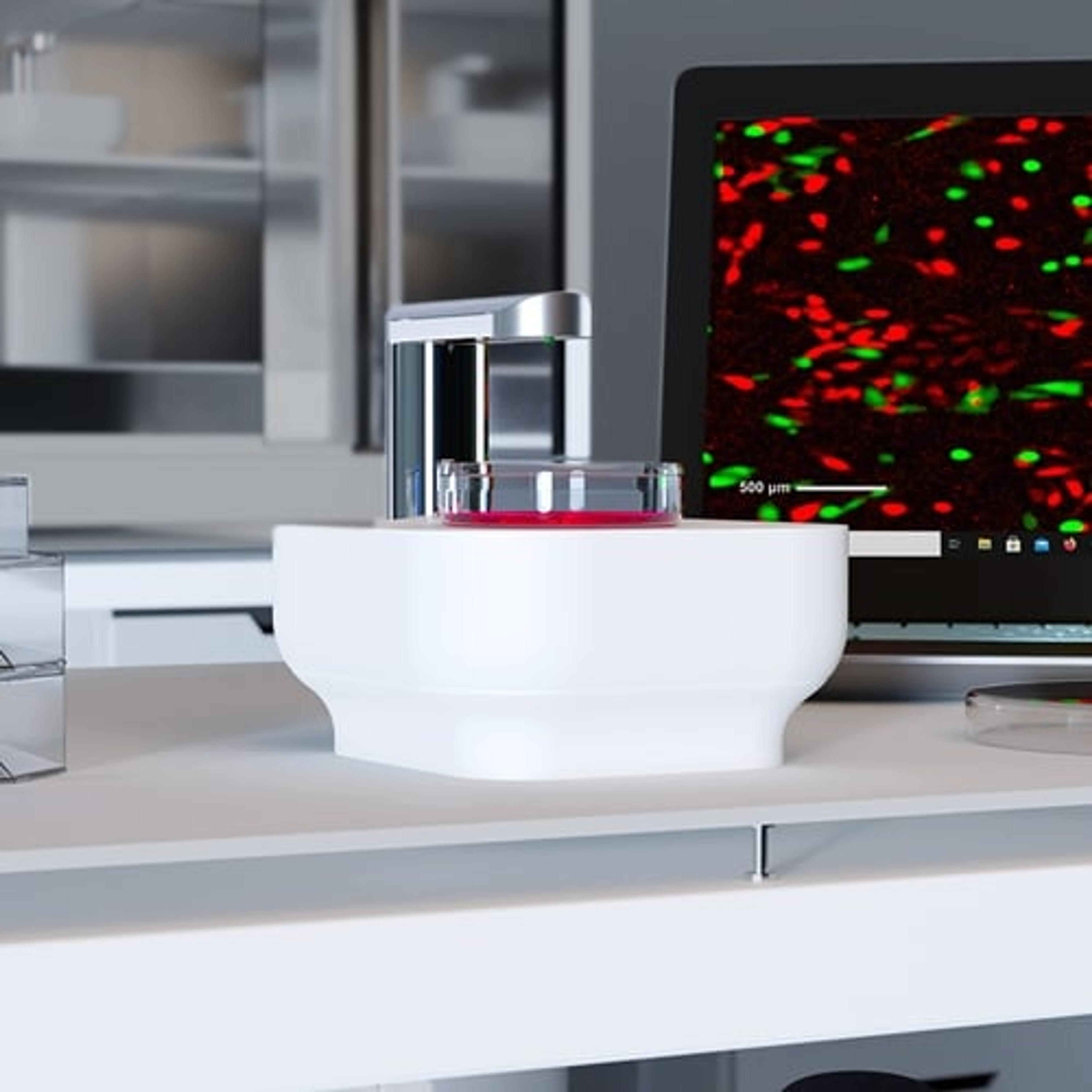
Single-well live-cell analysis with brightfield and fluorescent imaging from your incubator


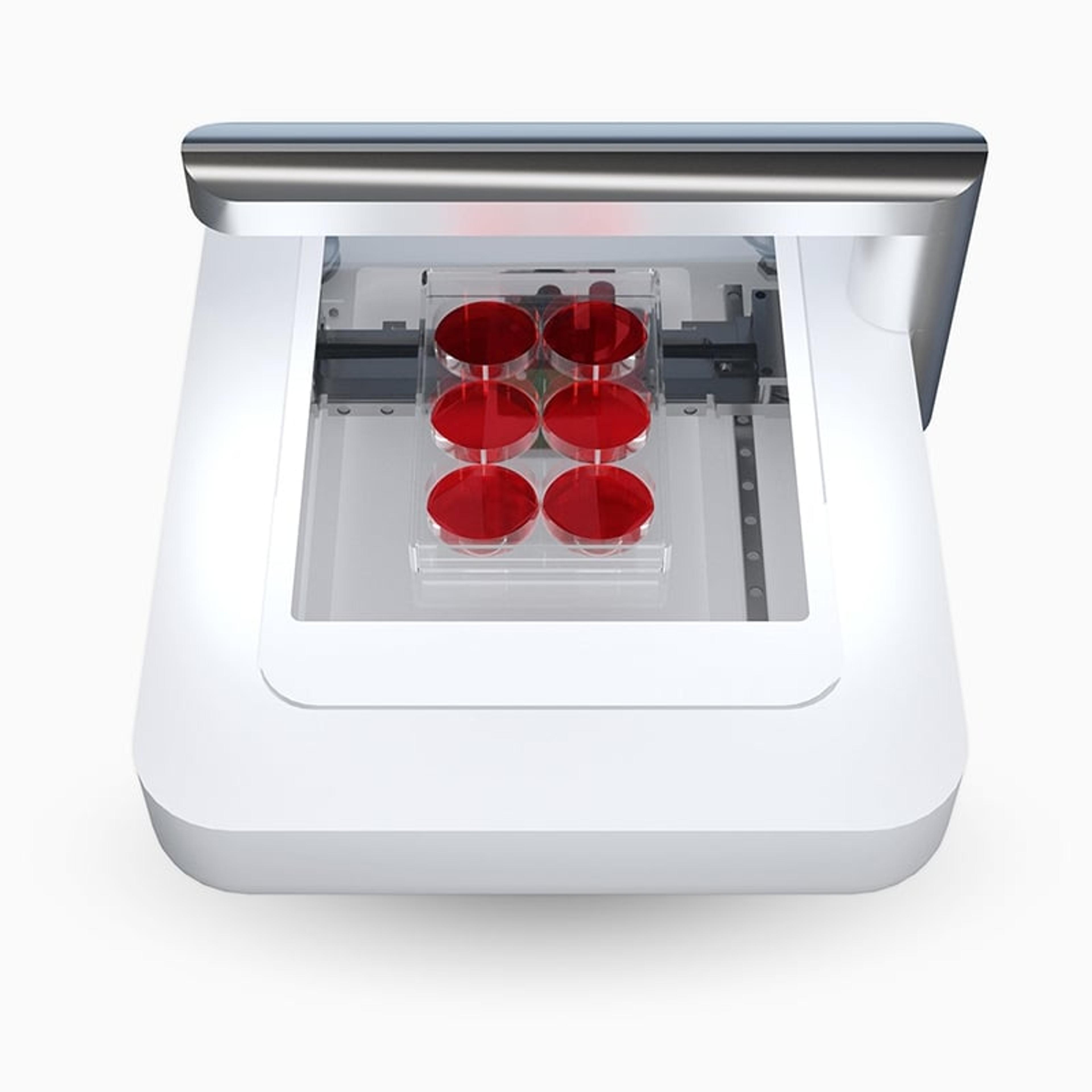
Analyze cells in their desired culture environment

Automatically calculate the number of fluorescent objects

High-quality images and real-time time-lapse movies


Receive your quote directly from the manufacturer.
I am a user of this instrument and it gives satisfactory results.
Cell imaging and calculating cell culture confluency
This is very useful in calculating cell culture confluency, in optimising the efficiency of cell culture assay, and also in analysing all aspects of stem cell culture.
Review Date: 28 Oct 2022 | Axion BioSystems
Useful live cell imaging in our lab
Live cell imaging
Elegant way to use for live cell imaging. For several experimental set-ups such as apop assay, confluency, cell trackers, and (induced) cardiomyocyte contraction assays in a qualitative and quantitative manner. Cytosmart develops novel software for these machines to make them applicable for many assays. Guidance and support are around the corner, they are always available for a call, zoom or a visit to stay in touch with the customer.
Review Date: 15 Aug 2022 | Axion BioSystems
Amazing instrument! Easy to setup and obtain publication quality fluorescence images!
Live cell tracking using microphysiological systems
The Lux3 FL system is an amazing, compact and easy to install fluorescence microscope! Microphysiological systems like organ-chips are excellent tools to study human physiology however visualizing real-time cellular interactions and cell migration processes over extended experimental time periods is difficult as traditional microscope require on-stage incubator attachments and use of microscopes over these extended time periods might also be inconvenient to other researchers. The Lux3 FL system is a perfect solution to this problem as it neatly fits into standard sized incubators and are easily compatible with contemporary organ-chip biotechnology. We have been using this system routinely in our lab to study cell migration and rearrangement using our organ-chip platforms and the results are amazing! The Lux3 FL system is very easy to setup and the user interface is surprisingly easy to understand while delivering notable functionality. The acquired images are publication quality and very easily editable on the cloud. The timelapse videos are amazing and can be directly used for publication purposes. CytoSmart also has amazing customer service and support! They are very helpful and prompt in troubleshooting and assistance with maintenance etc. Summing it all up, I love this instrument! It’s compatibility with organ-chips and ability to perform long term cell visualization studies are a big advantage of this product and deserve a five star rating!
Review Date: 27 Jun 2022 | Axion BioSystems
A great addition for any cell culture lab.
Live cell imaging, also in fluorescence mode
Simple to set up and very straightforward to use. Cells in our microfluidic chips can also be imaged with great ease, even underflow. A must-have for anyone keen on knowing what their cultured cells are doing when they think no one’s looking. No need to open the incubator, just check-in from behind your computer. The ability to image in fluorescence mode is the icing on the cake.
Review Date: 24 Nov 2021 | Axion BioSystems
Absolutely a must have instrument in a matter of saving both space and time.
Cell culture
Live cell culture fluorescence inside of an incubator, small and transportable product in absolute correlation to the value of money.
Review Date: 16 May 2021 | Axion BioSystems
Good for GFP-labeled live cell imaging.
Cell imaging
Good for GFP-labeled live cell imaging.
Review Date: 16 May 2021 | Axion BioSystems
Great instrument to monitor fluorescently labelled cells
Measure GFP and RFP in human cells before and after crispr assays
In our school we used the cytosmart FL system monitor GFP expression in green. This works very well, and we obtained nice results and we were also able to see how GFP K.O. cells develop after a Crispr experiment. For RFP it was more difficult as we would like to see gain in expression from GFP to RFP but we have no signal to start with, therefore we were not able to set the settings in a most optimal way to start with. However, we were very pleased to monitor our cells for many days in a row.
Review Date: 15 Dec 2020 | Axion BioSystems
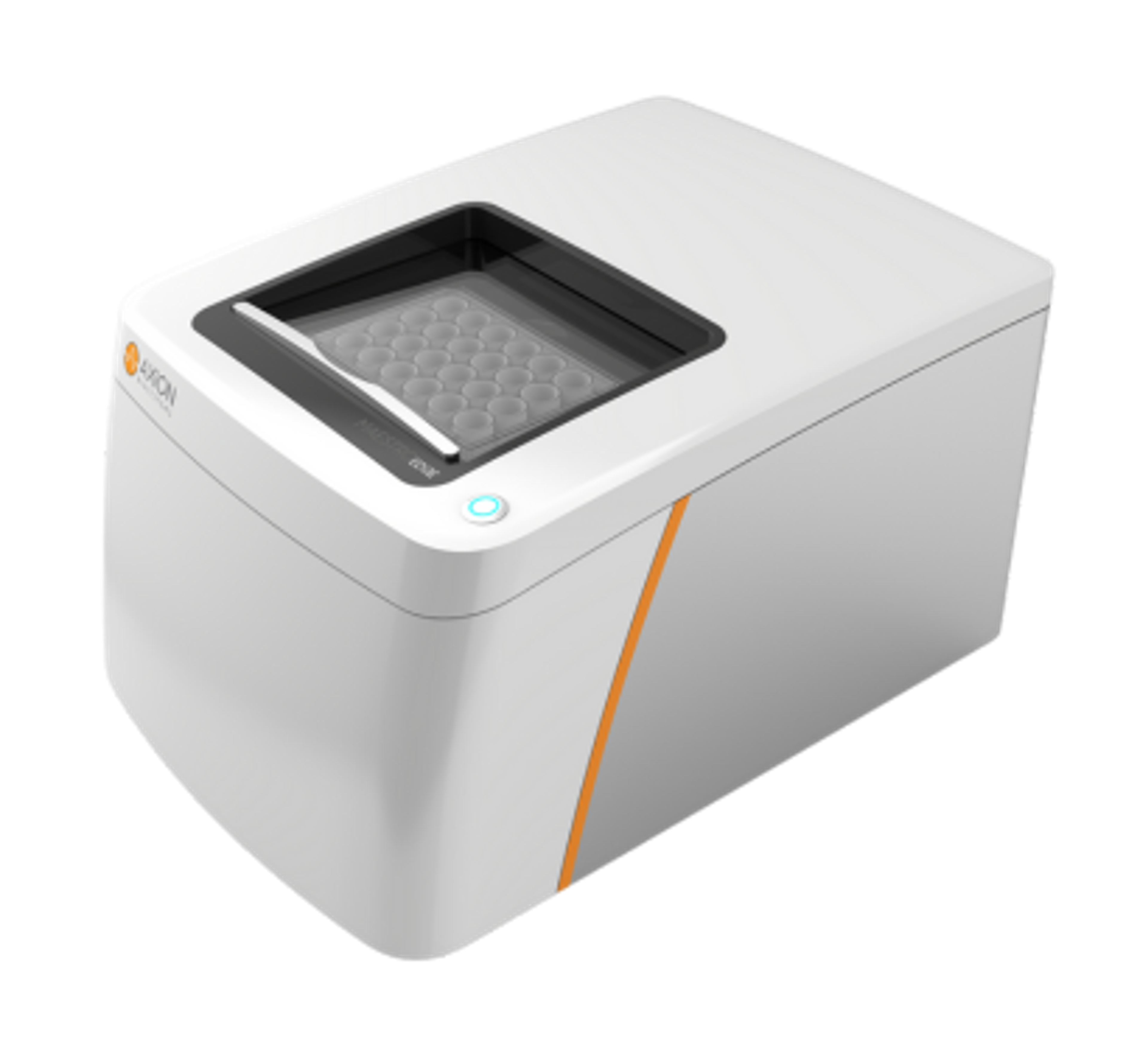
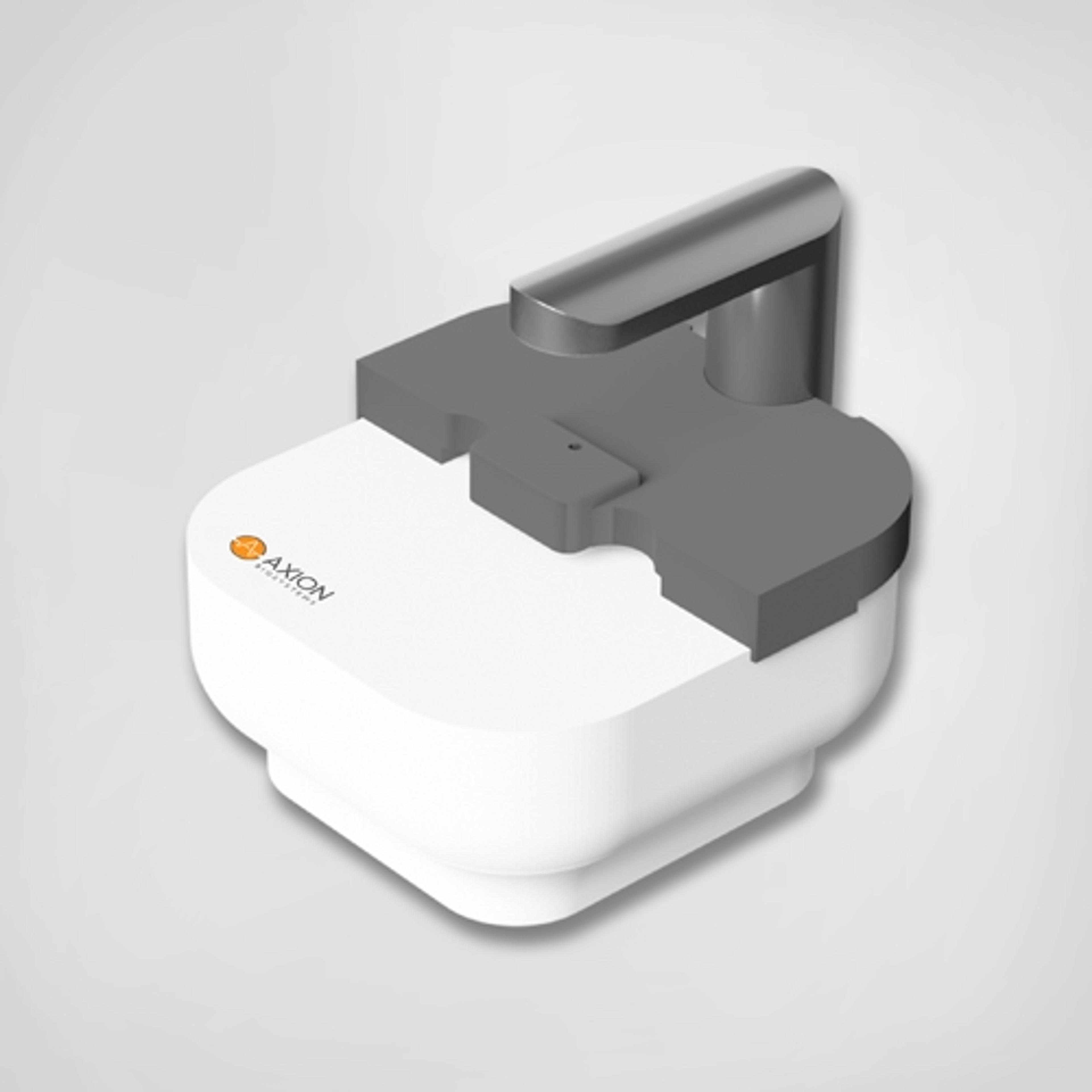
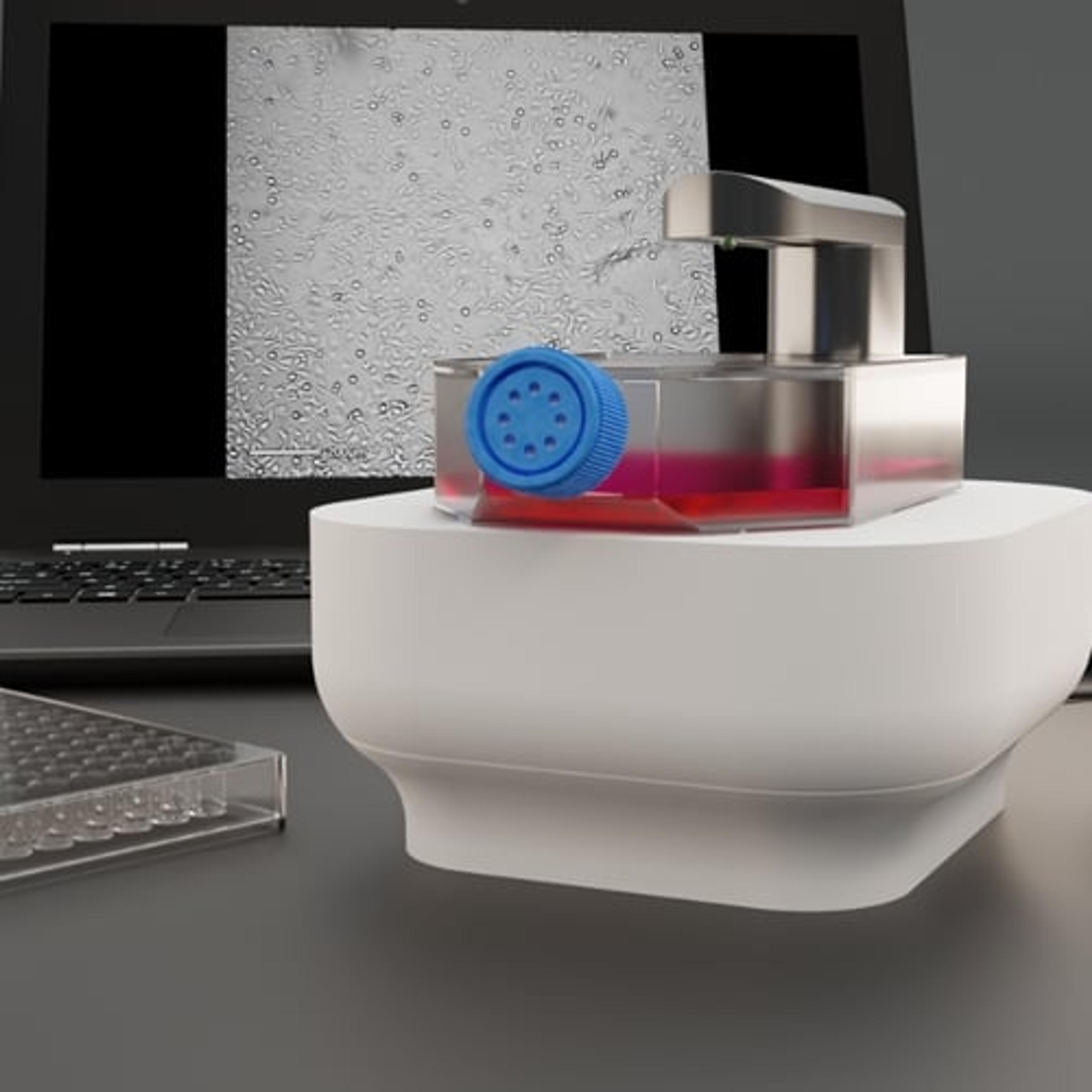
The Lux™ (Lux3™) is a miniaturized live-cell analysis platform designed to help researchers monitor cell cultures directly from the incubator using automated brightfield and fluorescence live-cell imaging. Visualize cell morphology and behavior in real-time with an easy-to-use system with fast and reliable data analysis.
KEY FEATURES
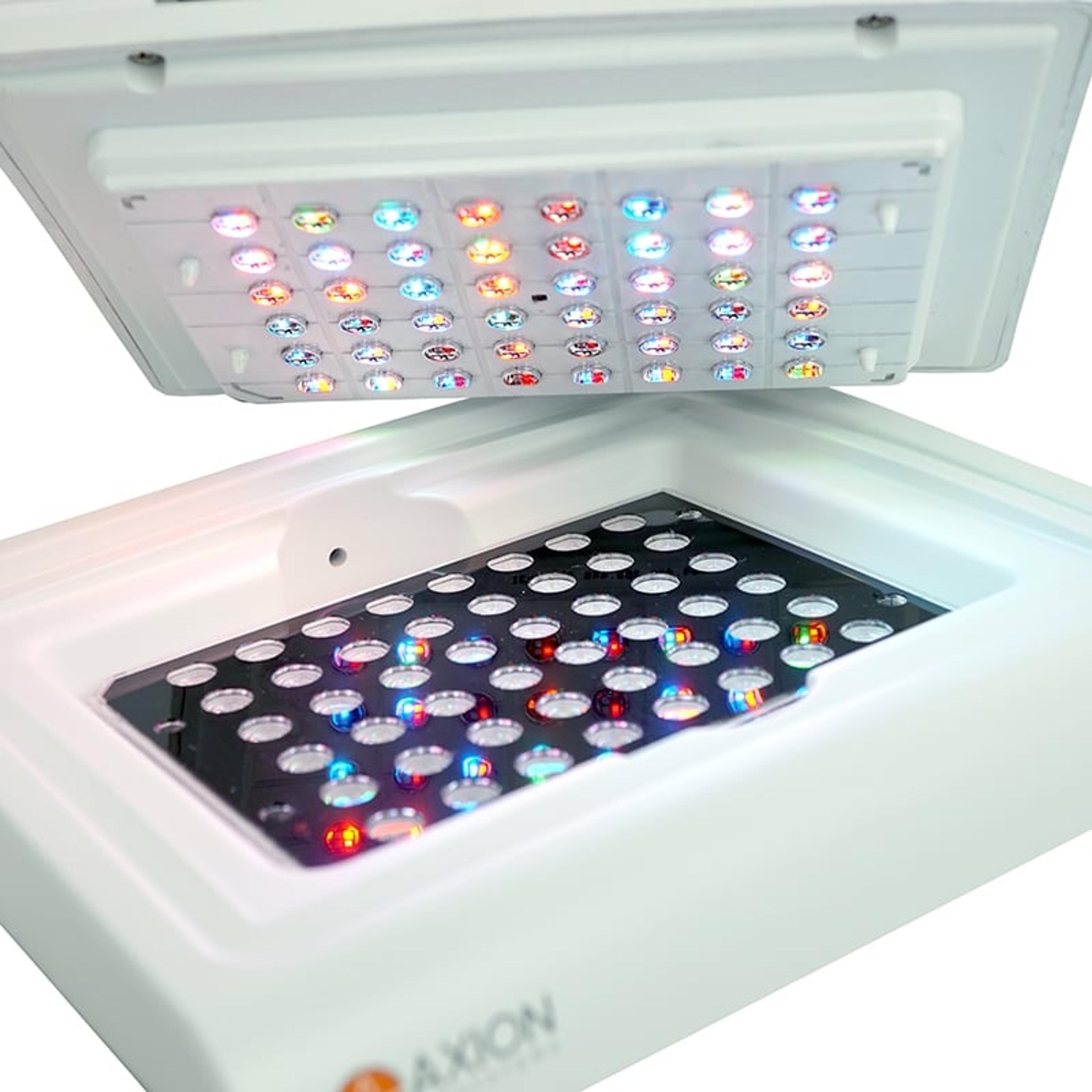
- Track complex biology in brightfield and fluorescence — From label-free to fluorescence-based cell tracking, the Axion Lux can help you to see and learn more.
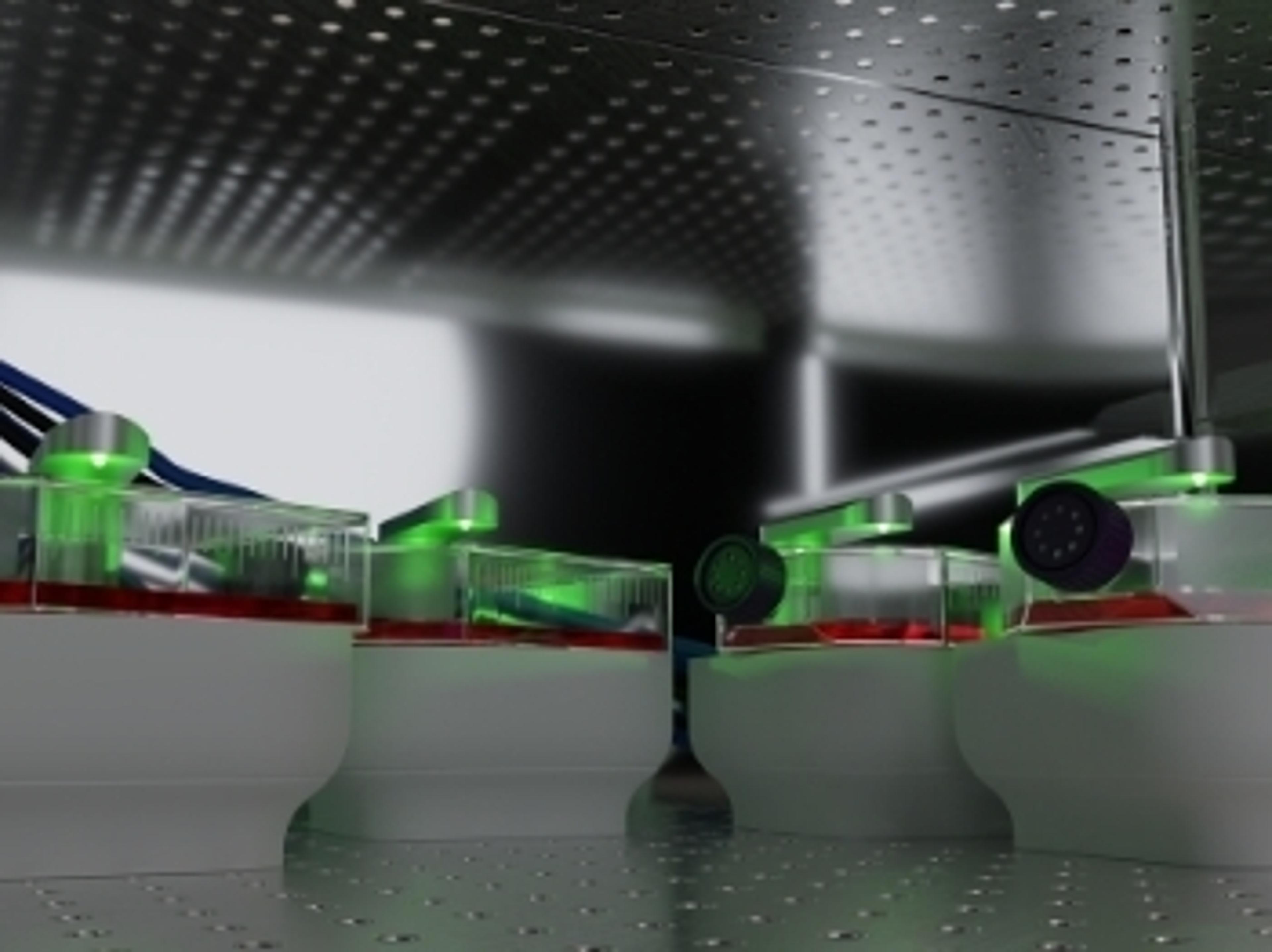
- Capture every moment, without crowding your incubator — The Lux operates within an incubator, automatically capturing timelapse images as your cells grow in an optimal environment. The compact device is perfect for labs with limited incubator space.
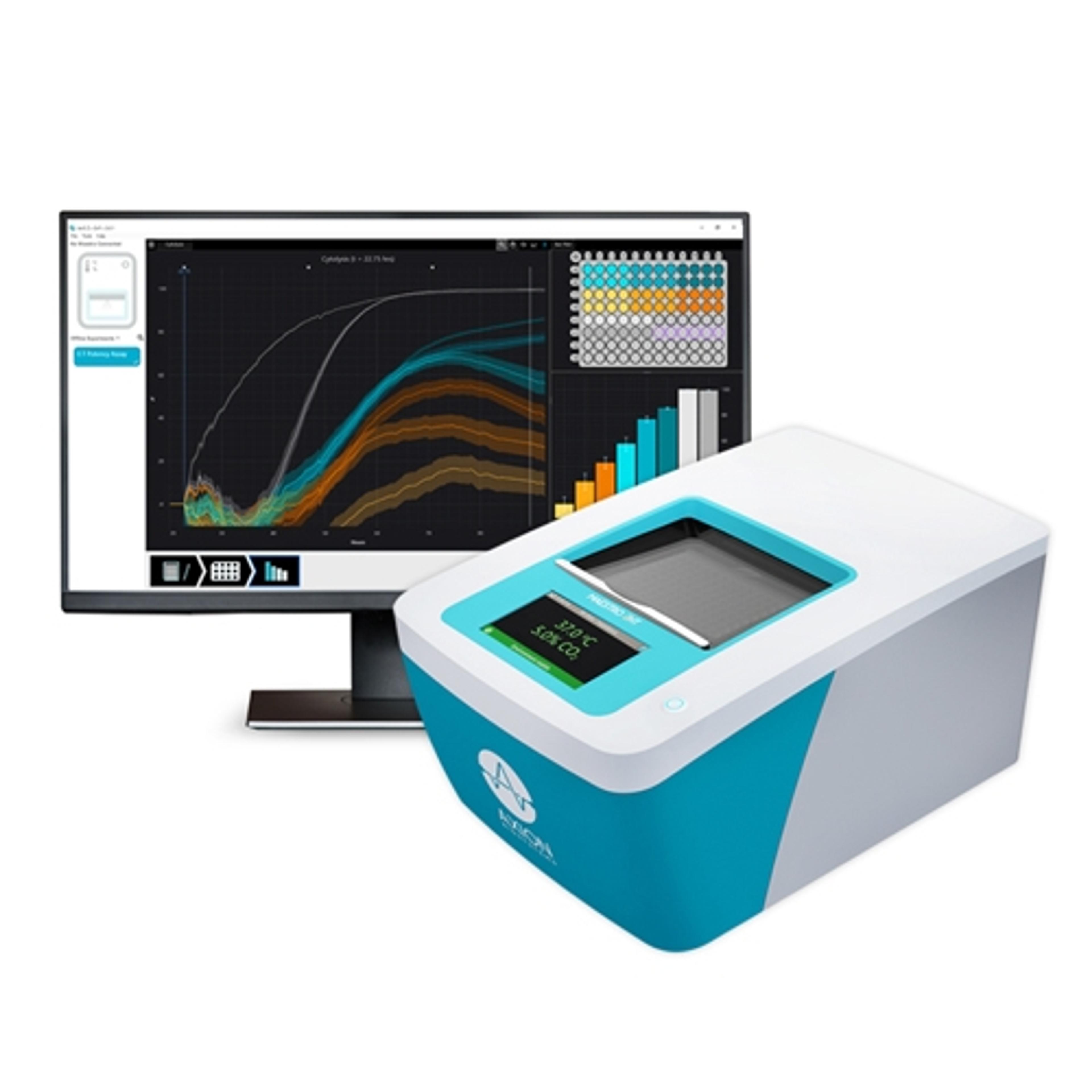
- Monitor your cells and analyze remotely — The AxIS Vue™ software allows you to monitor your cells and perform data analysis from your desktop.
- Get started quickly — A short training will get you up and running with the Axion Lux systems. The devices are easy to install and require no maintenance or calibration.
- Expand to meet your needs — Link multiple Lux devices together to simultaneously monitor multiple culture vessels.
SOFTWARE
The Lux platform has three available software modules to match your assay needs:
- Confluency Module – Know exactly when to passage your cells or track cell growth and death in your experiments with brightfield confluency.
- Fluorescence Module – Add fluorescent assay capabilities with fluorescence confluency, intensity, and object detection.
- Scratch Assay Module – Investigate the progression of wound closure over time.

CytoSMART introduces new, label-free live-cell microscopy solutions with high image quality
The CytoSMART Lux3 BR is a small brightfield microscope equipped with a high-quality 6.4 MP CMOS camera

Meet the winners of the 2021 Scientists’ Choice Awards for Life Sciences
Scientific, technological and communications excellence has been celebrated at the Virtual Cancer and Immunology Research Summit, with Logos Biosystems, INTEGRA Biosciences and Sartorius among those recognized