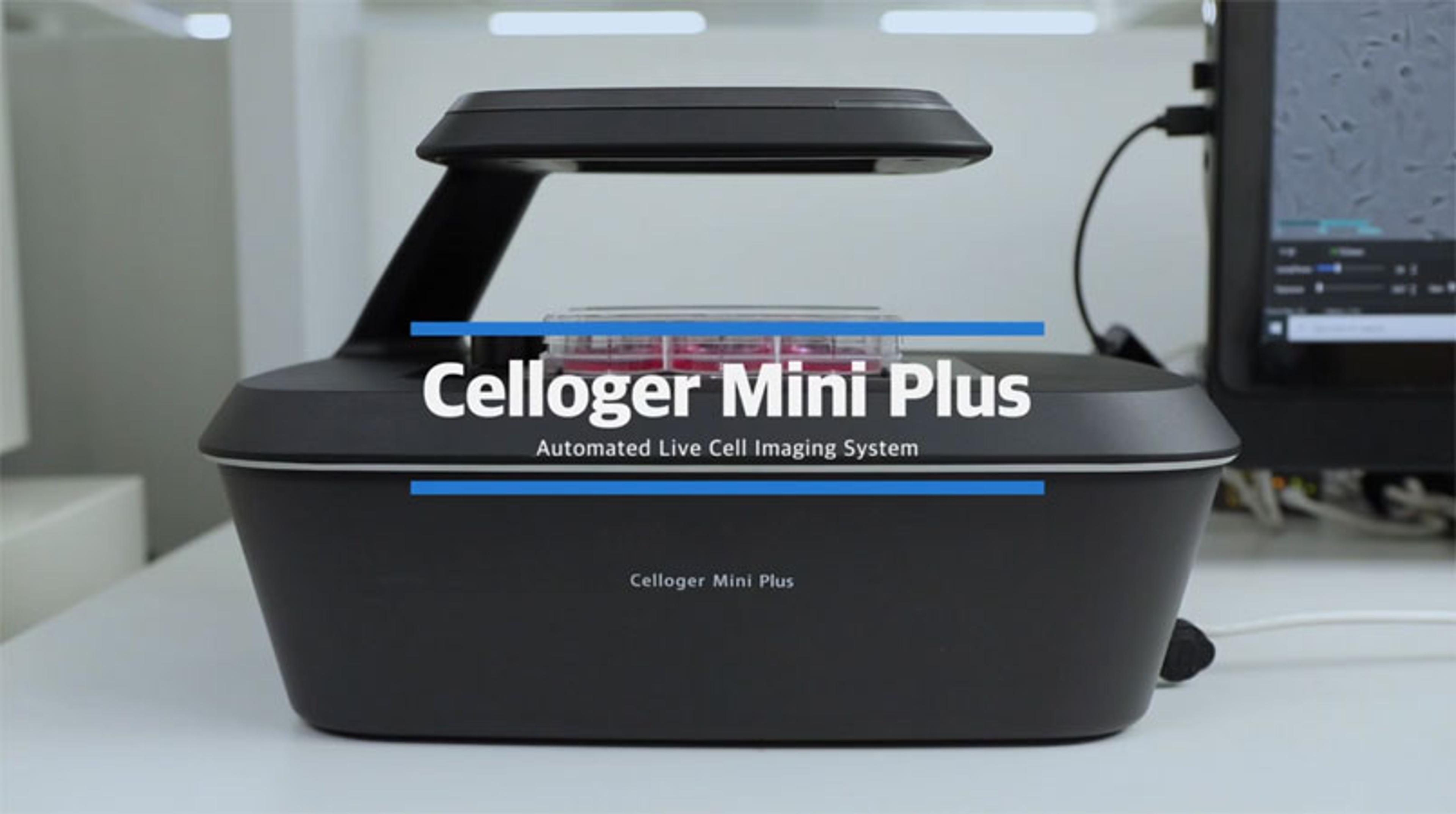
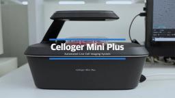
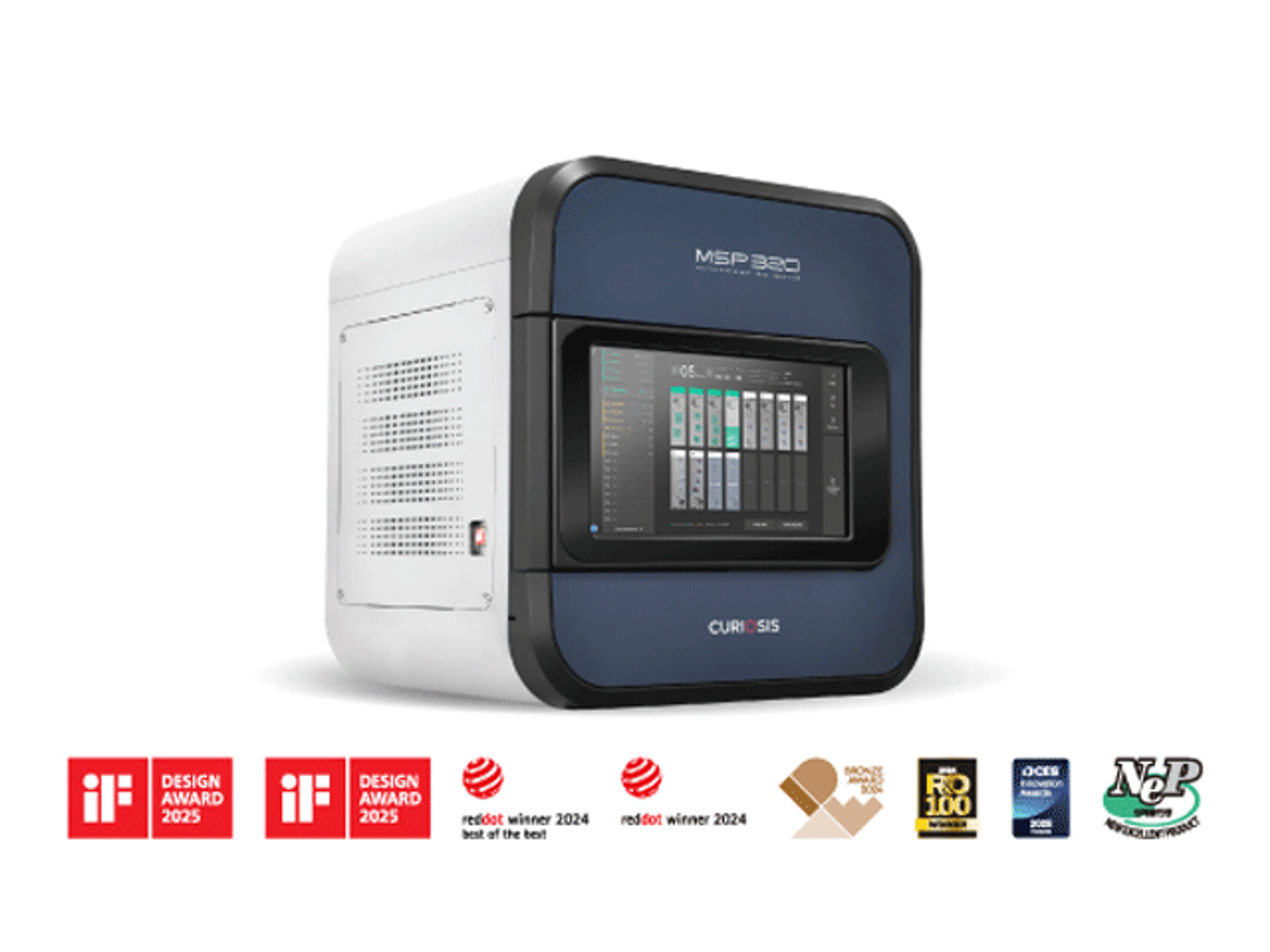
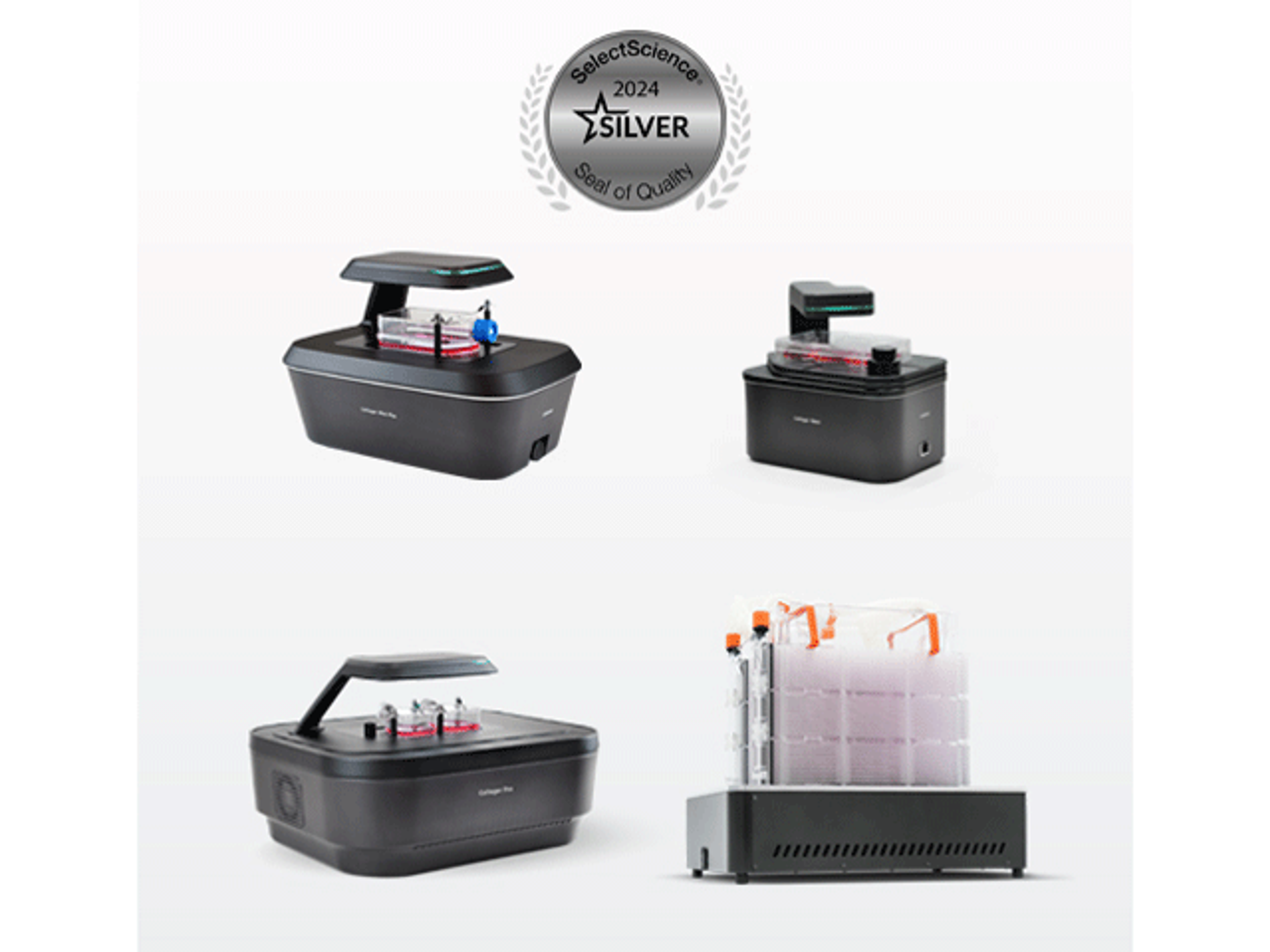
Celloger® Mini Plus, Automated Live Cell Imaging System from Curiosis
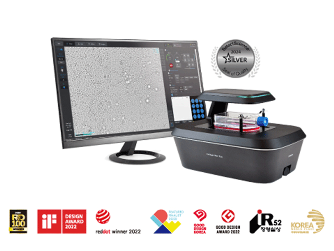
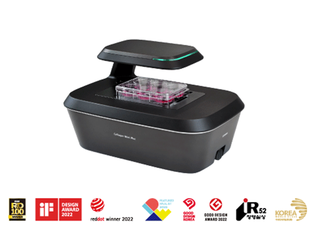
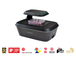
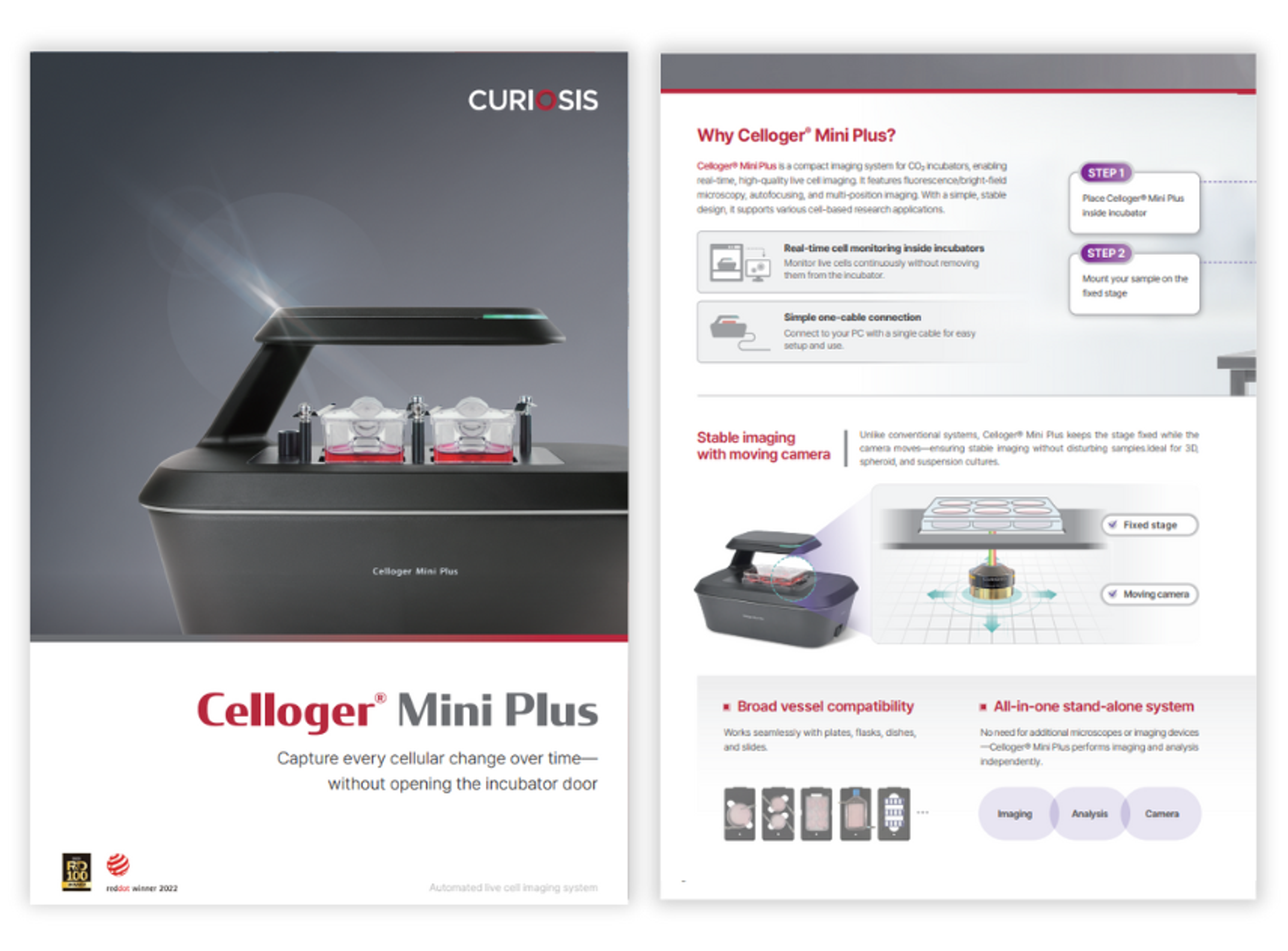
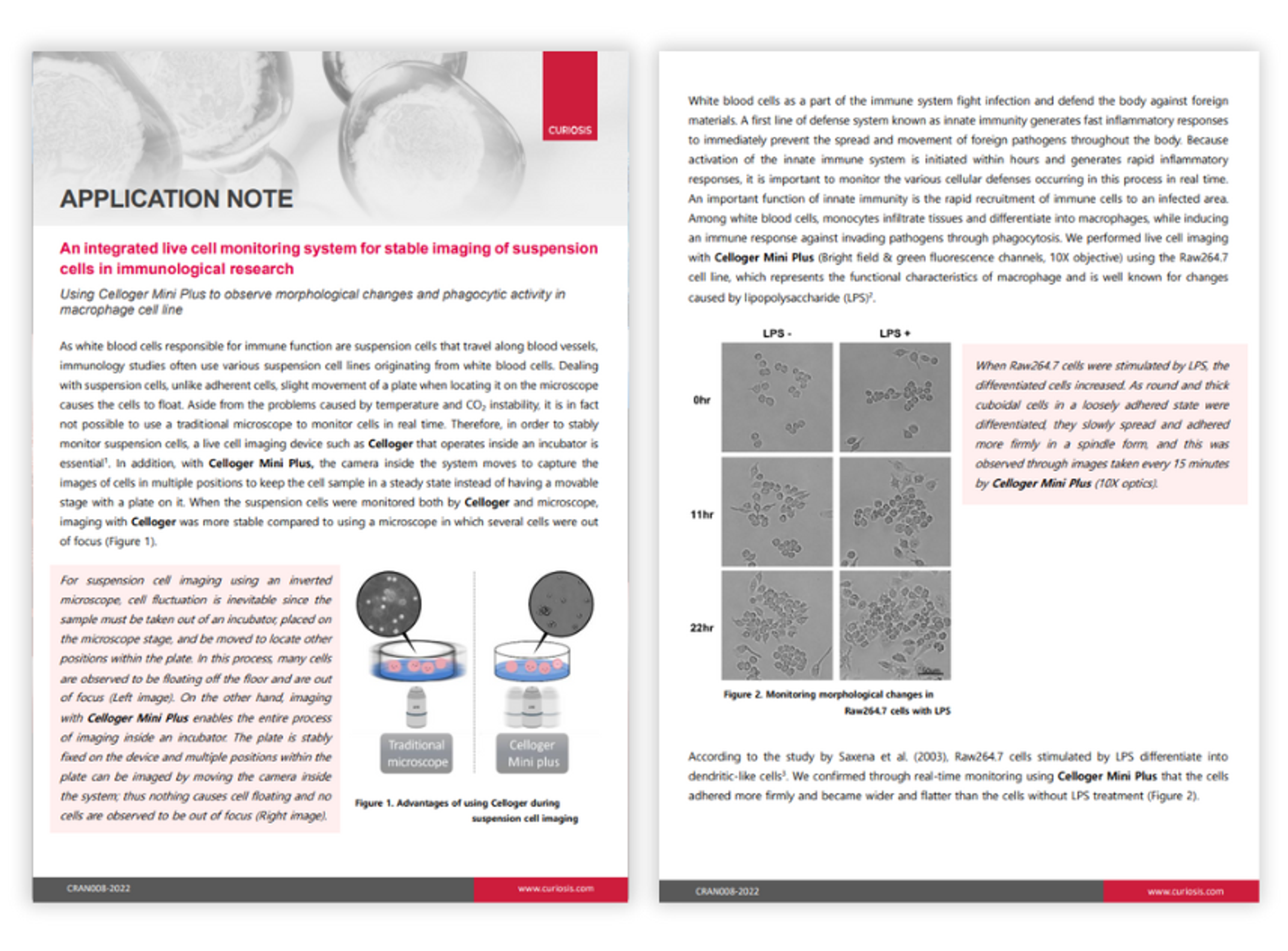
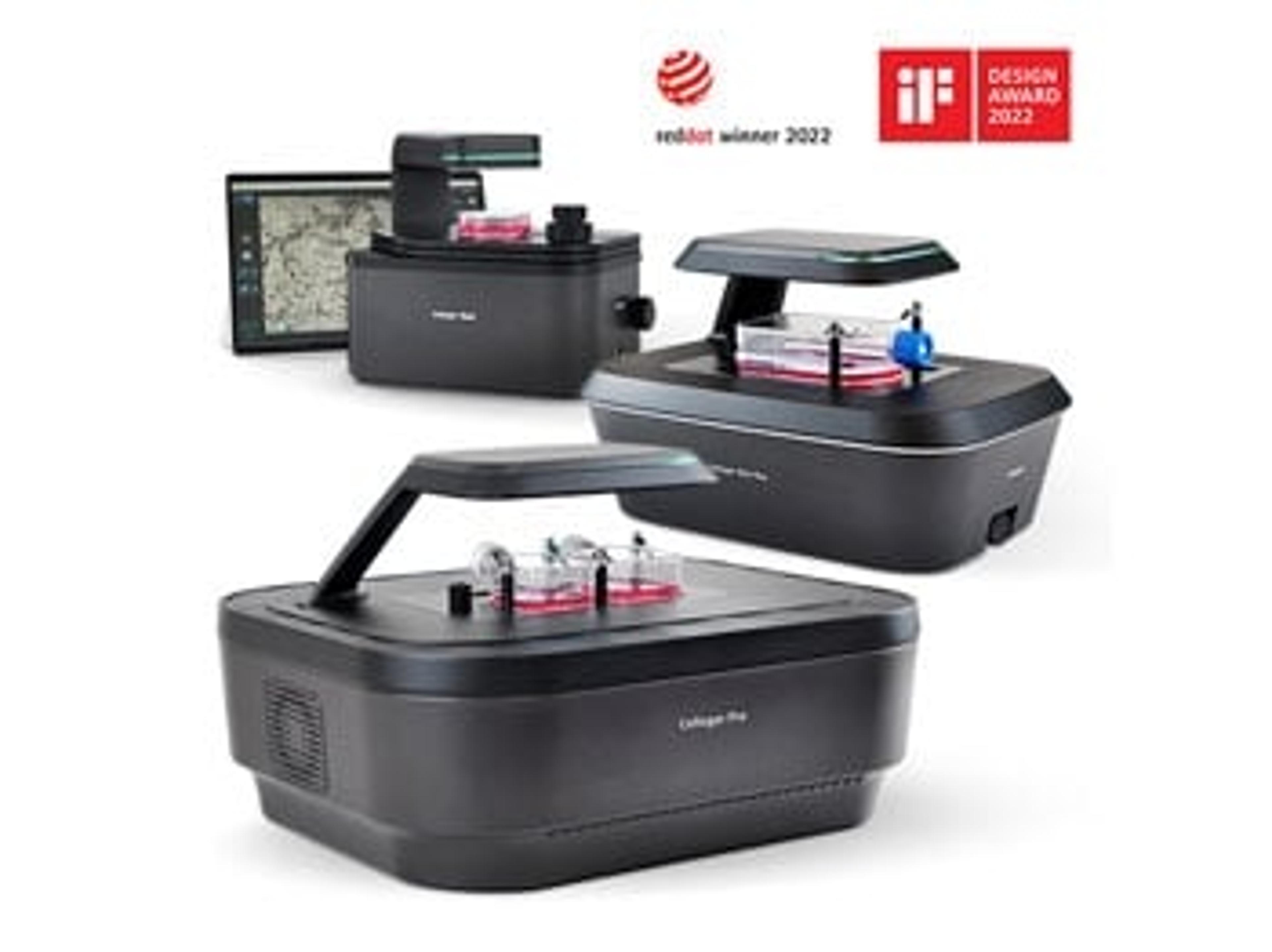
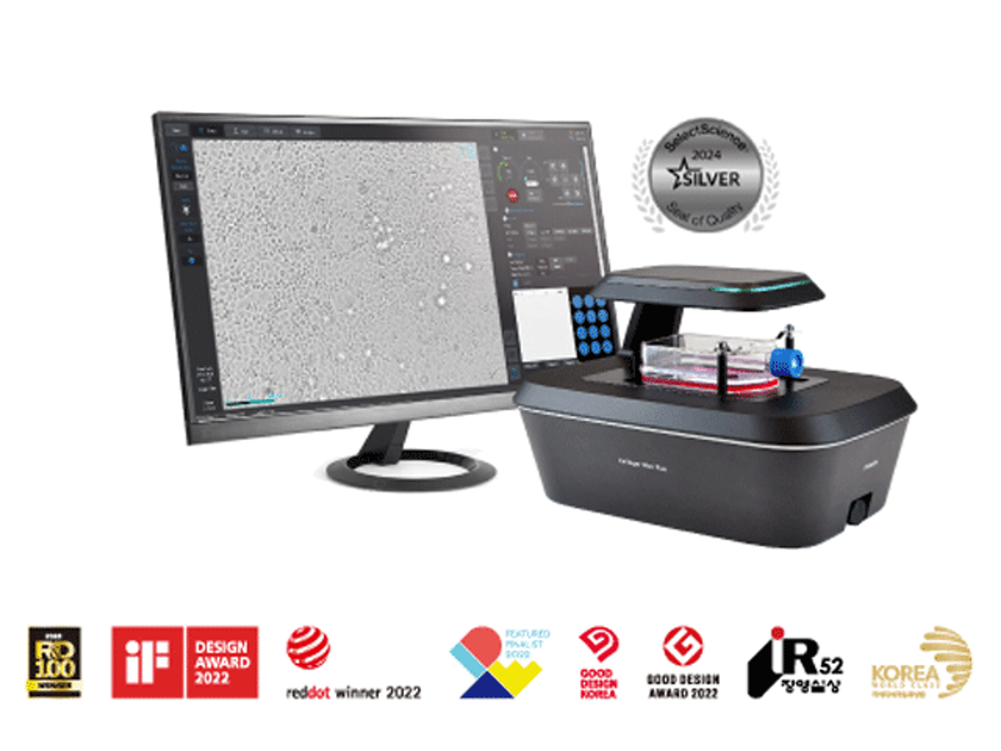
Celloger® Mini Plus is an automated live cell imaging system with fluorescence and brightfield microscopy. Celloger® Mini Plus makes it faster and easier to accumulate outstanding research results tailored to your research protocol.
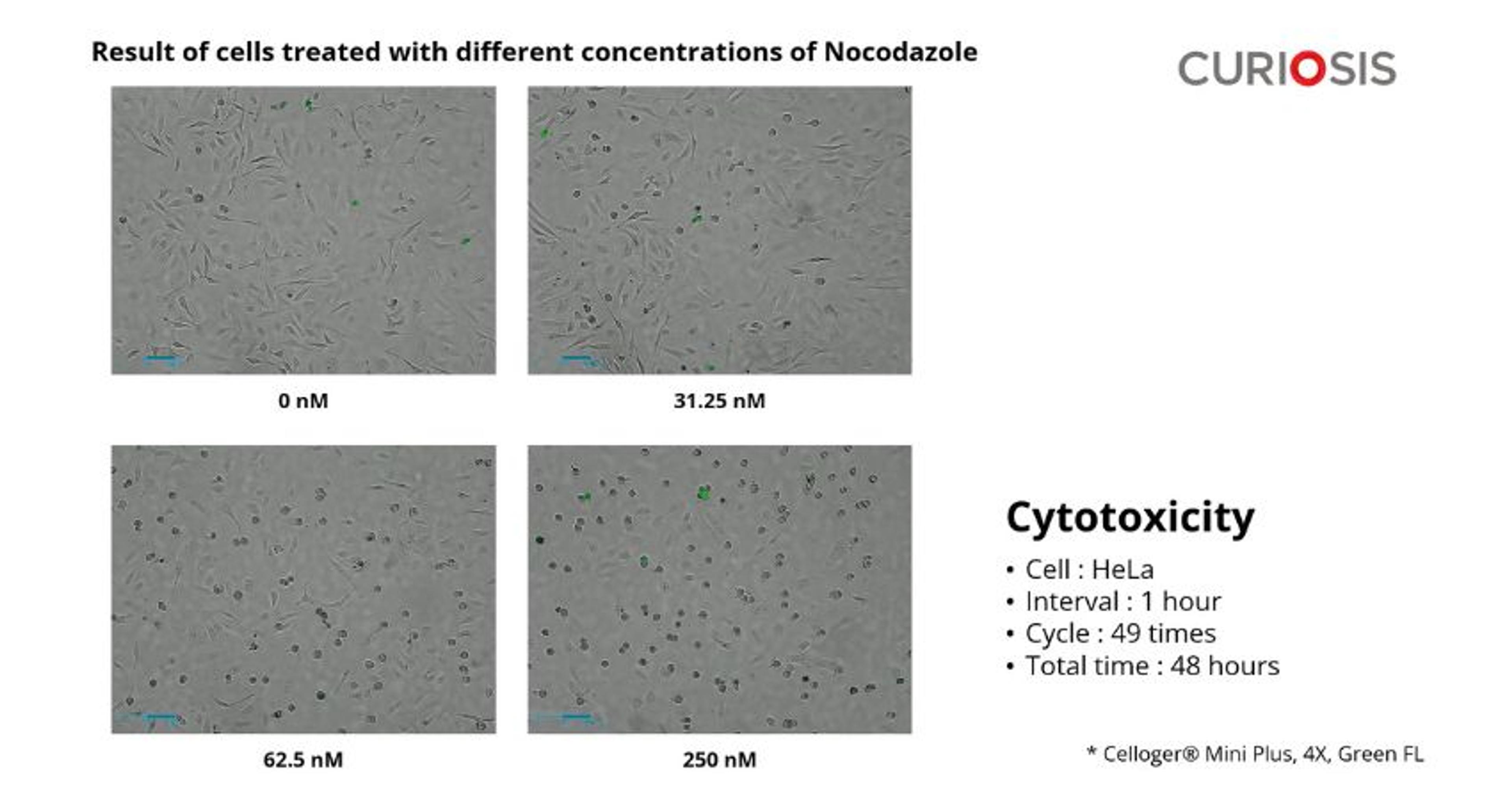
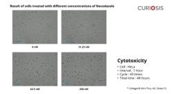
Using Celloger® Mini Plus for apoptosis experiments made it very easy to monitor cytotoxicity in real time.
Cell death research
Using Celloger® Mini Plus for apoptosis experiments made it very easy to monitor cytotoxicity in real time. It’s simple to use, so I could quickly see how the cells were responding without going through any complicated procedures.
Review Date: 25 Nov 2025 | CURIOSIS
Celloger® Mini Plus allowed reliable, stable monitoring of suspension cell growth over time.
Suspension cell growth monitoring
Celloger® Mini Plus allowed reliable, stable monitoring of suspension cell growth over time.
Review Date: 24 Nov 2025 | CURIOSIS
Data acquisition for both wound-healing assays and cell proliferation analysis went very smoothly, with no issues.
Proteostasis network
Data acquisition for both wound-healing assays and cell proliferation analysis went very smoothly, with no issues.
Review Date: 24 Nov 2025 | CURIOSIS
Overall, the system was very user-friendly and made it easy to get my work done without any stress.
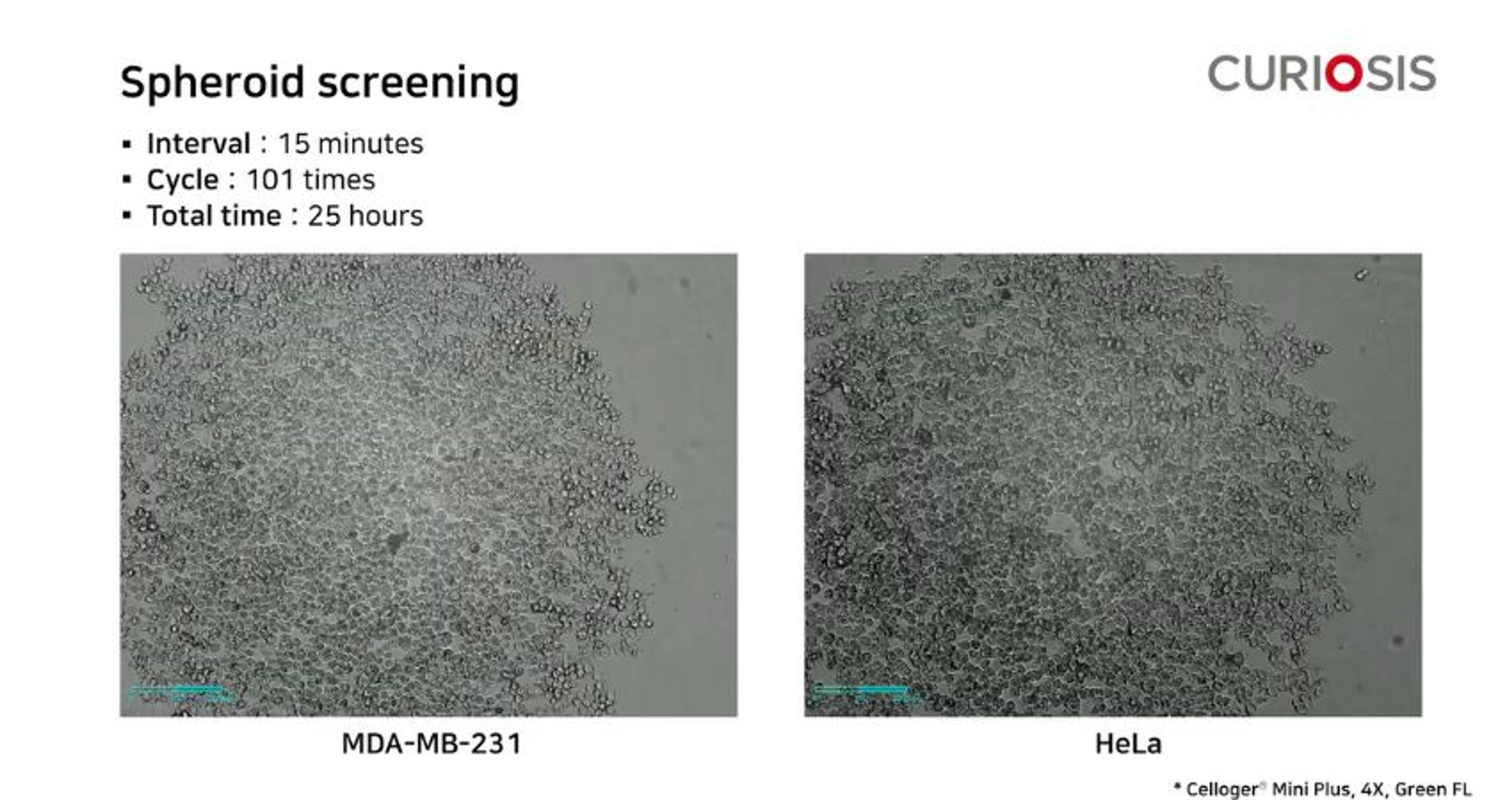
Spheroid monitoring
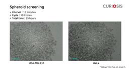
Overall, the system was very user-friendly and made it easy to get my work done without any stress. In particular, the coordinate copy/paste, centering, autofocus, and shooting-direction settings (row, column, one-way, or round-trip) were all very convenient, and the holder was easy to attach and detach while still feeling stable during use. I was also pleased with the clear 5-megapixel images. After image acquisition, the function that automatically combines individual images into a single view based on the multi-well layout was extremely useful. Being able to capture images without opening the incubator was a big advantage, as it minimized stress on both the spheroids and the user, and CO₂ consumption remained low. I didn’t notice any vibrations during operation—the system was quiet, and the spheroids didn’t move at all. Thanks to the short imaging time, I could also retake images quickly whenever needed, which made the overall workflow very efficient.
Review Date: 24 Nov 2025 | CURIOSIS
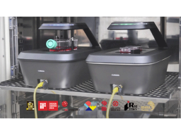
The instrument was compact and easy to install, so it fit well even in the tight space inside the incubator.
Cancer biology
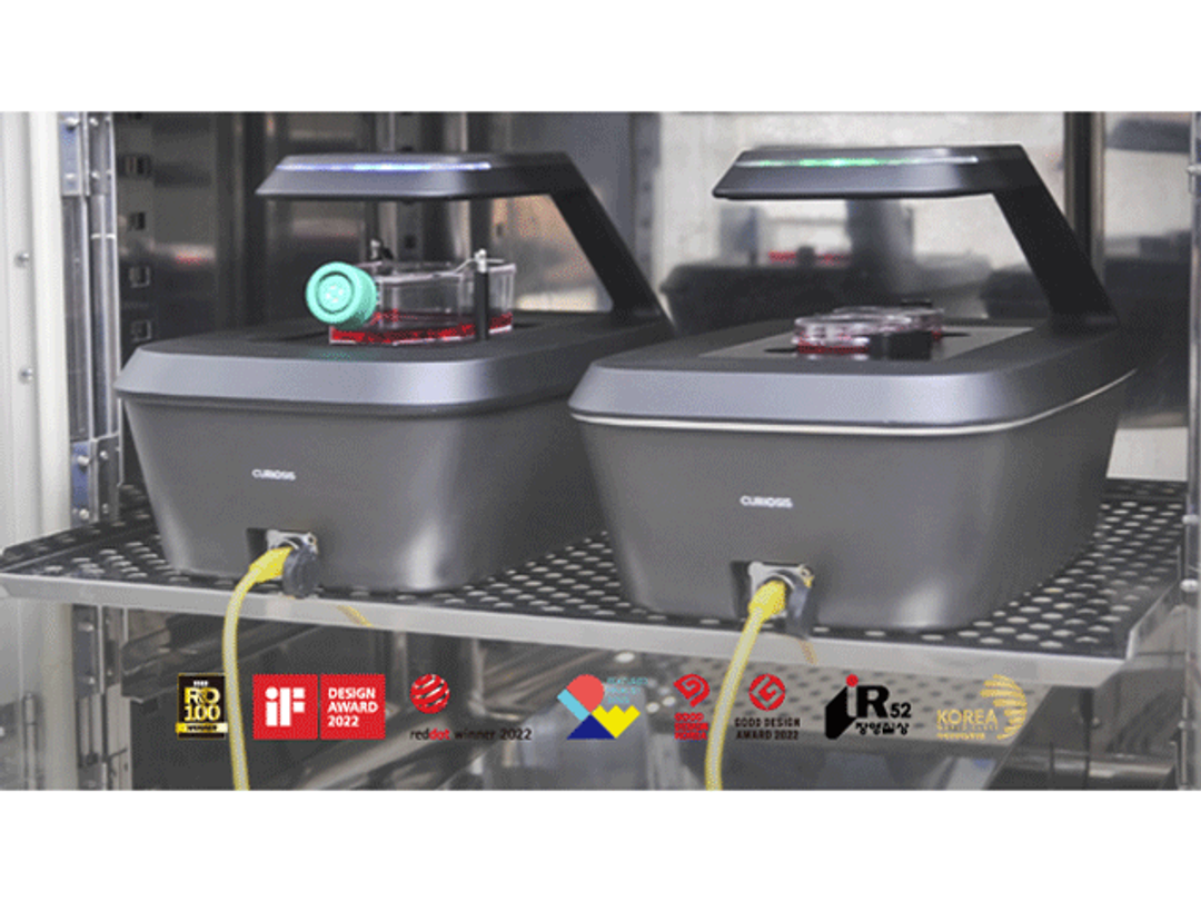
The instrument was compact and easy to install, so it fit well even in the tight space inside the incubator. It operated stably and was simple to maintain. It was also useful that the analysis software could generate results directly, without needing any separate analysis tools.
Review Date: 24 Nov 2025 | CURIOSIS
The ability to set multiple imaging positions within a single well was very helpful for increasing replicate numbers.
Cancer biology
The ability to set multiple imaging positions within a single well was very helpful for increasing replicate numbers. It was also a big advantage to monitor live cells in real time directly inside the incubator without disturbing the culture. By defining several points per well and capturing them in one acquisition, it was easy to collect more replicates efficiently.
Review Date: 24 Nov 2025 | CURIOSIS
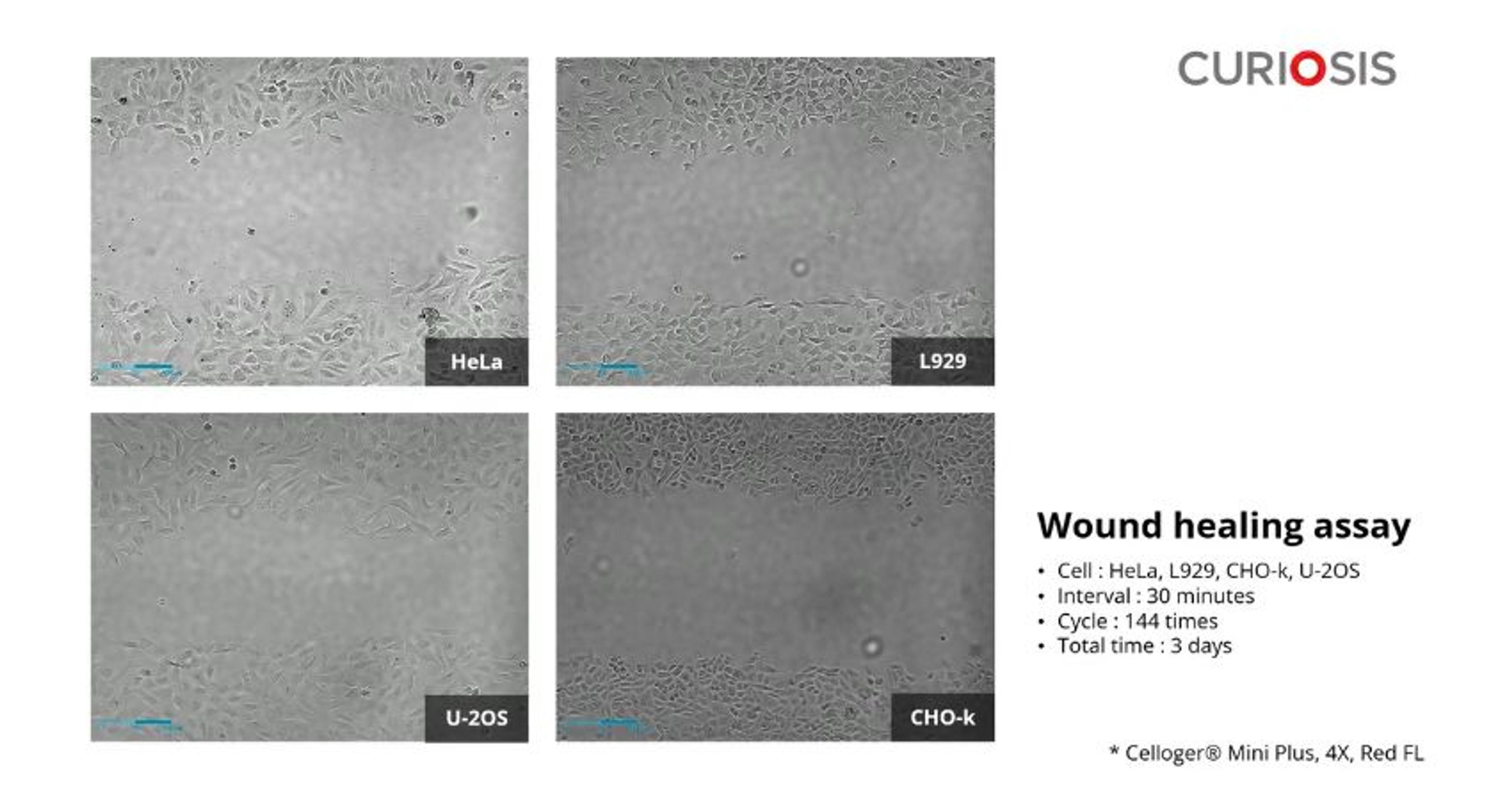
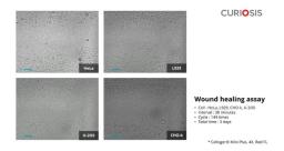
The analysis software offered convenient built-in tools for area measurement and automated wound-healing analysis.
Cancer biology
The analysis software offered convenient built-in tools for area measurement and automated wound-healing analysis. Because the captured images could be analyzed directly in the software, the overall workflow was much more efficient, and the flexible time-lapse scheduling made it easy to tailor experiments to different cell types and conditions.
Review Date: 24 Nov 2025 | CURIOSIS
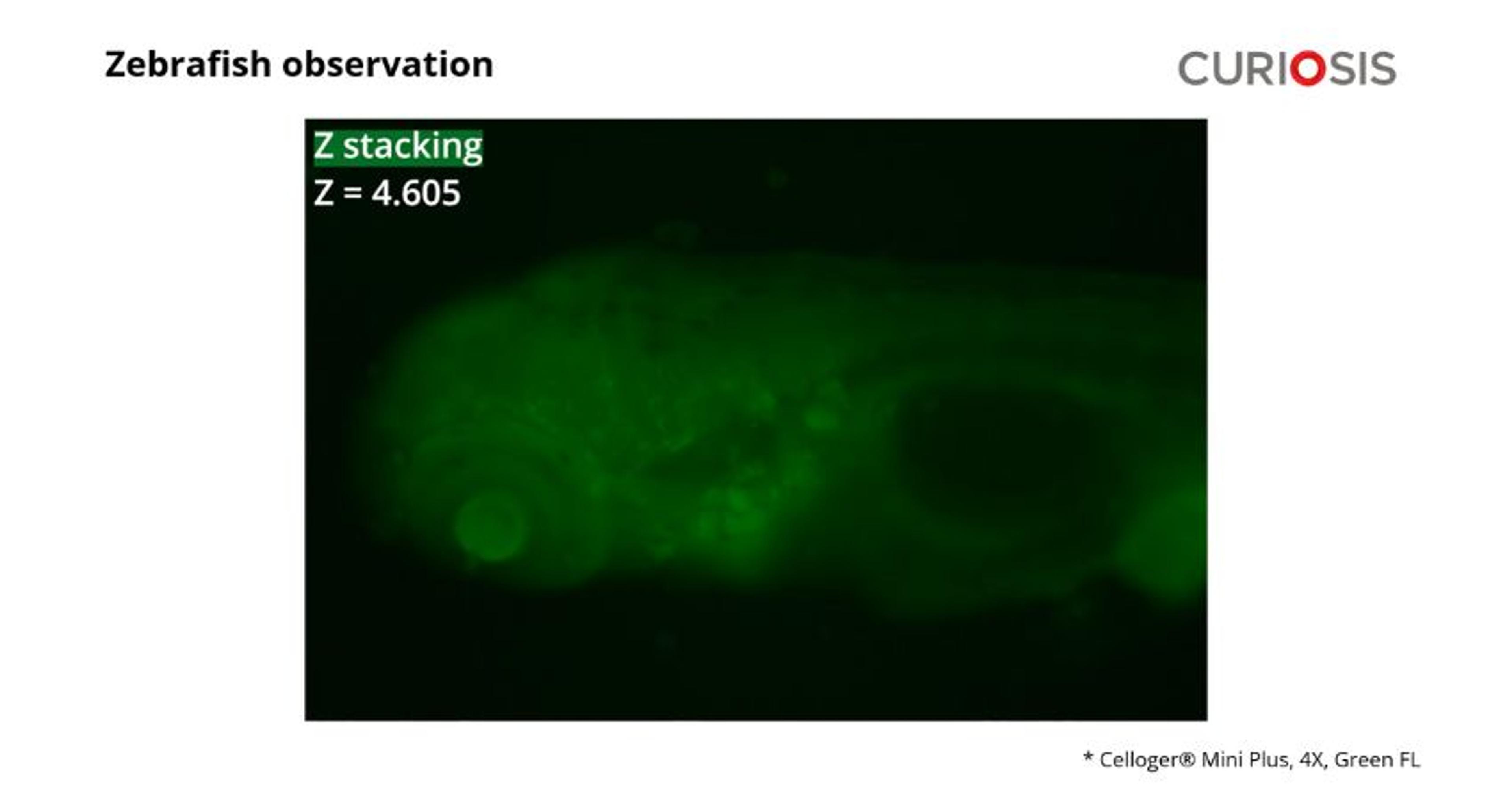
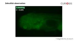
Z-stacking function was a strong feature, allowing the acquisition of images at multiple focal planes
Biochemistry
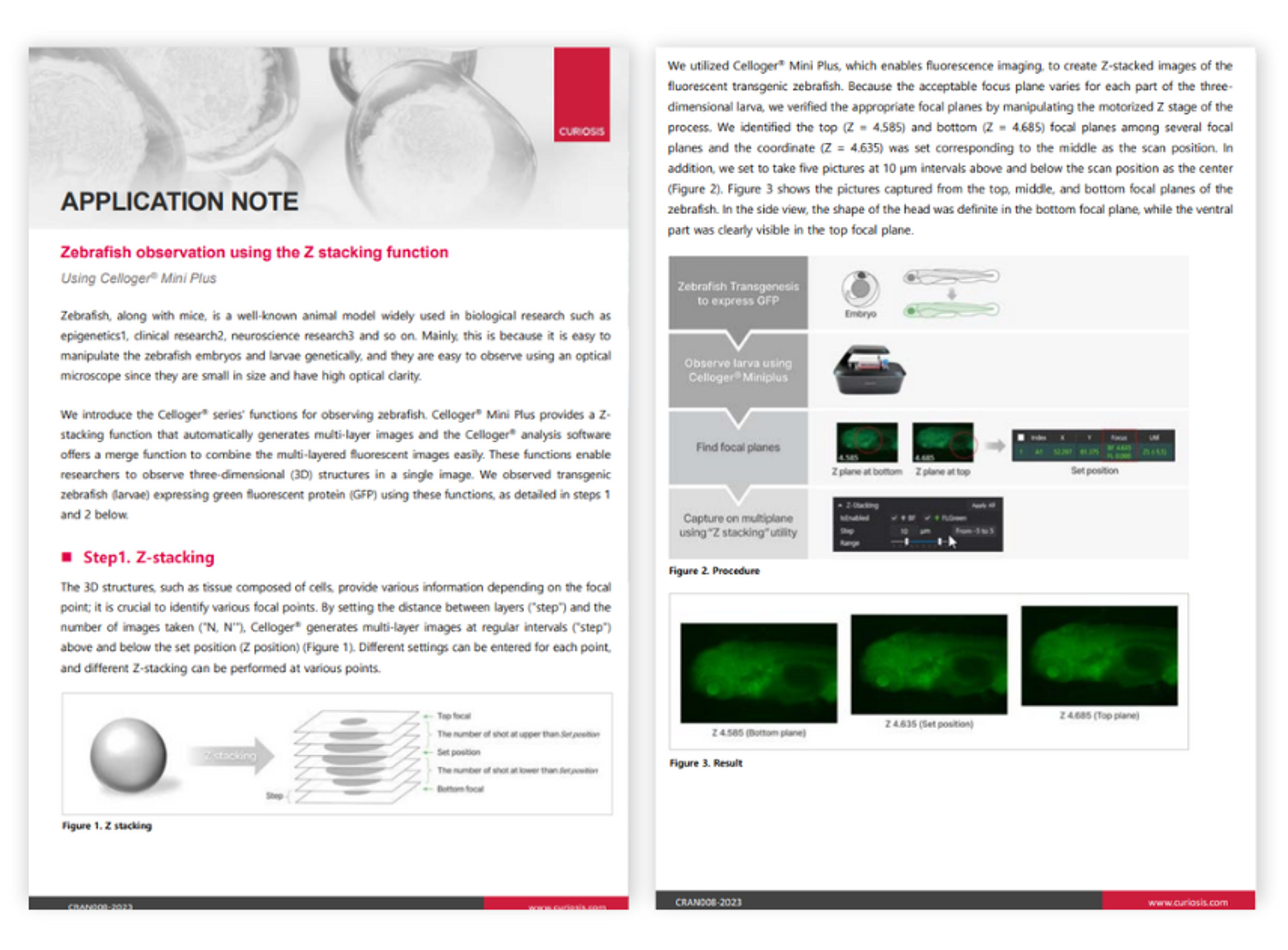
For COS7 cell research, the Z-stacking function was a strong feature, allowing the acquisition of images at multiple focal planes and combining them to generate enhanced 2D images with improved depth information. The image stitching function was also valuable, facilitating the analysis of large cell populations by merging multiple images into a single comprehensive view.
Review Date: 16 Apr 2025 | CURIOSIS
Compatibility with various culture vessels was one of the most appreciated features
Biochemistry
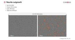
In conjunctiva cell research, compatibility with various culture vessels was one of the most appreciated features. The system supported flasks, dishes, and well plates, allowing for flexible experimental setups. The user-friendly software made it easy to track cell confluency, monitor growth curves, and perform data analysis. The time-lapse video function was especially useful for visualizing dynamic cellular changes over time, making it an excellent tool for cell behavior analysis.
Review Date: 16 Apr 2025 | CURIOSIS
One of the most satisfying aspects of using Celloger® Mini Plus was its real-time monitoring capability
Biochemistry
One of the most satisfying aspects of using Celloger® Mini Plus for pterygium cell research was its real-time monitoring capability. It allowed continuous observation of cell growth and changes without disturbing the culture environment, ensuring stable data acquisition. Additionally, the multi-position imaging function enabled the simultaneous analysis of multiple samples, significantly improving experimental efficiency. The autofocus function was also highly reliable, providing consistently sharp images without losing focus.
Review Date: 16 Apr 2025 | CURIOSIS
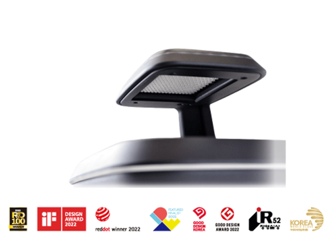
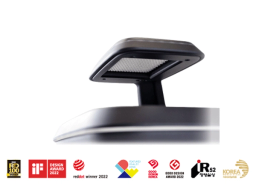
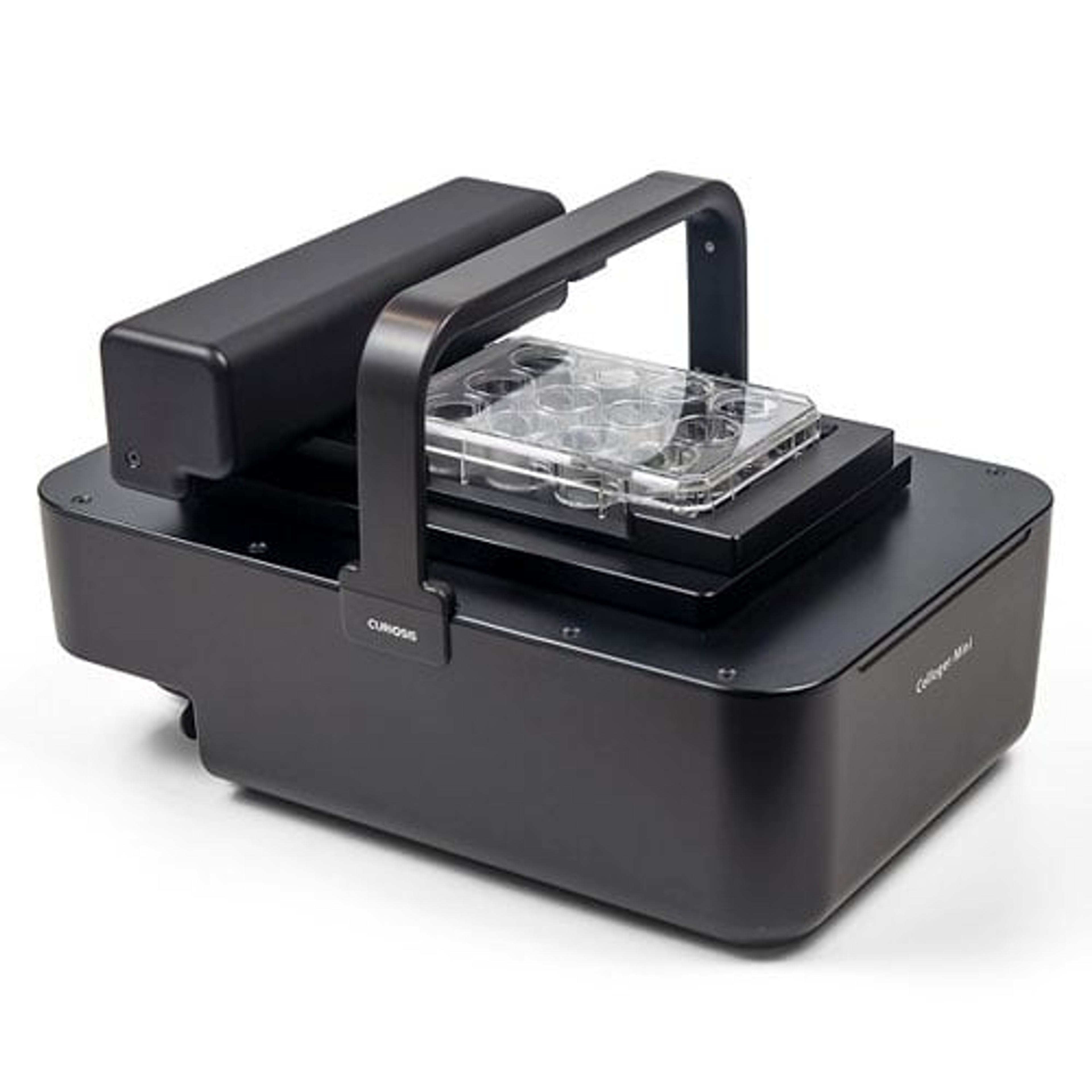
Celloger® Mini Plus is a live cell imaging system based on bright field and fluorescence(green/red) microscopy. The motorized stages move on X, Y, and Z axes that minimize vessel movement, allowing stable cell observation with no shaking. Researchers may obtain stable research results in line with individual research protocols through the use of functions that are optimized for cell research, such as automatic focusing, real-time multi-position imaging, etc.
Key features
- Compact size that easily fits into standard CO₂ incubators
- Fully motorized multi-position imaging up to 96 well plate
- Compatible with various cell and tissue culture vessel types
- Multiple focal planes can be captured through Z-stack imaging
- Intuitive UI/UX and easy to acquire confluency data
- Increased focus speed and reproducibility with reliable autofocusing function
- Image stitching possible to enable analysis of larger volume and sections
Applications
- Wound healing assay
- Cell migration
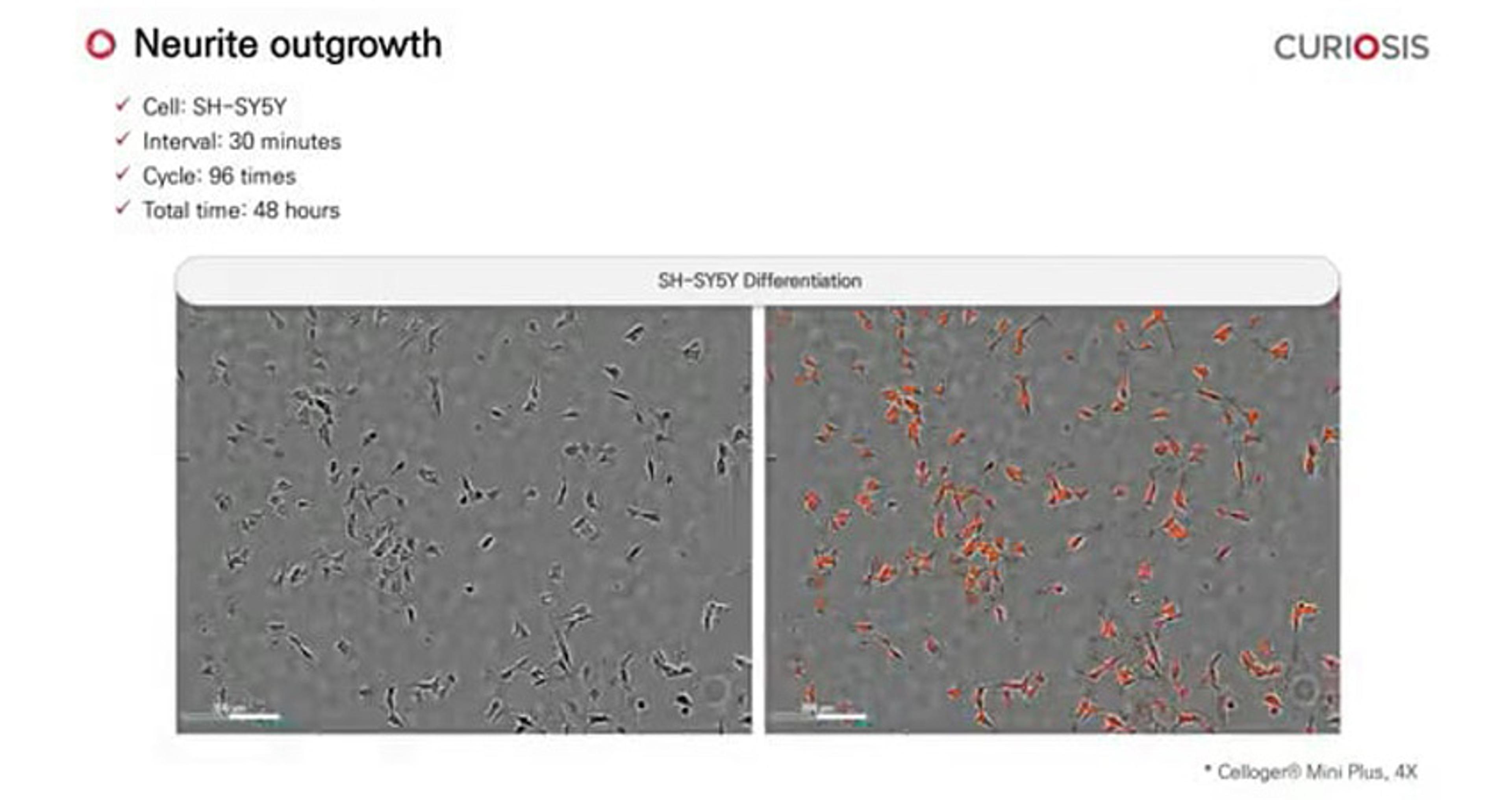
- Cell morphology
- Cell confluency
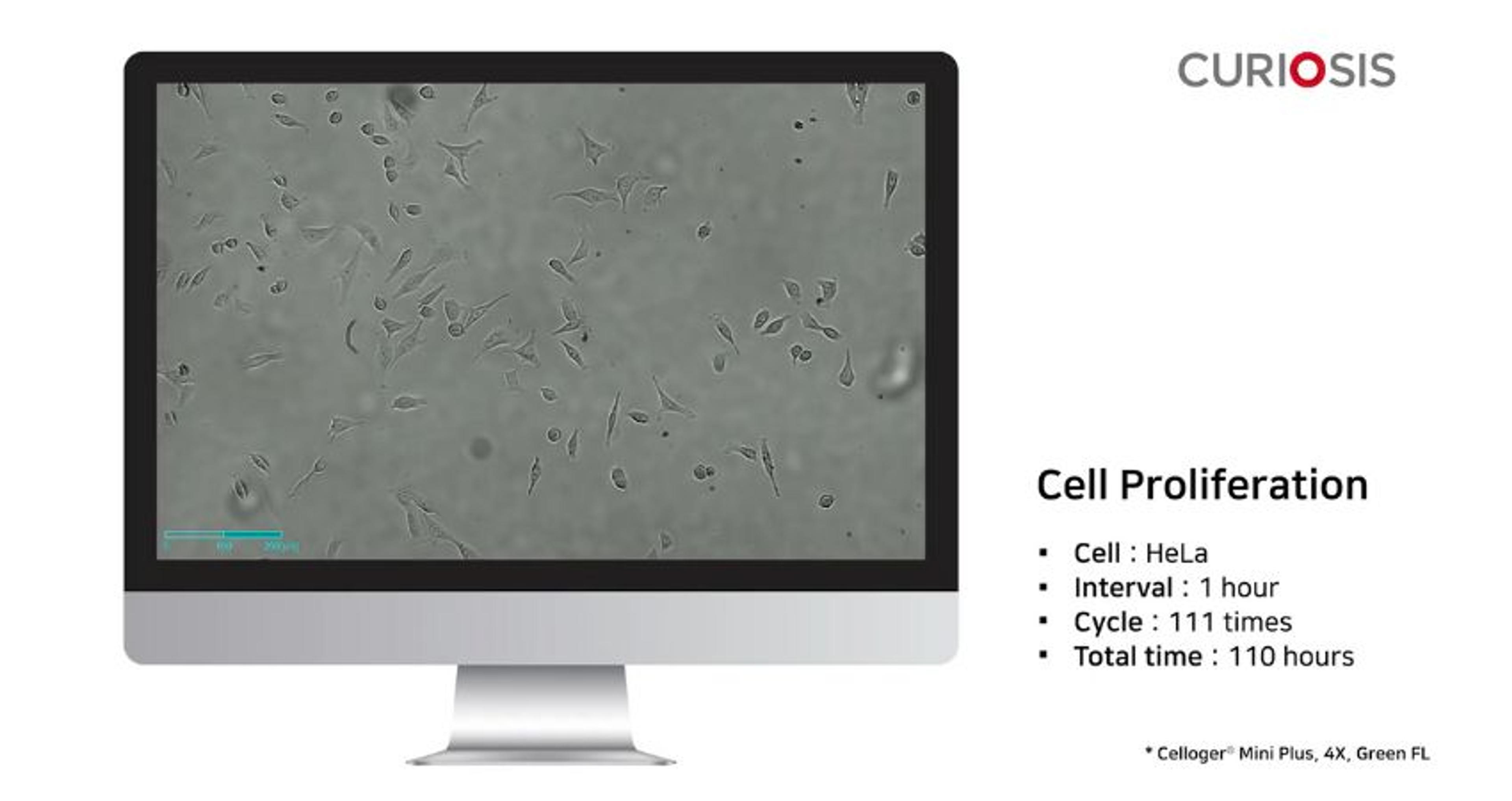
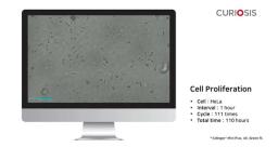
- Cell proliferation
- Cytotoxicity assay
- Transfection efficiency assessment
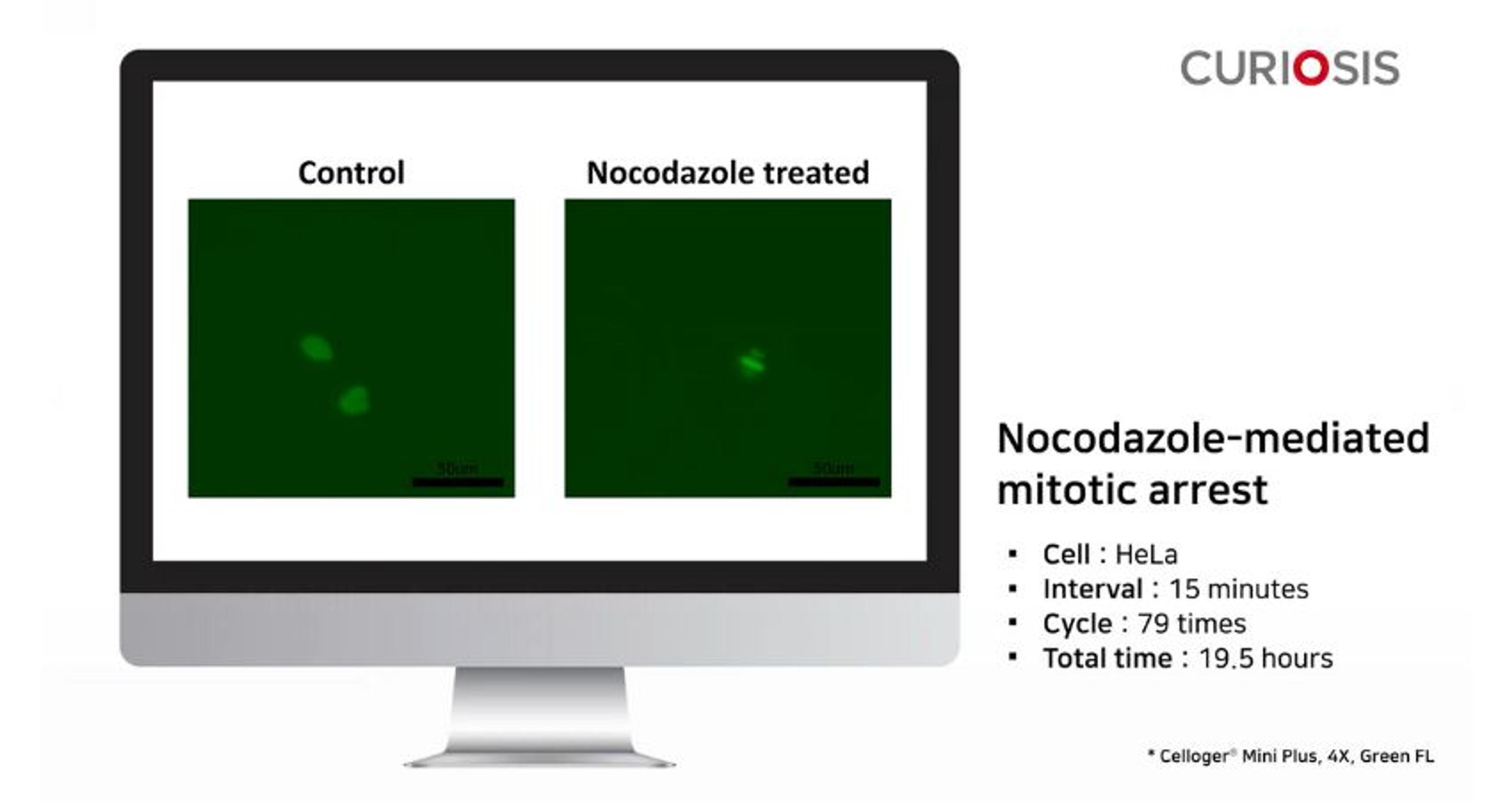
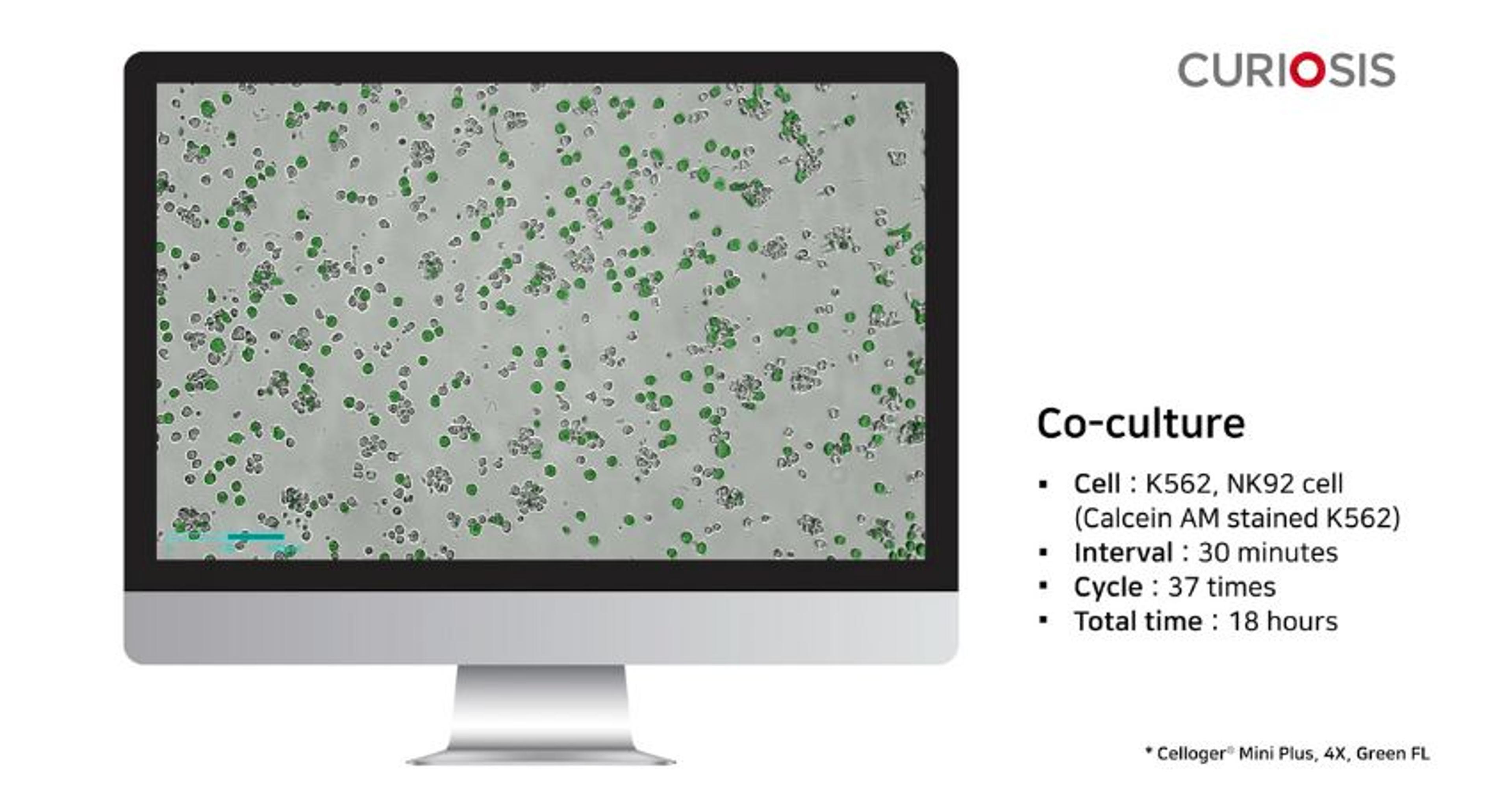
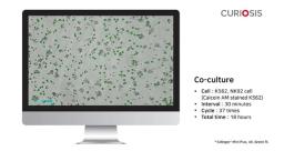
- Coculture monitoring
- Multipoint cell monitoring
Specification
- Dimension: 226 x 358 x 215mm
- Weight: 5.6kg / 12.3lb
- Objective: 4X / 10X
- Imaging modes: Brightfield, Fluorescence (Green/Red)
- Fluorescence: Green - Excitation (470/40x), Emission (510lp) / Red - Excitation (525/30x), Emission (570lp)
- Light source: LED
- Stage: Motorized XYZ

