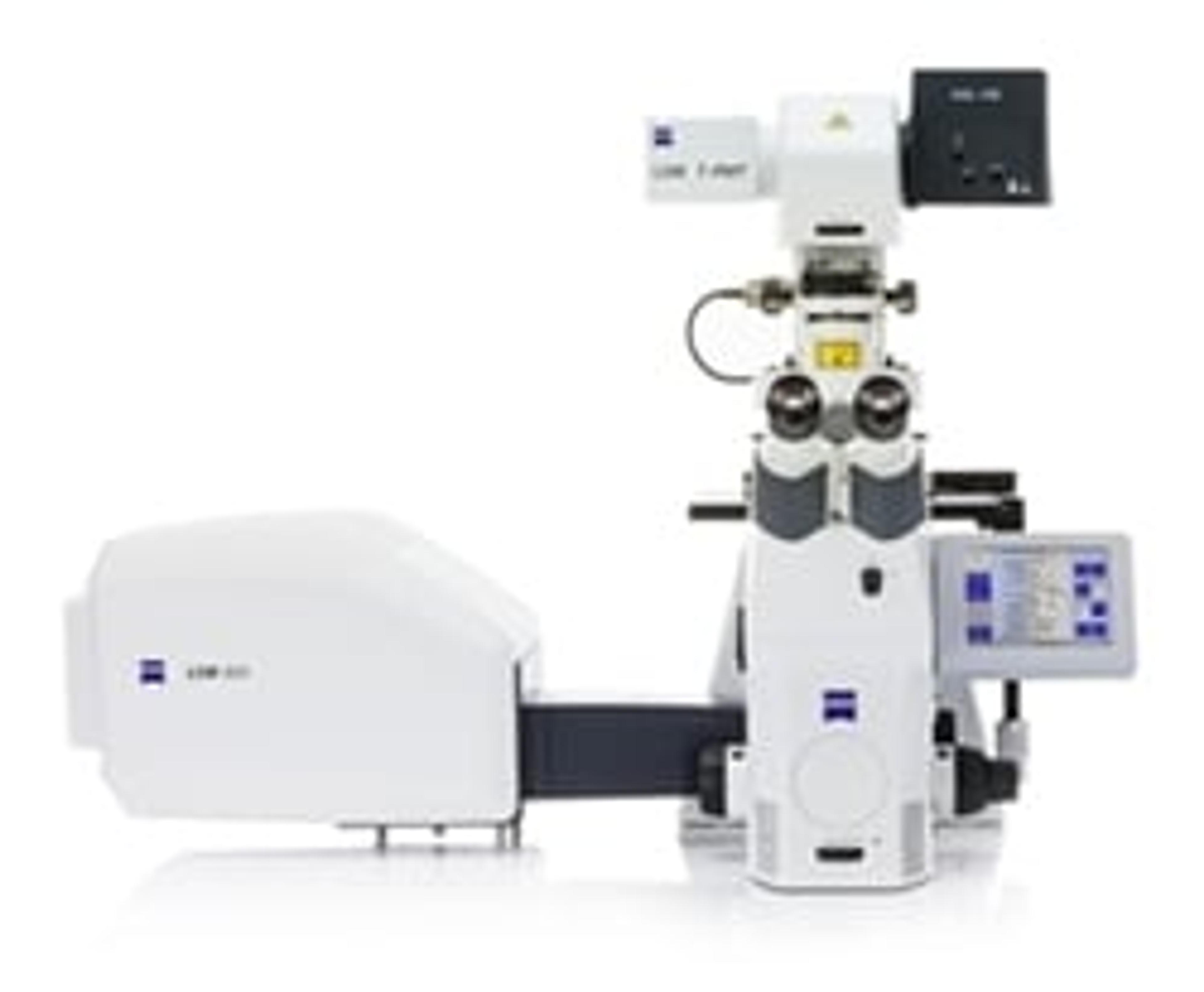
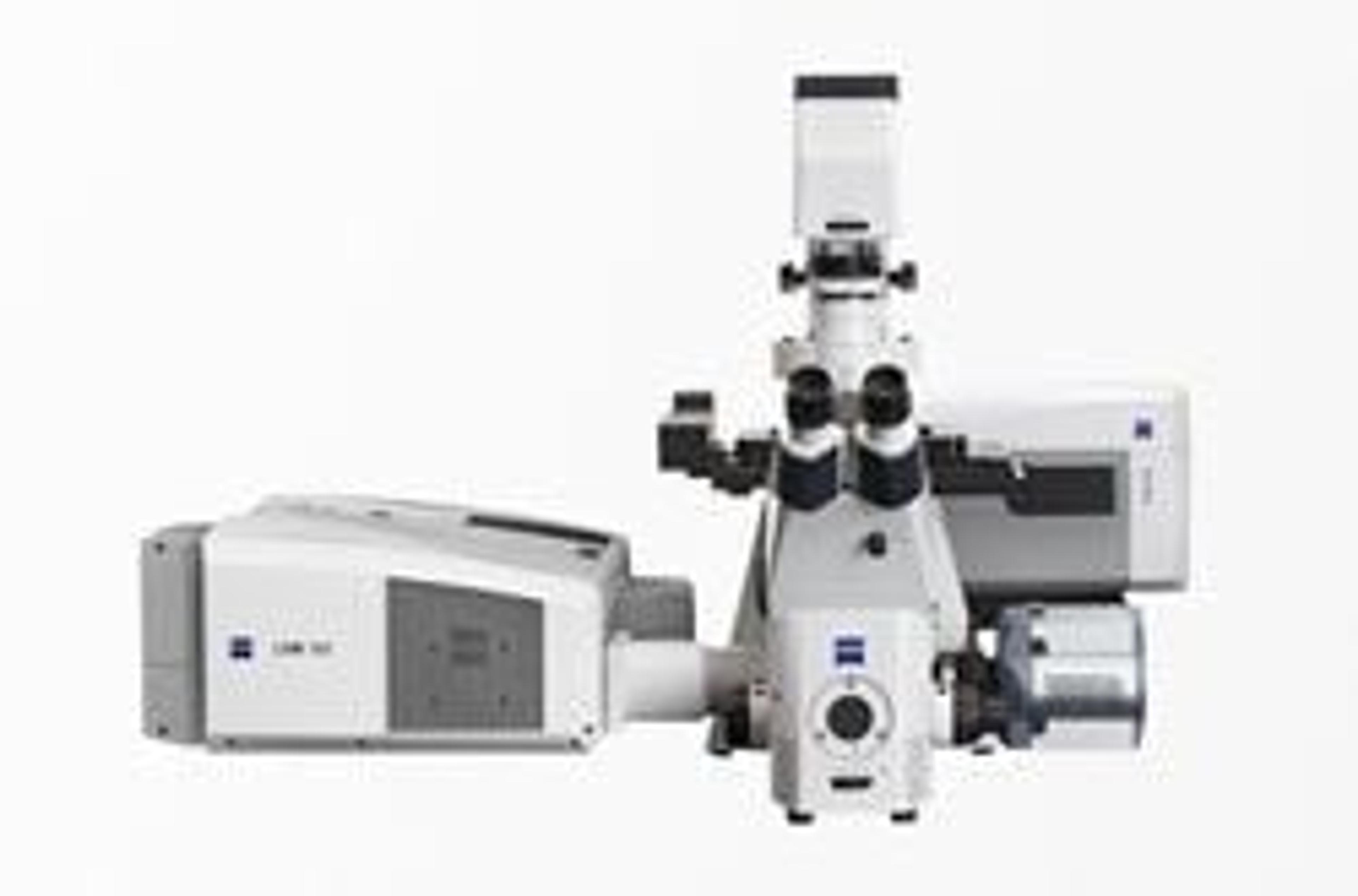
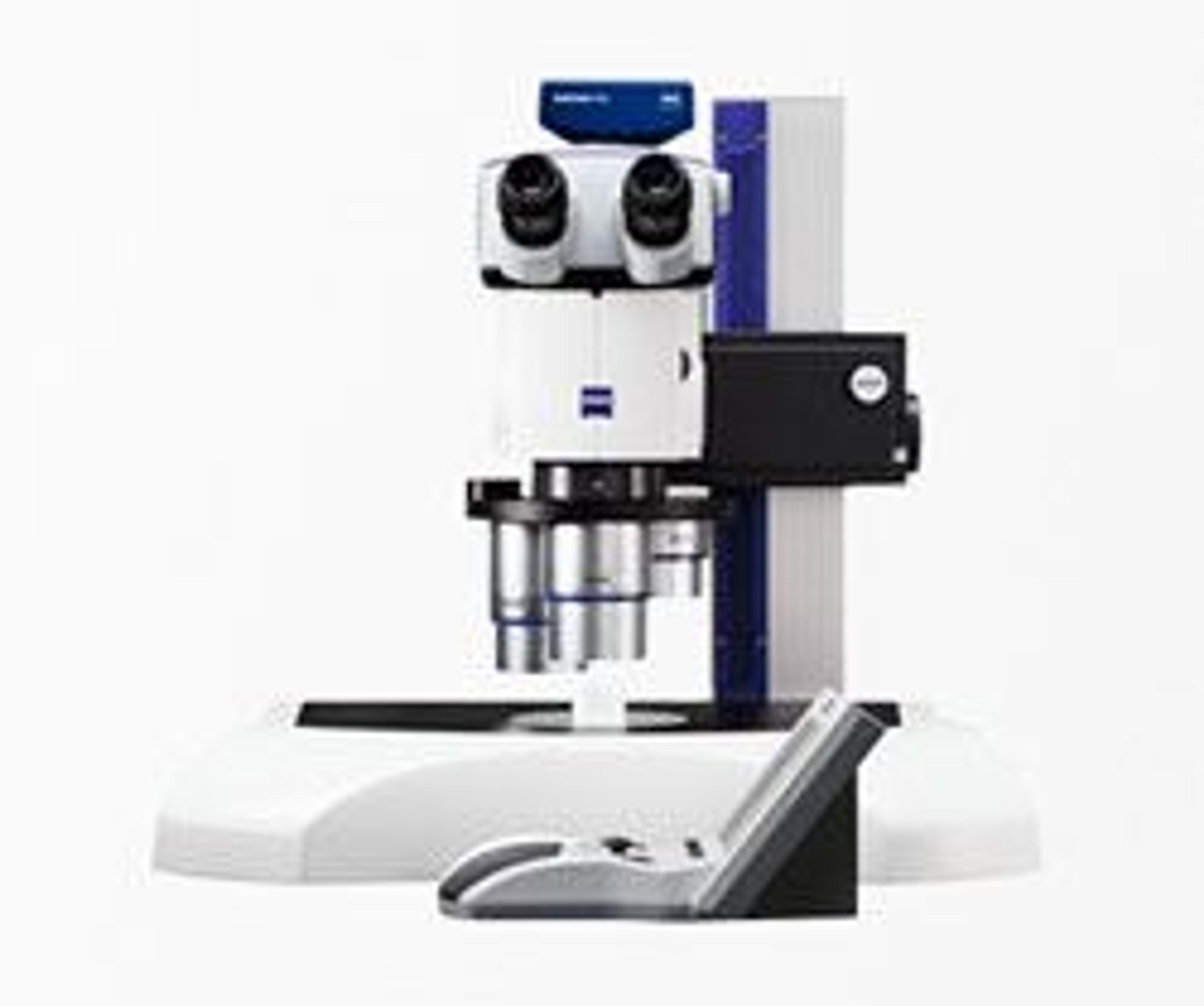
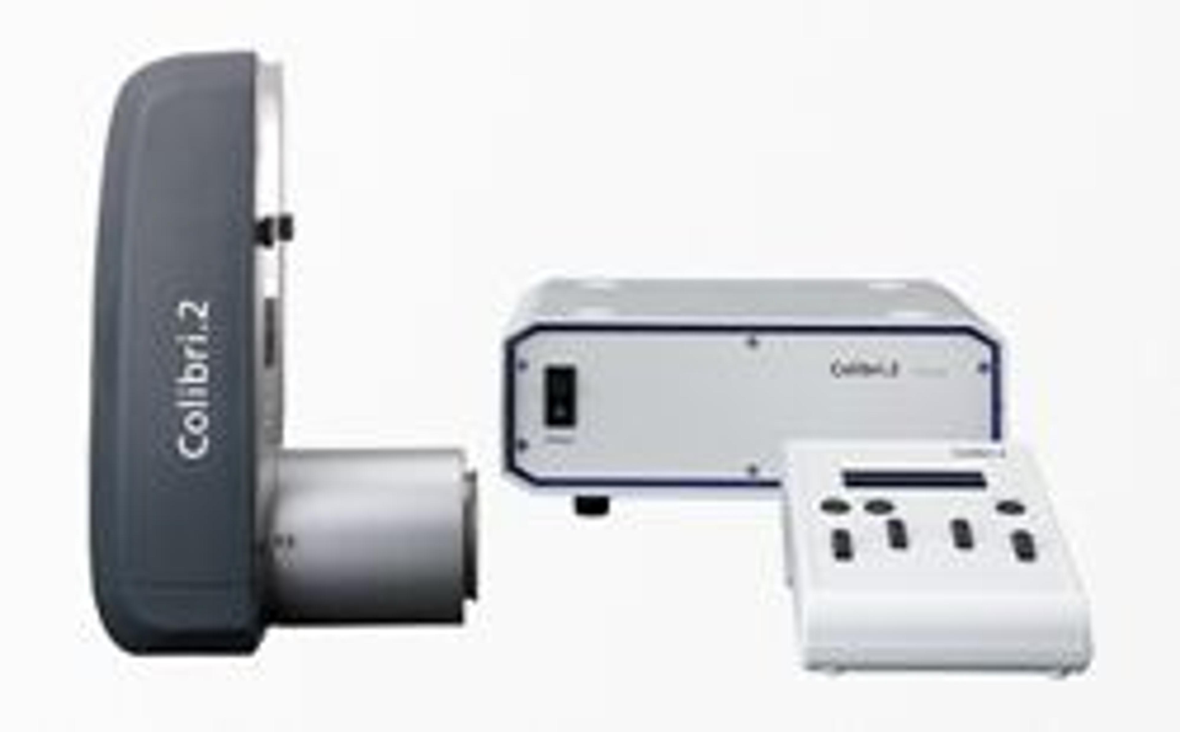
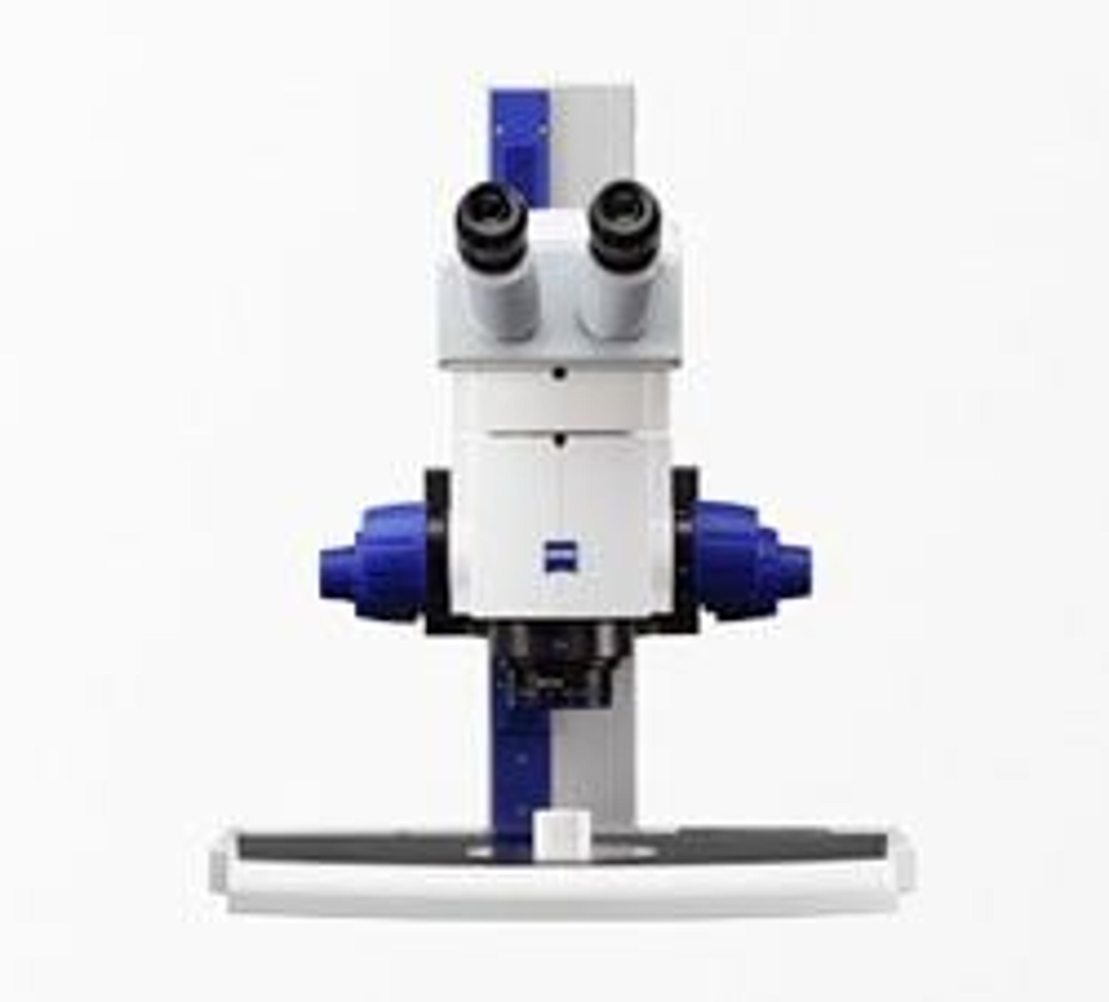
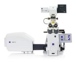
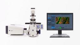
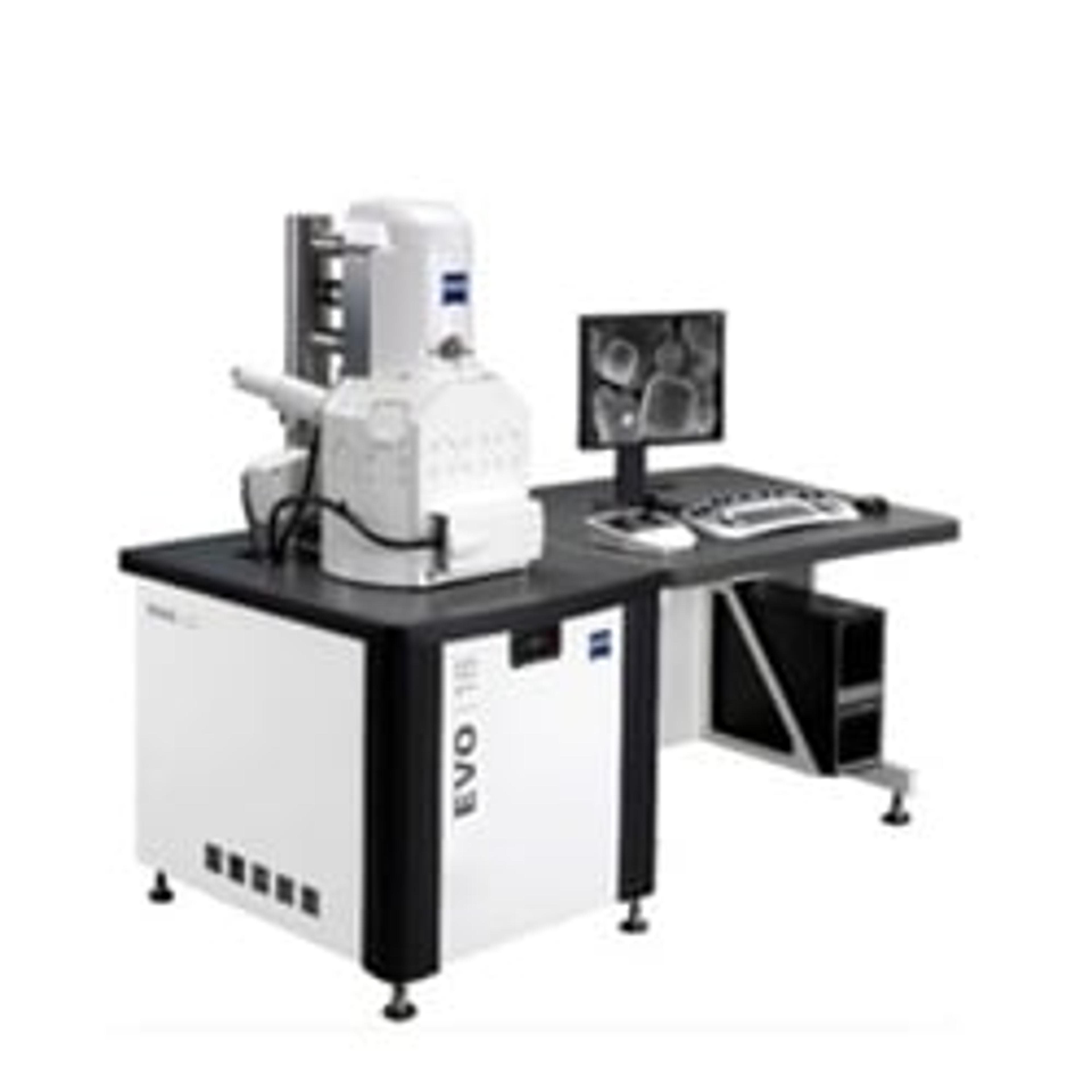
ZEISS LSM 800 with Airyscan
Your Compact Confocal Power Pack.


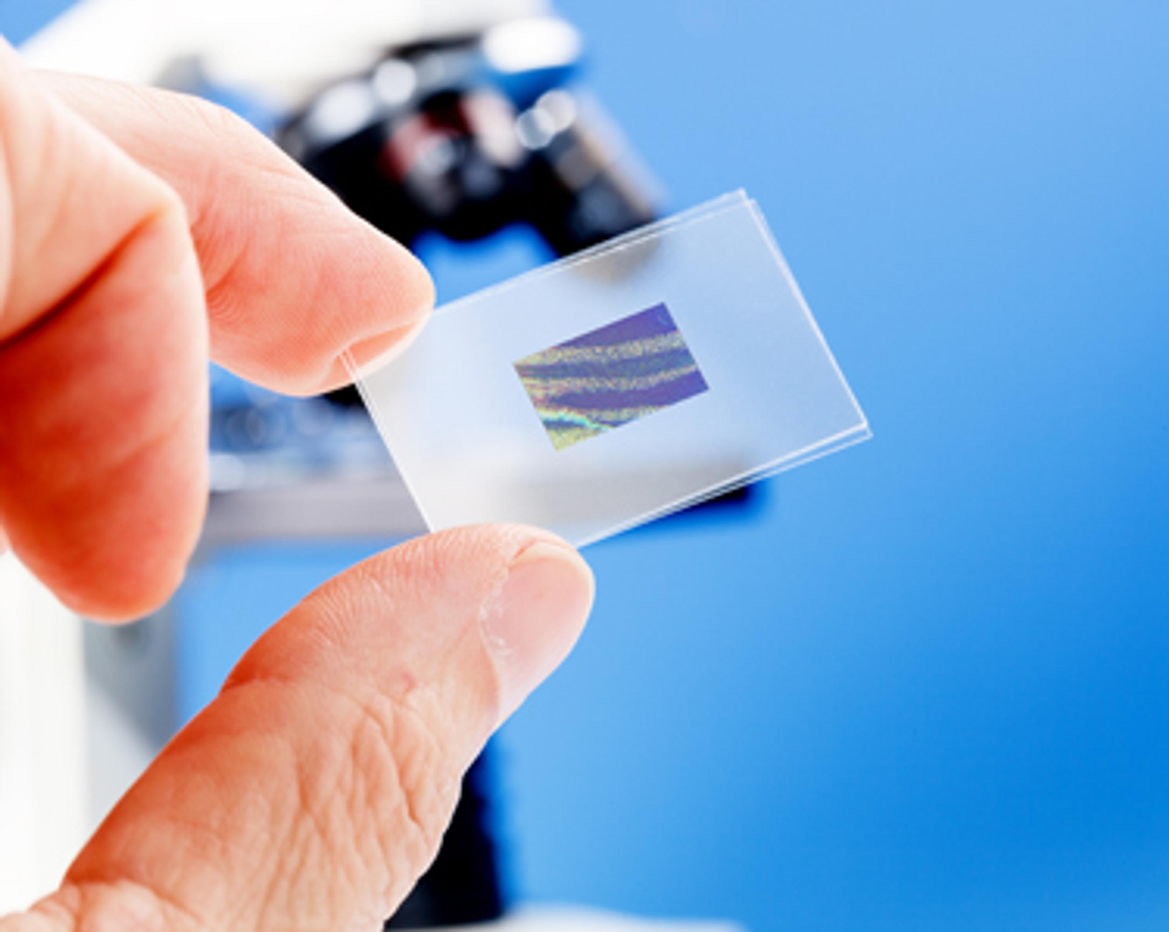
Receive your quote directly from the manufacturer.
Comprehensive image - stimulate imagination.
3D Imaging for life & material science
Great life science images with any recommended fluorophores/dyes for quantitative image analysis - clear 2D and defined colocalization data. Uncompromised image quality that fits well with my testing laboratory and robust platform to grow more for R&D - likely Airyscan, FRAP, and FRET. Recently, we upgraded to expand on material science analysis for surface areal (S) and roughness profile parameters (R). Integrated well with Confomap software from MountainMap and Apochomat lens - minimal image setup, less outlier, and most important traceable with relevant optical surface analysis standard material.
Review Date: 19 Mar 2021 | ZEISS Research Microscopy Solutions
Offers a dramatic improvement in basically all functionality of a confocal microscope.
Translational and basic research in neuroscience
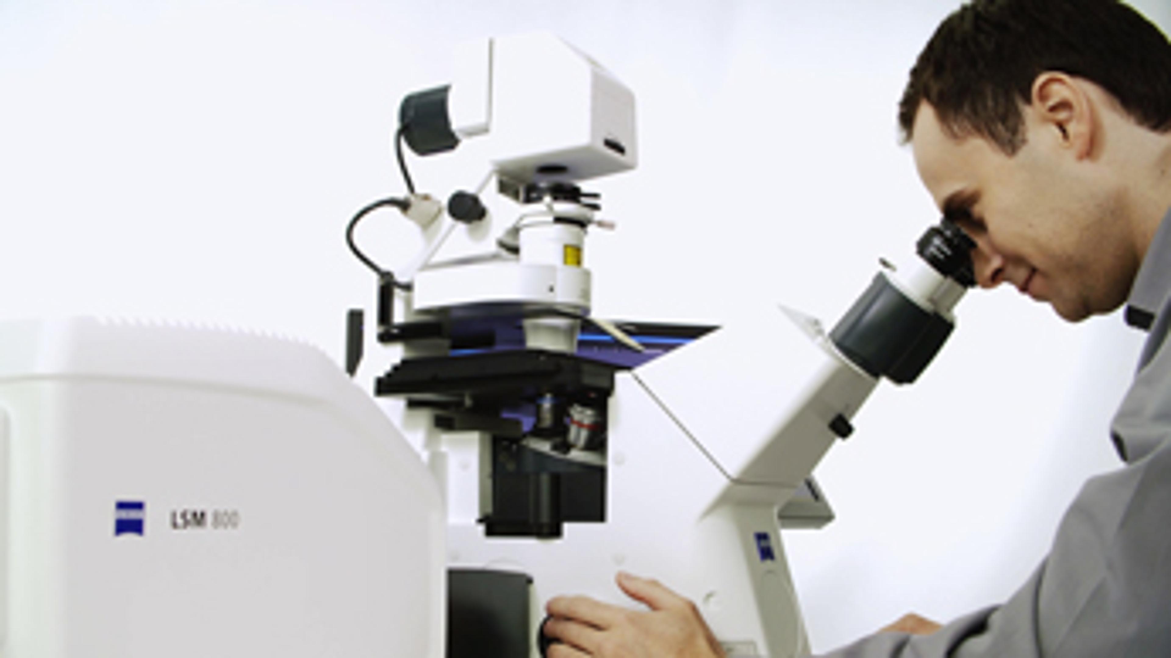
The LSM 800 from Zeiss is our latest microscope in the microscope core facility. When using the microscope you realize that it offers a dramatic improvement in basically all functionality of a confocal microscope compared to the version before. In basically no time most users of confocal microscopes turned towards the LSM 800. Both it's ease of use and the impressive improvements in resolution and sensitivity convinced us right away.
Review Date: 18 Feb 2019 | ZEISS Research Microscopy Solutions
Great result, recommend purchasing this one.
Analysis for live dead bacteria
It was easy to use and gives excellent magnification and resolution.
Review Date: 31 Aug 2018 | ZEISS Research Microscopy Solutions
Airyscan allows us to study localization of protein deep inside the root.
Arabidopsis thaliana - root cell imaging
The system enables higher quality resolution of image.
Review Date: 27 Apr 2016 | ZEISS Research Microscopy Solutions
Best product ever. Makes work in the lab more effective.
Cell imaging
Very high quality results with outstanding resolution. Absolute recommendation for any colleague.
Review Date: 22 Apr 2016 | ZEISS Research Microscopy Solutions
Must buy item for the individual laboratory or core facility.
Biological sample
It is an innovative product with excellent image quality and very good price.
Review Date: 22 Apr 2016 | ZEISS Research Microscopy Solutions
Great product.
Cell Biology & Physiology
Great results, very good value for money when comparing to other manufacturers, would definitely recommend it to others.
Review Date: 22 Apr 2016 | ZEISS Research Microscopy Solutions
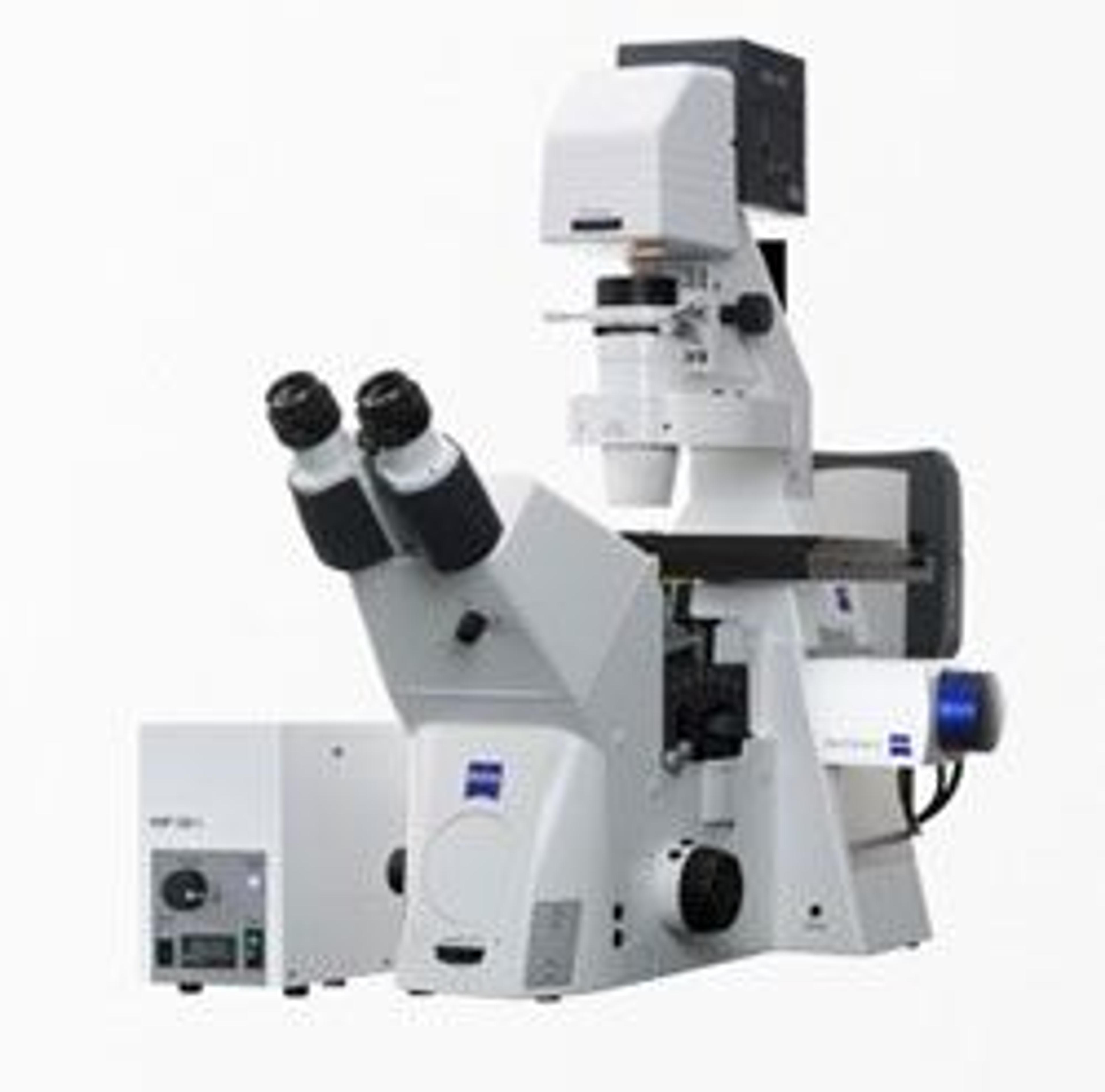
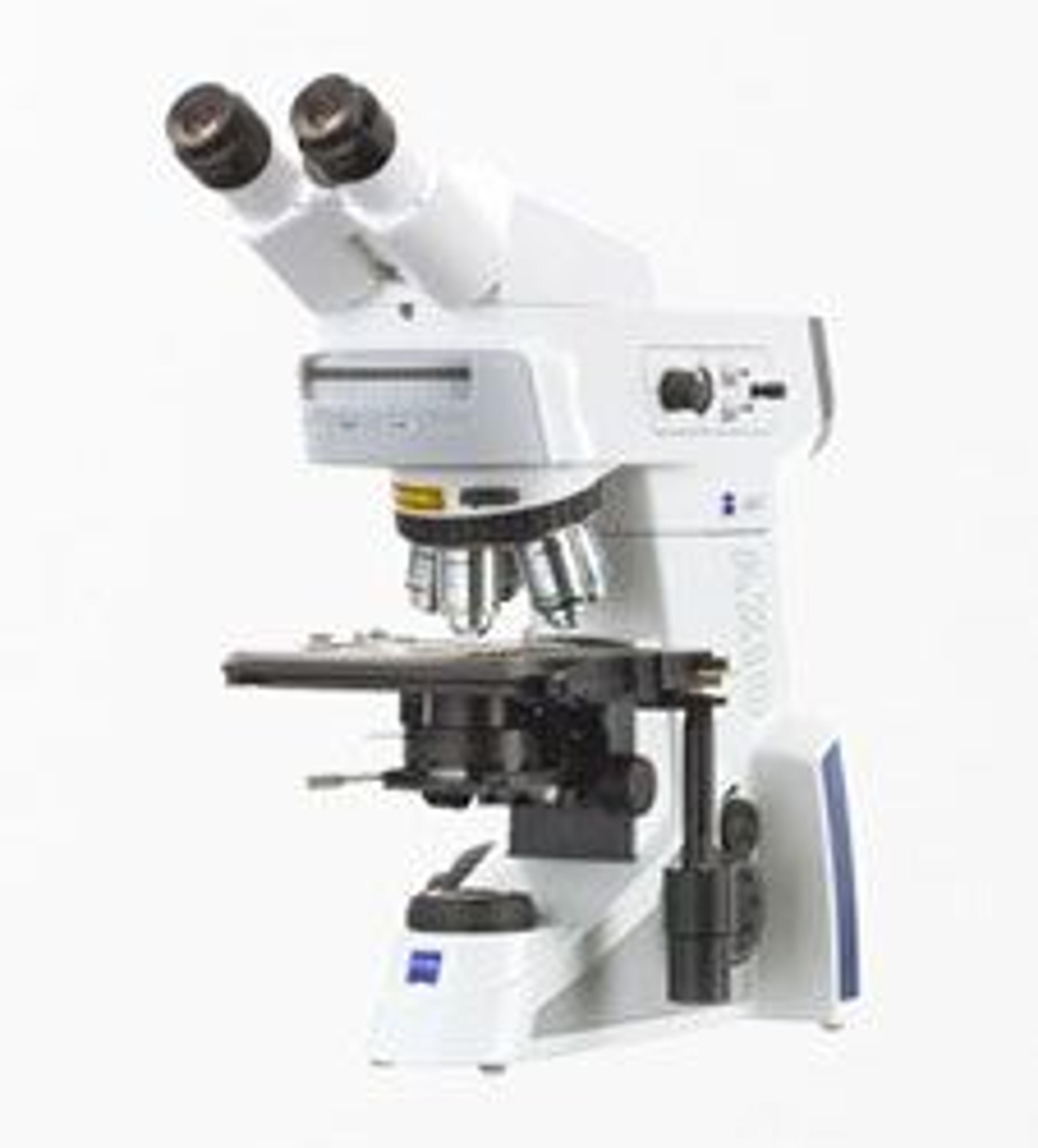
Confocal imaging demands the very best imaging quality. With LSM 800 you are choosing a flexible and compact confocal laser scanning microscope, complete with highly sensitive GaAsP detector technology and fast linear scanning.
Add Airyscan, the revolutionary detection concept from ZEISS, and you will gain 1.7× higher resolution in all three dimensions – resulting in a 5× smaller confocal volume. And you will be pushing sensitivity beyond the limits of all conventional confocals.
LSM 800 is your entry into the world of high-end confocal imaging. Simply decide which options your system needs today, then upgrade in the future as your needs grow.

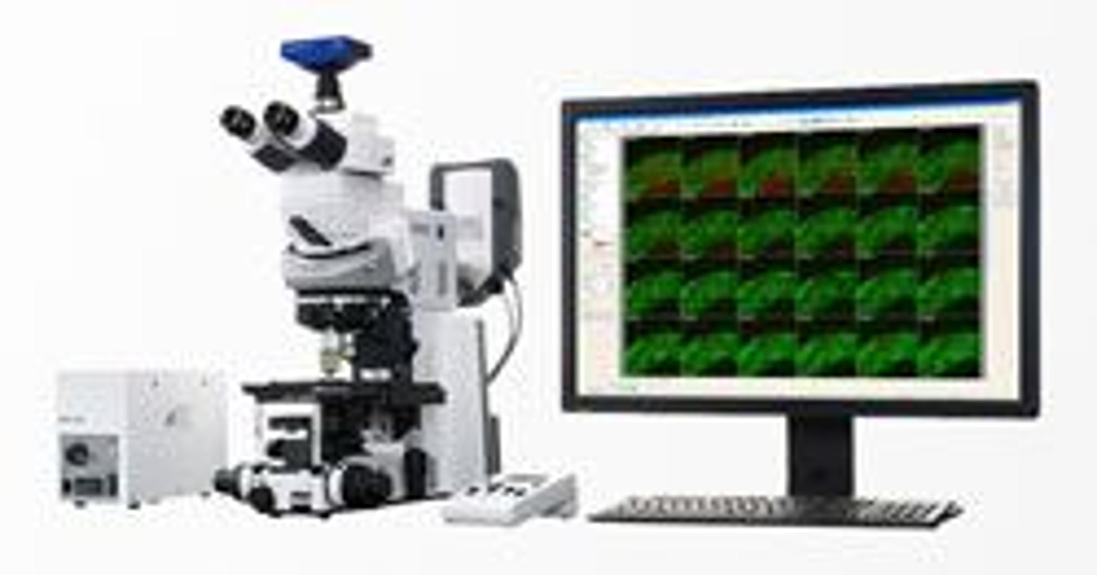
Confocal Imaging with Improved Signal-to-Noise Ratio and Superresolution
Confocal imaging has grown to become the standard choice for most fluorescence microscopy applications. The optical sectioning ability of a confocal imaging system has been brought about by placing a field stop, the pinhole. This application note looks into the details of the recent development and market introduction of the Airyscan detector from ZEISS. Where traditional pinhole and detector design have been reworked to offer greatly improved resolution and signal-to-noise ratio (SNR).
Cryo-Confocal Imaging with Airyscan Improving Resolution and Signal-to-Noise in Cryo-Fluorescence Microscopy
This application note describes how the ZEISS Airyscan can improve resolution and Signal-to-Noise in cryo-fluorescence microscopy, it allows the user to record high-quality cryo- fluorescence data even without immersion optics, thanks to its novel confocal detection scheme.
ZEISS LSM 880 with Airyscan: Introducing the Fast Acquisition Mode
In this application note ZEISS introduce the features and benefits of the Airyscan detector concept for confocal laser scanning microscopy (LSM). With this feature additional light and spatial information can be collected, improving the spatial resolution and signal to noise ratio.
Confocal Imaging with Improved Signal-to-Noise Ratio and Superresolution
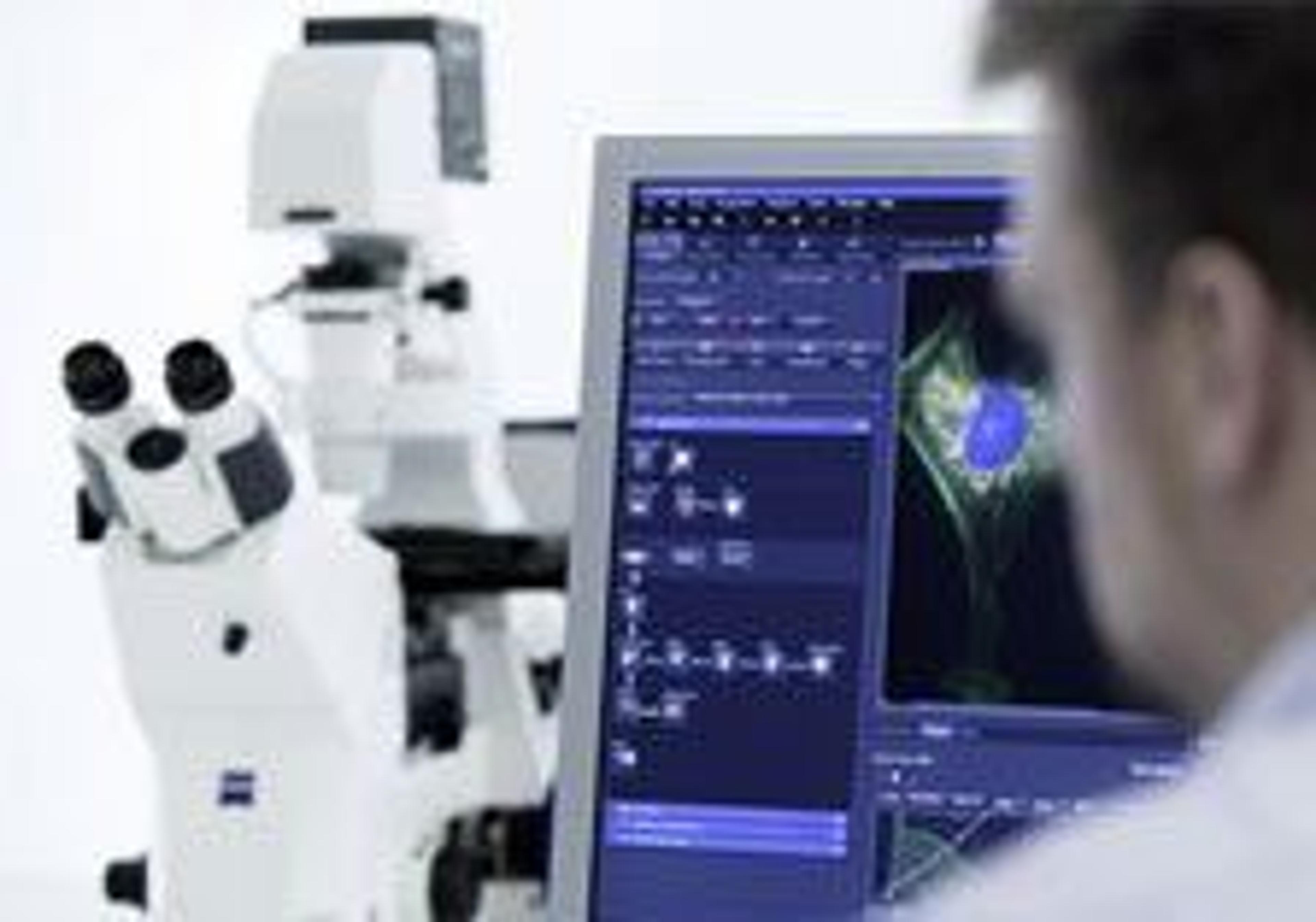
This application note explains how the Airyscan detector hardware is implemented and describe how the resolution and SNR increase is achieved and how it compares to traditional laser scanning microscopy imaging. Over the last 25 years the technique of confocal imaging has grown to become the standard choice for most fluorescence microscopy applications. The increase in utilization of confocal imaging systems in basic biomedical research can be attributed to the ability of a confocal imaging system to produce optically sectioned images with high contrast while providing acquisition versatility to address many sample and application demands. Over time, novel approaches and options to increase image contrast and instrument versatility have been developed but creating the optical section has not changed.
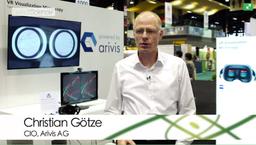
Virtual Reality Visualization of Complex Data Sets Acquired on ZEISS Microscopes
In this video, Christian Götze, Head of Development at arivis AG, explains a new approach to look at the large data acquired with microscopes. ZEISS and arivis partnered to create a prototype which allows you to render and visualize terabytes of volume data with the Oculus Rift virtual reality headset. You get a deeper insight into very complex brain structures and intuitively understand your data. You can virtually step into your sample, fly through a brain, past brain cells and analyze the image from every angle, and from inside.
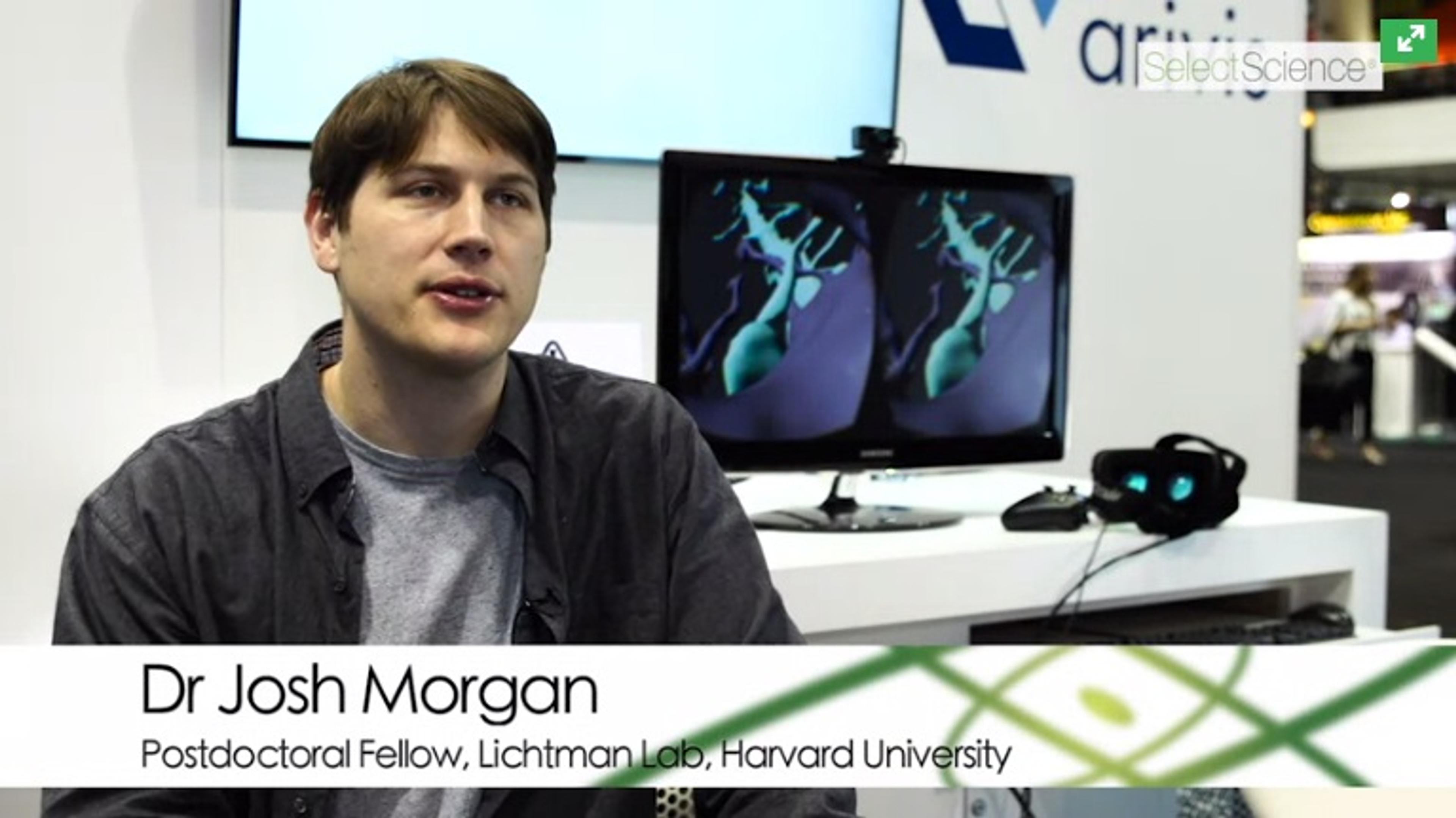
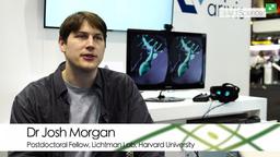
How Large Scale Electron Microscopy is Revealing Animal Behavior at Harvard University
Discover how Dr Josh Morgan, Postdoctoral Fellow in the Lichtman Lab at Harvard University, is trying to understand how the nervous system works by looking at how neurons organize their synapses with one another. All behavior in animals depends on cells talking to each other via specific connections, sending out long processes and forming synapses with each other. Understanding the pattern of those connections is one way of understanding animal behavior. Using large scale electron microscopy to reconstruct neural circuits allows you to see every cell and synapse in a circuit and find how they’re wired to one another.
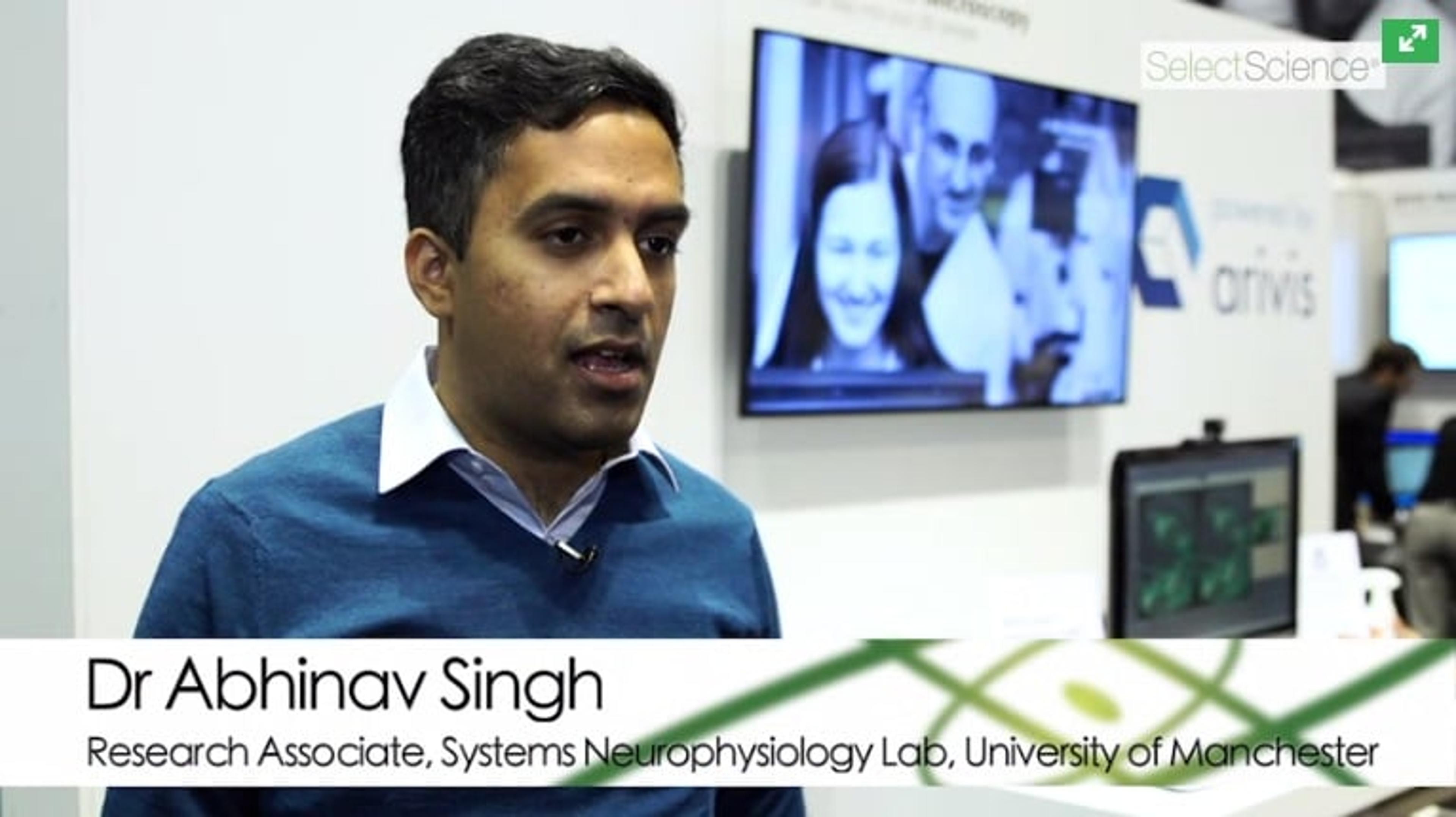
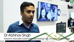
Solving the Problem of Neural Population Coding at Manchester University
Watch this video to hear Dr Abhinav Singh, Research Associate in Systems Neurophysiology Lab at University of Manchester explain the problem of population coding. When information about the external world is received via sensory signalling, it is transformed by our brain. Exactly how this transformation happens is unknown. Dr Singh is looking into how spikes in the pre-frontal cortex encode information about rule learning, and also discusses the fantastic viewpoint virtual reality offers in data analysis.
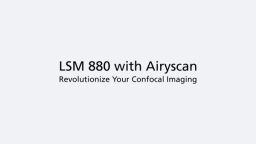
Revolutionize Your Confocal Imaging with LSM 880 with Airyscan
Watch this video to discover how Airyscan technology from ZEISS allows better signal-to-noise, resolution and image collection speed. Image areas lost in traditional confocal microscopy with super resolution - even single molecules!
Extend Your System Further with Airyscan Technology from Zeiss
Watch this video to hear Dr. Ralf Engelmann, Product Manager for LSM880 and Airyscan at ZEISS, explain how the LSM880 allows you to extract more information from your samples, allowing higher throughput and in turn, allowed you to image more samples. Extend the sensitivity of your instrument further with Airyscan technology, which allows imaging of areas lost in traditional confocal microscopy with super resolution - you can even image single molecules!
ZEISS Wins Life Science Scientists' Choice Award®
SelectScience presents the Scientists’ Choice Award® for Best New Life Science Product of 2014 to ZEISS Microscopy. The ZEISS LSM 880 with Airyscan was voted as the winning product by scientists around the world, and the award was presented at AACR, in Philadelphia, USA.
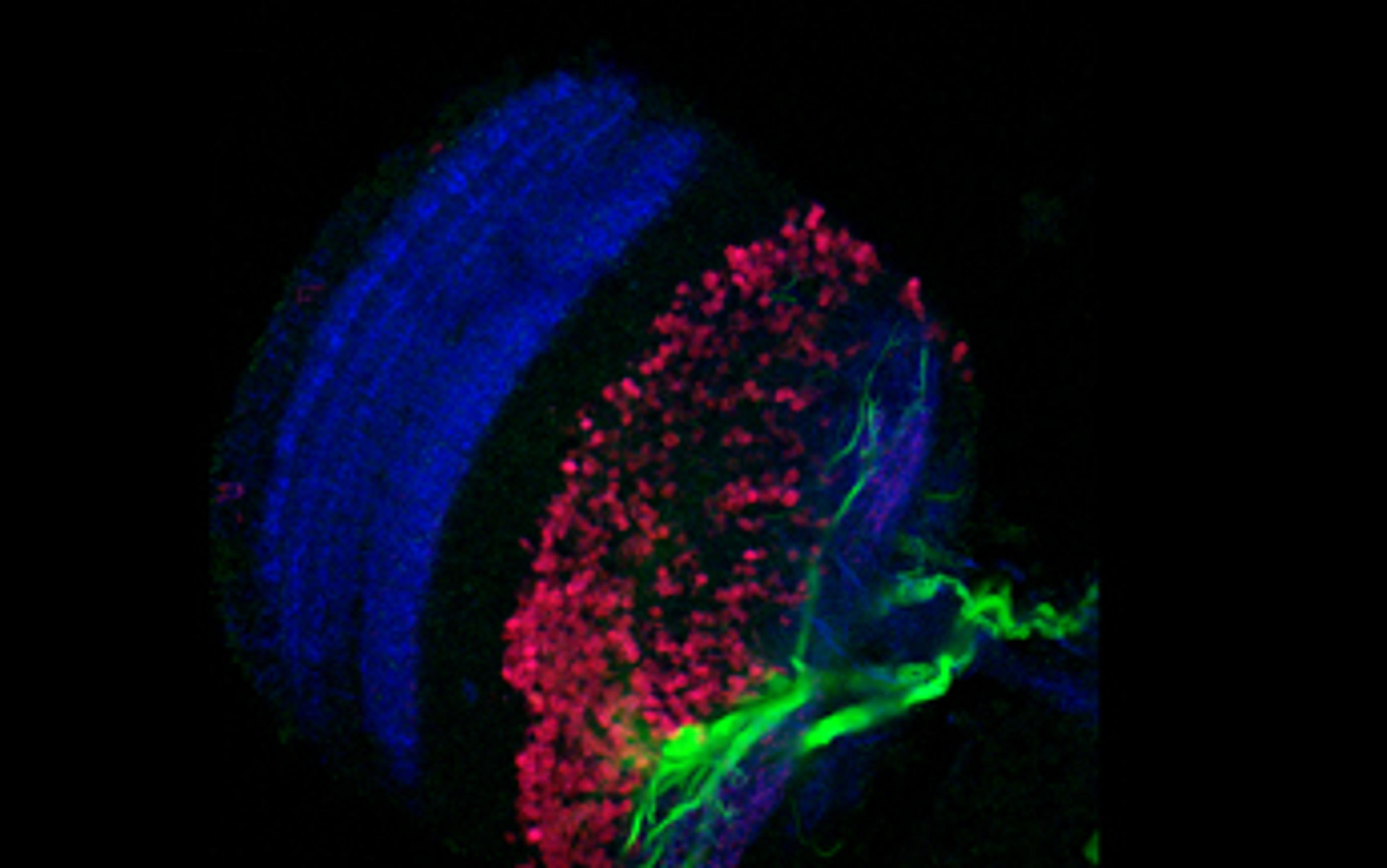
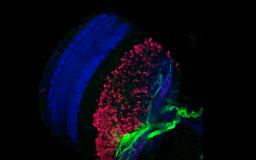
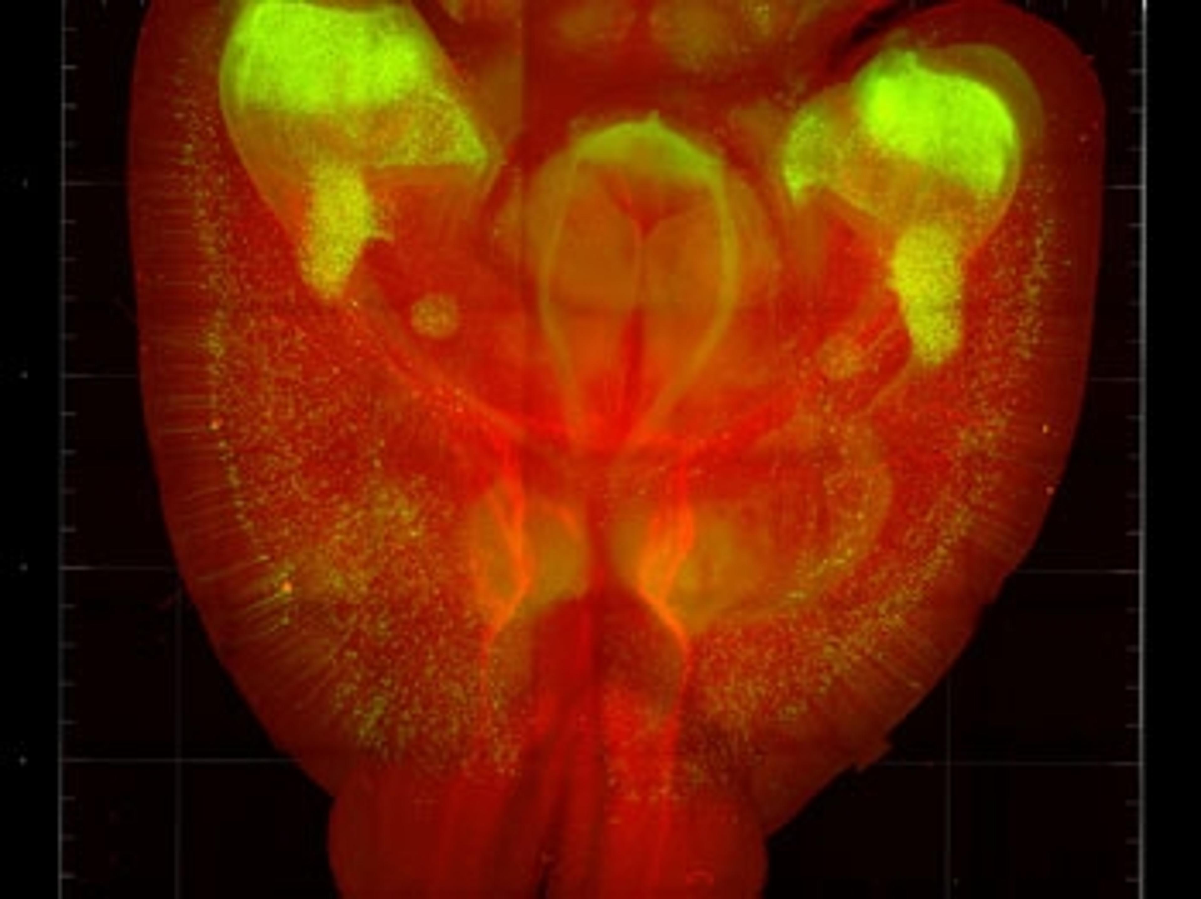
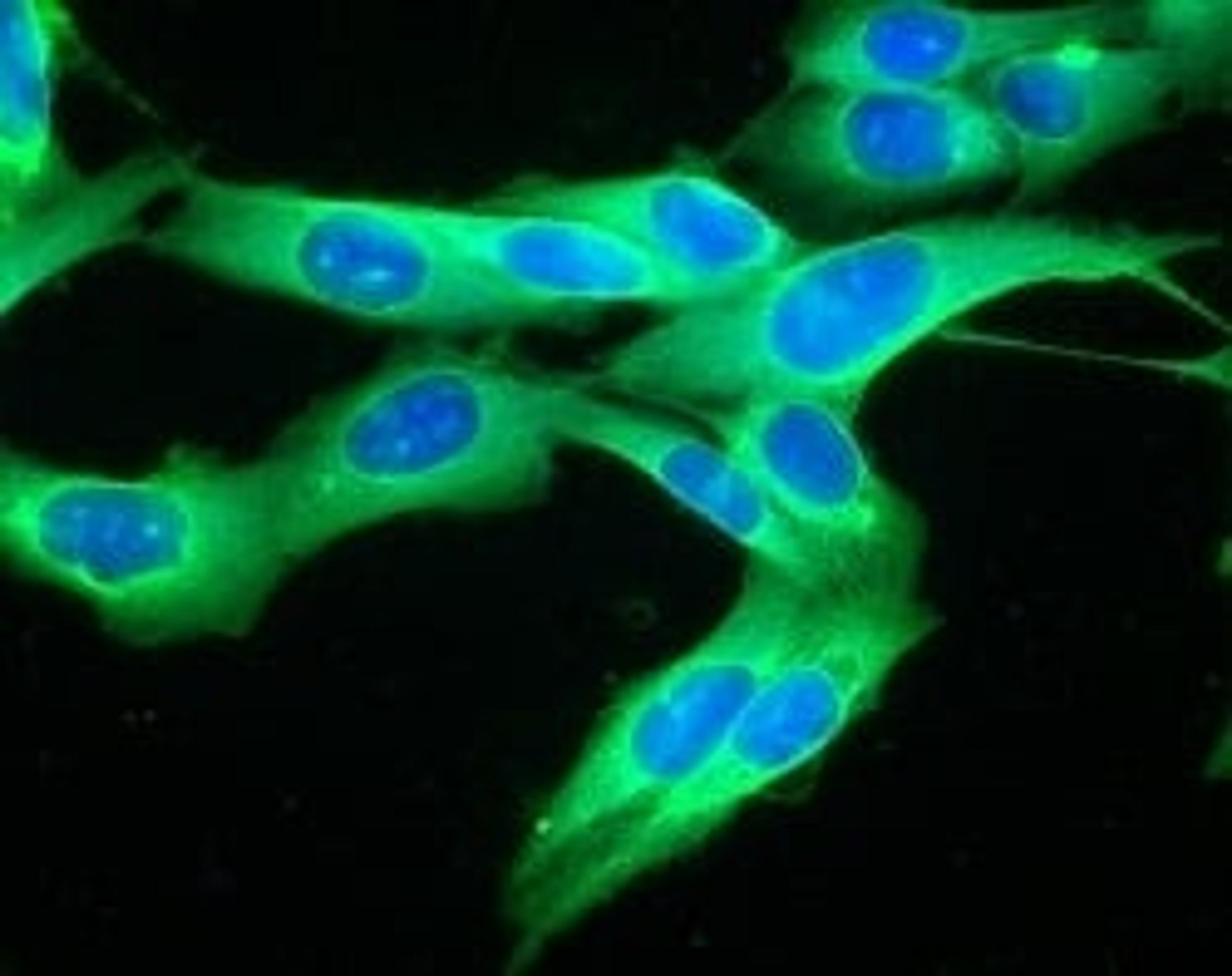
Drosophila Brain Imaging with the ZEISS LSM 800 with Airyscan
This video shows live imaging of a Drosophila brain with triple antibody staining, using the ZEISS LSM 800 with Airyscan. Airyscan gives 1.7 times higher resolution and higher sensitivity than any classic confocal. With the LSM 800, you choose a flexible and compact confocal laser scanning microscope that provides uncompromised image quality.
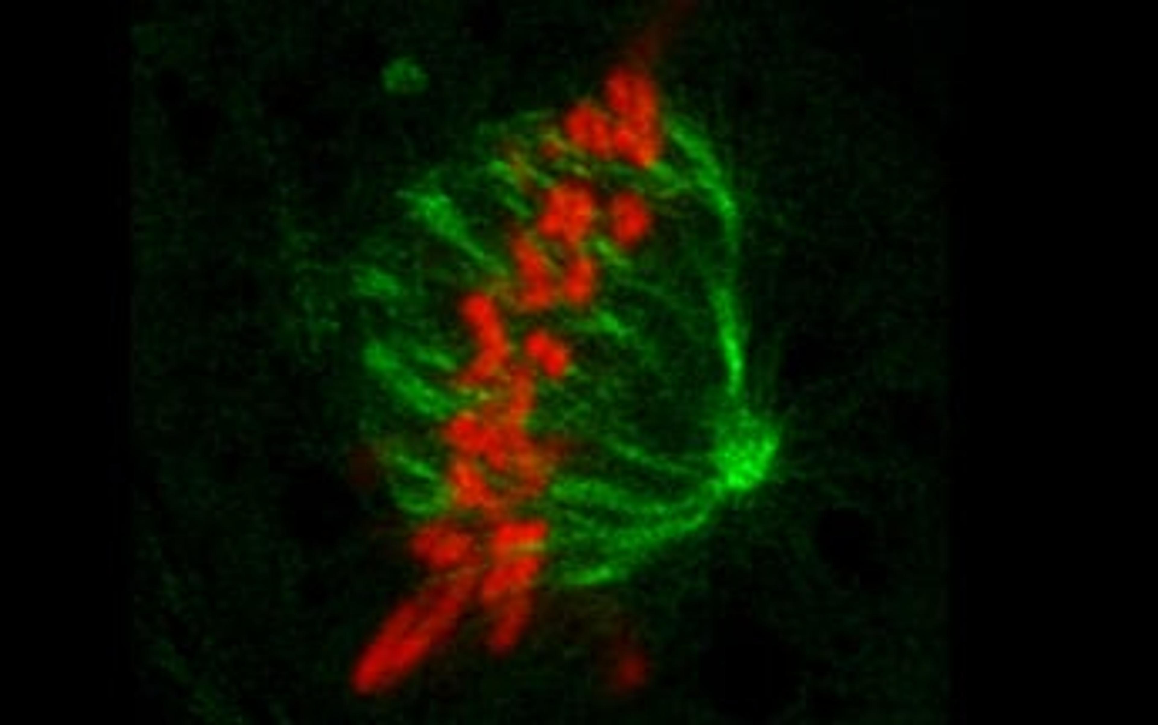
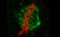
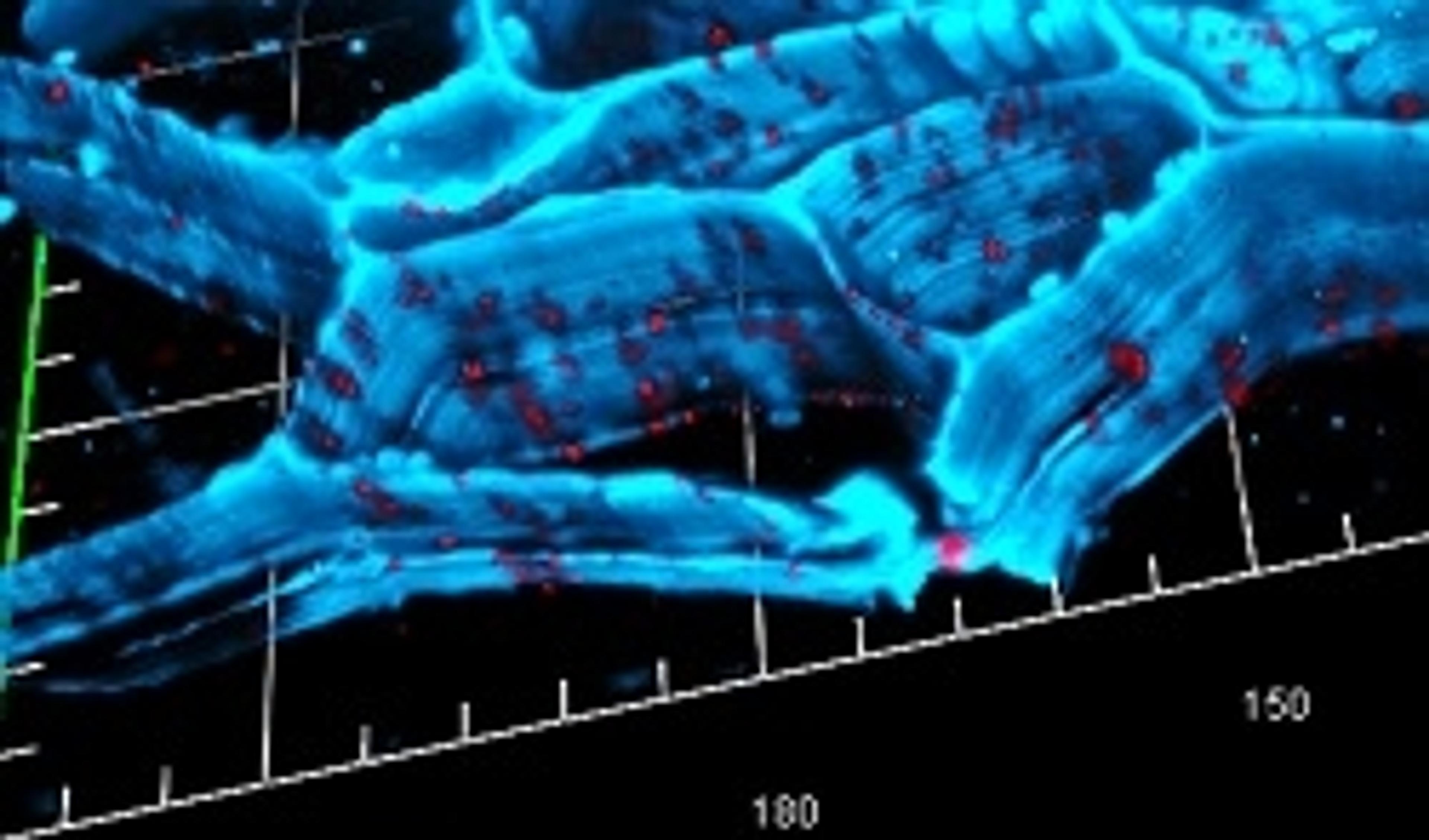
Kidney Cell Mitosis Imaging with the ZEISS LSM 800 with Airyscan
This video shows cell division of LLC-PK1 (Pig Kidney Epithelial) cells, imaged with the ZEISS LSM 800. Using Airyscan gives 1.7 times higher resolution and higher sensitivity than any classic confocal. With the LSM 800, you choose a flexible and compact confocal laser scanning microscope that provides uncompromised image quality.
Introducing the LSM 800 with Airyscan from ZEISS
A good image often forms the basis for fascinating discoveries. The LSM-800 from Zeiss is a compact confocal which gives you uncompromising image quality. Watch this video to discover how efficient and easy it is to operate the LSM-800.
Automated Microscope for Gentle and Fast Confocal 4D Imaging
Enhancing ZEISS Celldiscoverer 7 with ZEISS LSM 900 for optical sectioning
Solutions for the Visualization and Analysis of Big Image Data in Life Sciences
ZEISS and arivis AG partner to provide leading 3D imaging systems

ZEISS Opens New Microscopy Customer Center
From 3D Light to 3D Electron Microscopy: Highlights from the EMBL Workshop
Discover the latest news from this conference on correlative microscopy

Exclusive Videos from analytica 2016
Watch exclusive interviews filmed at the 25th analytica international trade fair for laboratory technology, analysis and biotechnology

Top 5 Webinars to Improve Your Imaging Techniques
Learn about the latest imaging technologies and innovative applications
From Phenotype to Genotype: Unlocking the Power of Correlative Microscopy for Cell Biology
SelectScience® spoke to Dr Peter O'Toole, Head of Imaging and Cytometry, Department of Biology at the University of York, about the technology serving his department
New Confocal Microscope ZEISS LSM 800 for Materials Research and Failure Analysis
All essential light microscopy contrasting methods and high precision 3D topography in a single instrument
Free SelectScience Webinar: New Acquisition and Detection Modes with ZEISS Airyscan
Register your place for this exciting event and Q&A session