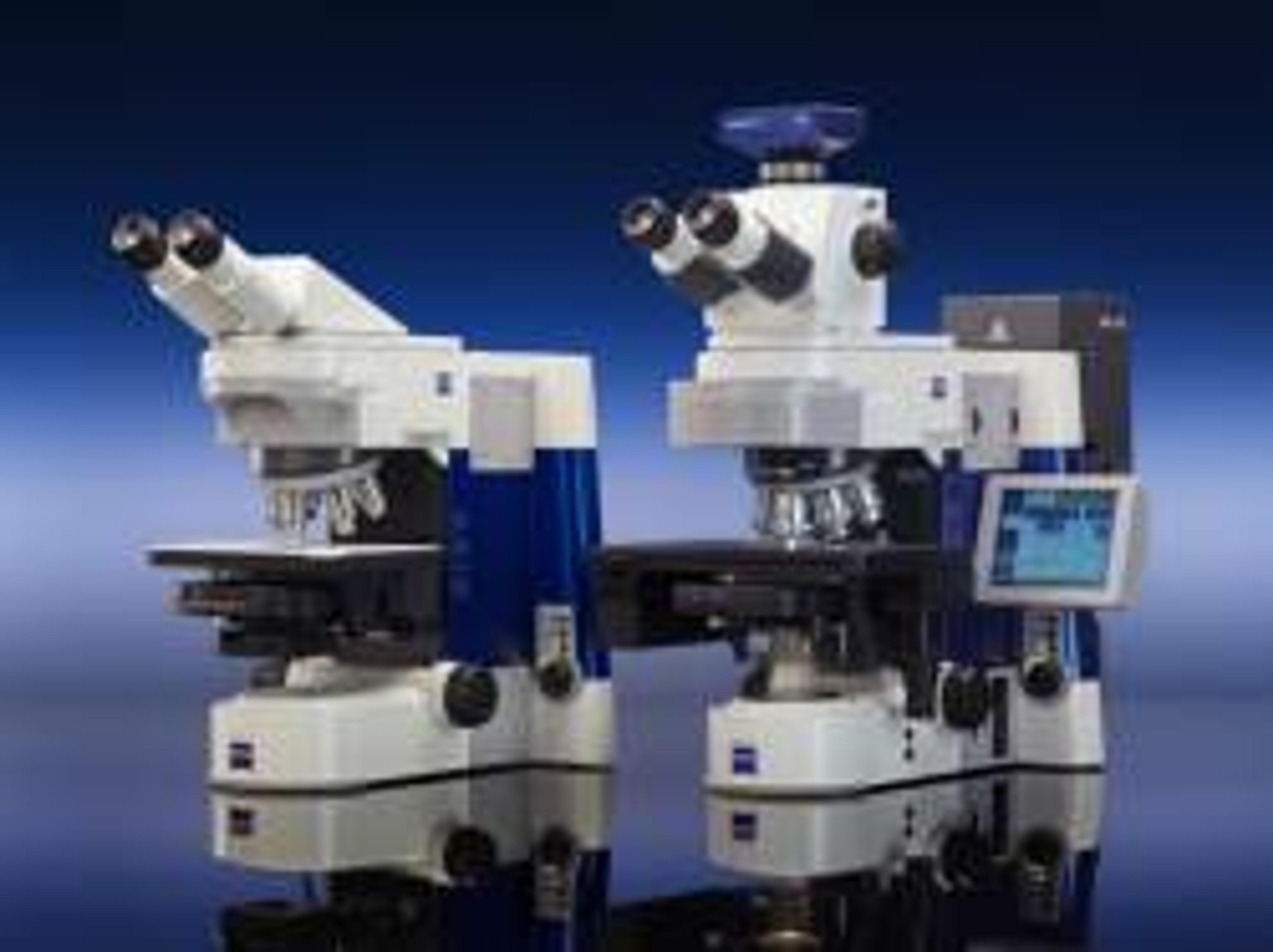
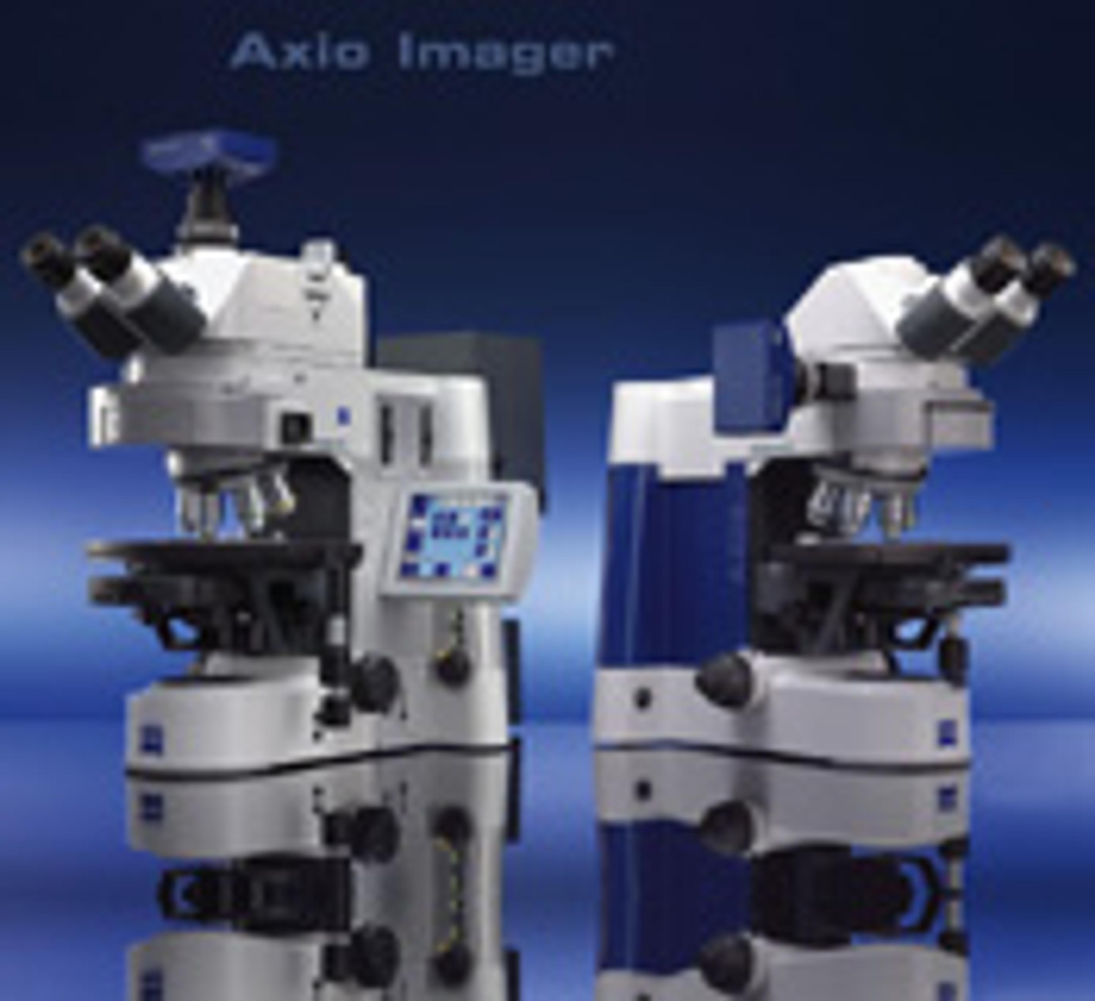
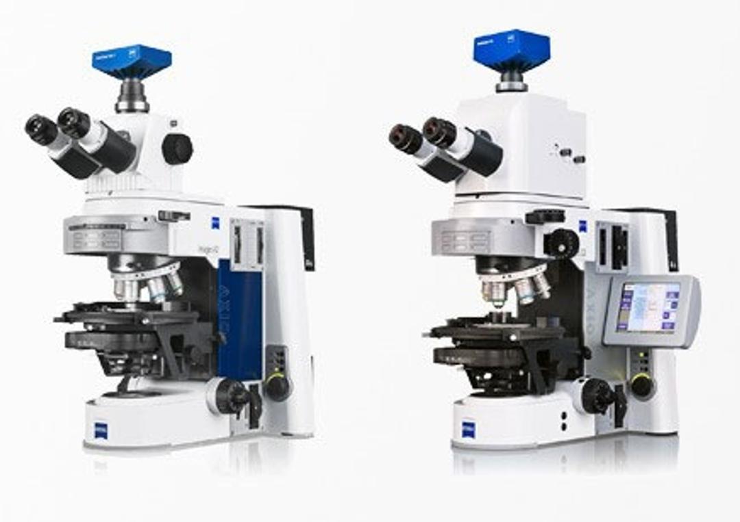
ZEISS Axio Imager - Modular System for Digital Fluorescence Microscopy
Axio ImagerAn innovative modular system for digital fluorescence microscopy, featuring advanced flexibility and application versatility. Featuring: New IC2S objectives (Infinity Contrast & Colour Corrected System) - optimise image quality and maximise contrast Special fluorescence filters - reduce exposure and image acquisition time for superior 3D imaging. 'Intelligent Stand' - automatically recognises added compone…


Receive your quote directly from the manufacturer.
Excellent performance under a variety of conditions
Clinical telemedicine
Top quality results in all areas
Review Date: 21 Nov 2022 | ZEISS Research Microscopy Solutions
Would recommend to colleagues
Fluorescence Optical Imaging
Highly useful for optical imaging of polymers, biological samples, etc. Versatility of microscope makes it quite useful for different applications. Ergonomic to use, also allows multiple adjustments to be made to suite specific user. Friendly GUI.
Review Date: 7 Aug 2018 | ZEISS Research Microscopy Solutions
Very good image quality
Staining of bacteria, plant parts and fungi
The product is very good. We have used for four years, no problem detected yet to date. Ease of handling and good image quality
Review Date: 9 Sept 2016 | ZEISS Research Microscopy Solutions
Fluorescense Imaging
An excellent and reliable automated microscope. Good software integration and imaging quality, especially with colibri LED illumination. Highly recommended!
Review Date: 21 Aug 2015 | ZEISS Research Microscopy Solutions
Cell Imaging
I use the Z2 version. It is easy to use, the slides go in the holder easily. Automated- 8 slides at a time. Karyotyping software is easy with full manual provided.
Review Date: 8 Oct 2012 | ZEISS Research Microscopy Solutions
FISH/Karyotyping
Intuitive interface- very good integration of camera view in software.
Review Date: 8 Oct 2012 | ZEISS Research Microscopy Solutions
This is a basic scope that works really well for IHC and other mounted imaging
Review Date: 8 Nov 2009 | ZEISS Research Microscopy Solutions
Easy to use and produces very good pictures.
Review Date: 11 Jan 2008 | ZEISS Research Microscopy Solutions
Review Date: 11 Jan 2008 | ZEISS Research Microscopy Solutions
Axio Imager
An innovative modular system for digital fluorescence microscopy, featuring advanced flexibility and application versatility.
Featuring:
- New IC2S objectives (Infinity Contrast & Colour Corrected System) - optimise image quality and maximise contrast
- Special fluorescence filters - reduce exposure and image acquisition time for superior 3D imaging.
- 'Intelligent Stand' - automatically recognises added components. "Contrast manager" ensures simple changes between contrasting techniques.
- Integration: standard interfaces permit communication via USB and TCP/IP
- Vibration-free Imaging Cell - isolated within the stand for stable observation and unparalleled precision.
- Apochromatic fluorescence beam path - ensures optimum colour correction
- Active stray light elimination - significantly higher contrast
- Fast, motorised reflector turrets - hold either six or ten filter modules.
- HBO lamp: convenient, self-aligning homogeneous illumination
- LCI Plan-Neofluar objectives - live cell imaging
- Flexibility - two freely configurable stand variants: Z1 (motorized) and D1(manual) or preconfigured stands M1 (motorized) and A1 (manual).
Rapid image acquisition with outstanding quality in up to 6 dimensions means more signal in less time.

Multiscale Analysis of Bacteria Population in Legume Root Nodules with "Shuttle & Find"
Correlative light and electron microscopy (CLEM) combines the overview information of fluorescence light microscopy (FLM) with structural details in scanning electron microscope (SEM) for high-content imaging. Hence, CLEM is very useful for investigating infection and colonization of the legume hosts by their rhizobial symbionts. This white paper illustrates a methodology involving the shuttle & find interface of CLEM to enable fast and reliable analysis of the bacterial infection of legume root nodule cells.
Visualizing the Architecture of Cells and Tissues
Laser scanning microscopes (LSMs) have proven extremely flexible in 3D imaging in a non-destructive manner. Most difficulties encountered in biological imaging are caused by intrinsic sample characteristics that compromise 3D imaging by limiting penetration depth and cause image distortions. Discriminating fluorescent labels from autofluorescence is another common challenge in plant and animal tissue samples. this application note describes how these problems can be overcome using Zeiss' LSM technology.
New Light Source & Slider Module for Fluorescence Microscopy
Watch this interview at ASCB 2010 to learn more about new technology from Carl Zeiss. The ApoTome.2 uses structured illumination to produce optical sections without the need for expensive equipment such as lasers. The Colibri.2 LED Light Source for live cell imaging eliminates UV bleedthrough and dramatically reduces phototoxicity compared to traditional Hg or Xe light sources. The LED technology allows precise and ultra fast intensity control individually per channel and exposure.