Visualizing the Architecture of Cells and Tissues
17 Nov 2017Laser scanning microscopes (LSMs) have proven extremely flexible in 3D imaging in a non-destructive manner. Most difficulties encountered in biological imaging are caused by intrinsic sample characteristics that compromise 3D imaging by limiting penetration depth and cause image distortions. Discriminating fluorescent labels from autofluorescence is another common challenge in plant and animal tissue samples. this application note describes how these problems can be overcome using Zeiss' LSM technology.
Related products
Request Quote for All Products
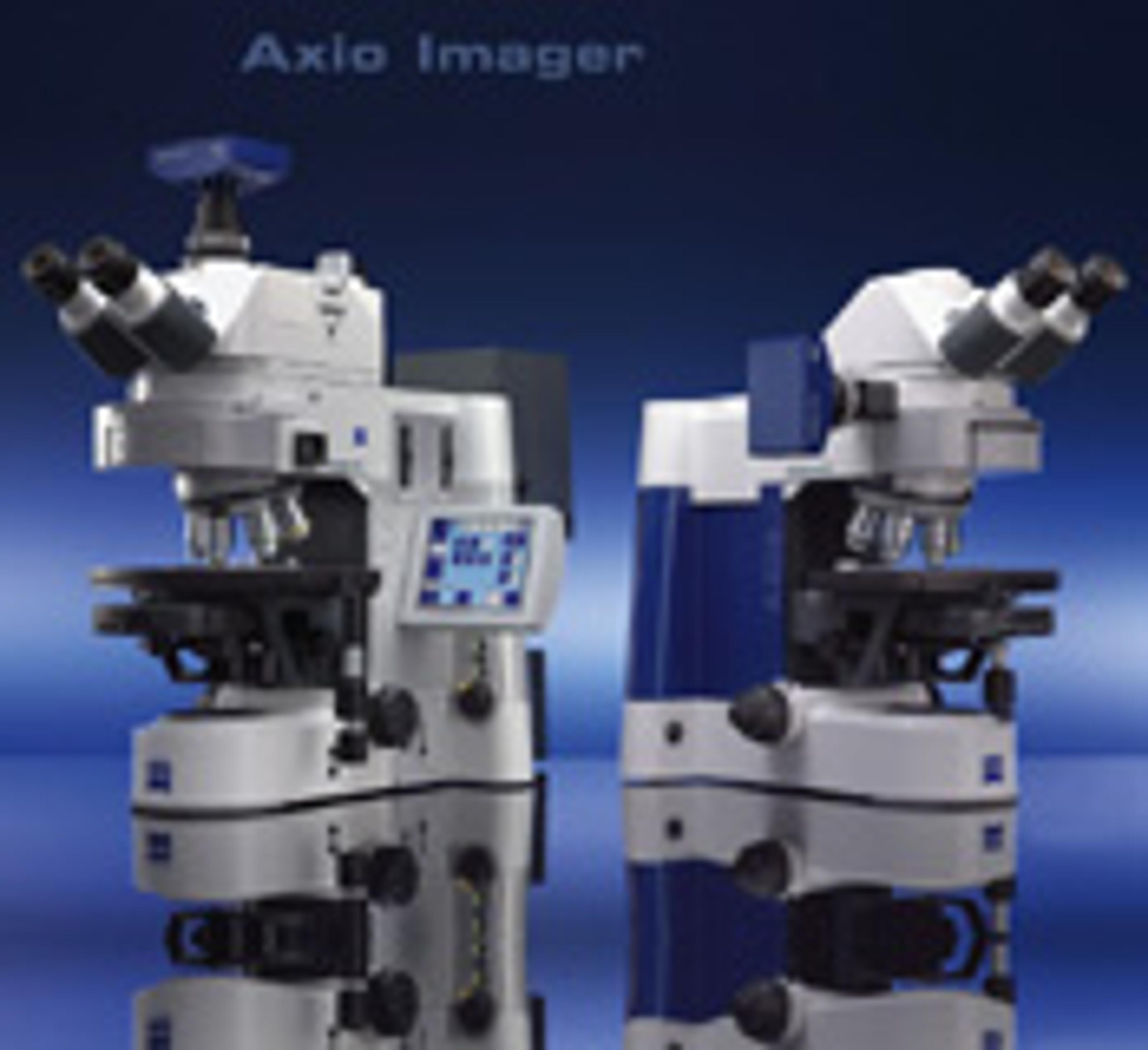
ZEISS Axio Imager - Modular System for Digital Fluorescence Microscopy
ZEISS Research Microscopy SolutionsAxio ImagerAn innovative modular system for digital fluorescence microscopy, featuring advanced flexibility and application versatility. Featuring: New IC2S objectives (Infinity Contrast & Colour Corrected System) - optimise image quality and maximise contrast Special fluorescence filters - reduce exposure and image acquisition time for superior 3D imaging. 'Intelligent Stand' - automatically recognises added components. "Contrast manager" ensures simple changes between contrasting techniques. Integration: standard interfaces permit communication via USB and TCP/IP Vibration-free Imaging Cell - isolated within the stand for stable observation and unparalleled precision. Apochromatic fluorescence beam path - ensures optimum colour correction Active stray light elimination - significantly higher contrast Fast, motorised reflector turrets - hold either six or ten filter modules. HBO lamp: convenient, self-aligning homogeneous illumination LCI Plan-Neofluar objectives - live cell imaging Flexibility - two freely configurable stand variants: Z1 (motorized) and D1(manual) or preconfigured stands M1 (motorized) and A1 (manual). Rapid image acquisition with outstanding quality in up to 6 dimensions means more signal in less time.