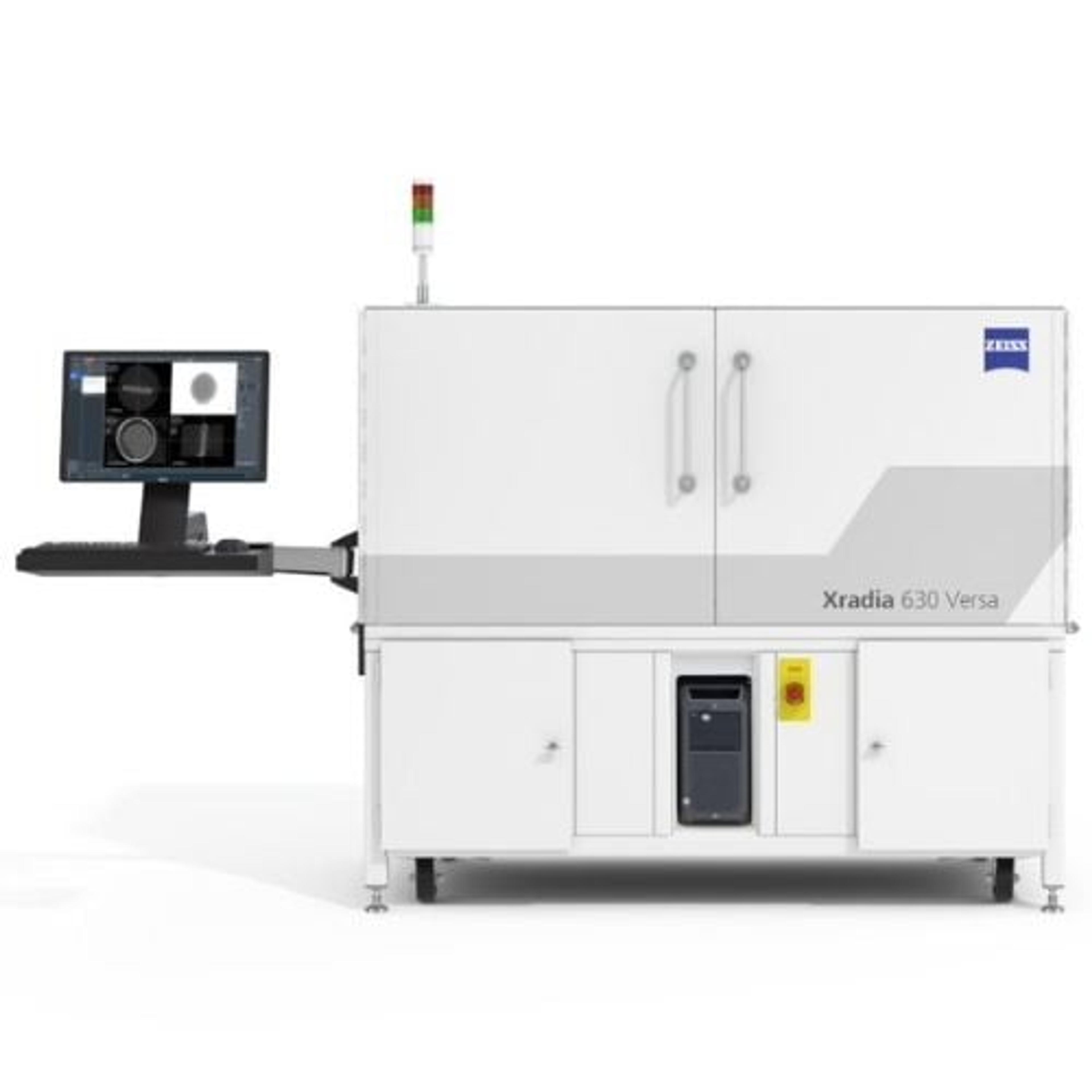
ZEISS Xradia Versa X-ray Microscopes
Discover More with Non-destructive 3D X-ray Imaging at Submicron Resolution


Receive your quote directly from the manufacturer.
Great Product
Faster Sub-Micron Imaging of Intact Samples
True spatial resolution of 500 nm with a minimum achievable voxel size of 40 nm High resolution across a broad range of sample types, sizes, and working distances. In situ imaging for non-destructive characterization of microstructures in controlled environments and over time upgradeable and extendible with future innovations and developments. High throughput with good image quality.
Review Date: 26 Mar 2022 | ZEISS Research Microscopy Solutions
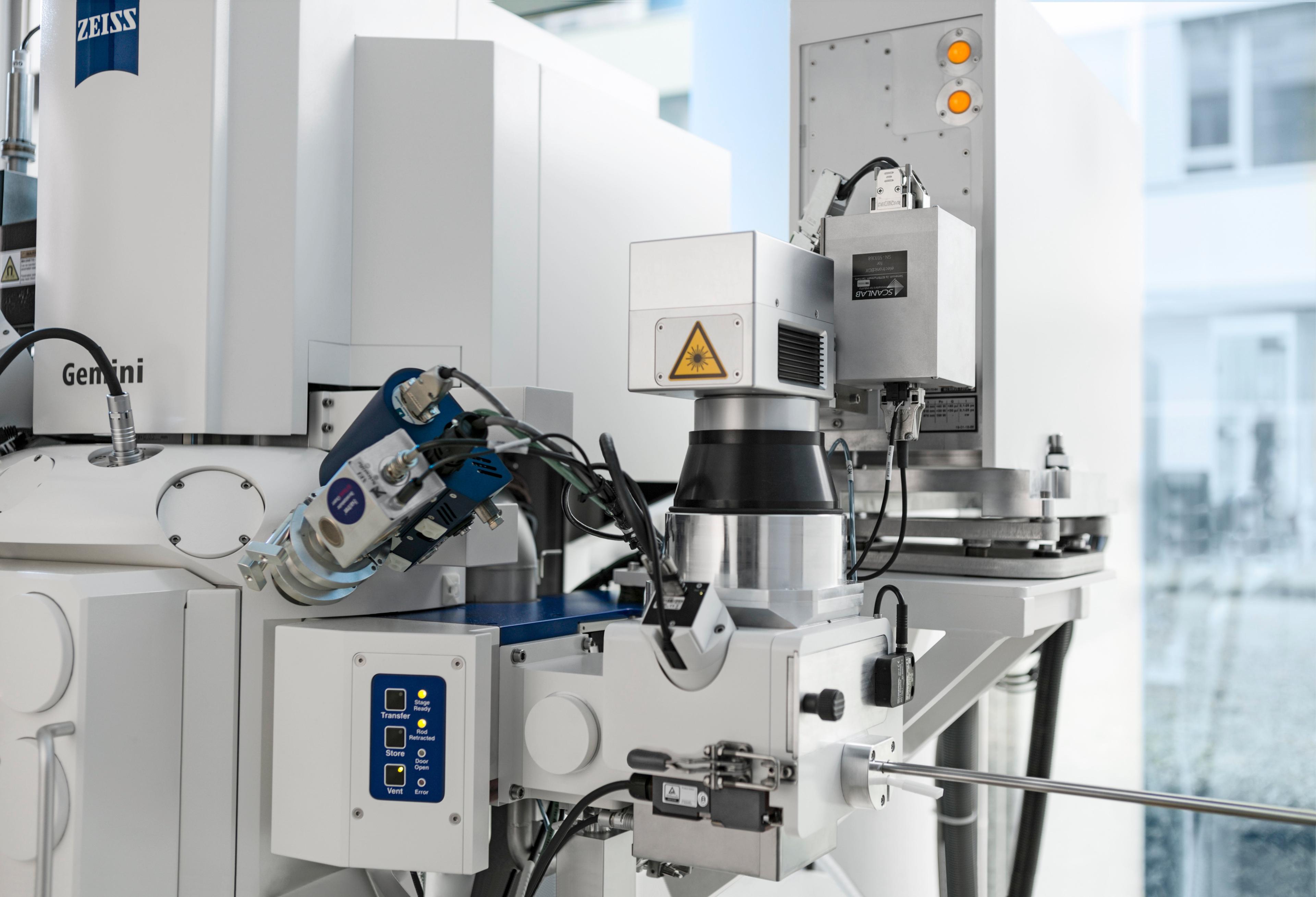
Extremely versatile ZEISS Xradia Versa 3D X-ray microscopes (XRM) provide superior 3D image quality and data for a wide range of materials and working environments. Xradia Versa XRM feature dual-stage magnification based on synchrotron-caliber optics and revolutionary RaaD™ (Resolution at a Distance) technology for high resolution even at large working distances, a vast improvement over traditional micro-computed tomography. Non-destructive imaging preserves and extends the use of your valuable sample, enabling 4D and in situ studies.
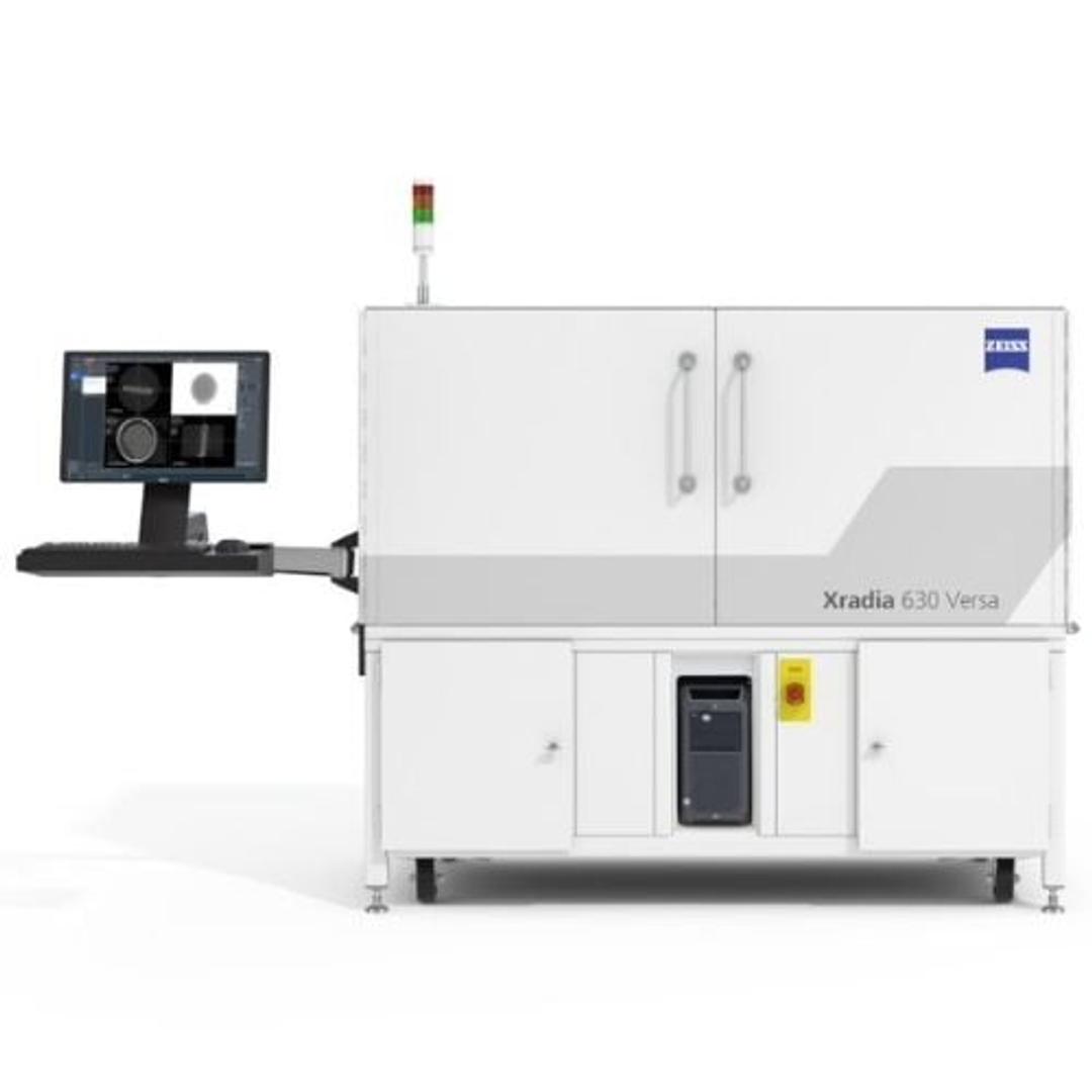
- Expanded access, productivity, capability - ZEISS Xradia 630 Versa
- Wide range of capabilities - ZEISS Xradia 620 Versa
- Faster submicron imaging - ZEISS Xradia 610 Versa
- Flexibility and ease of use - ZEISS Xradia 510 Versa
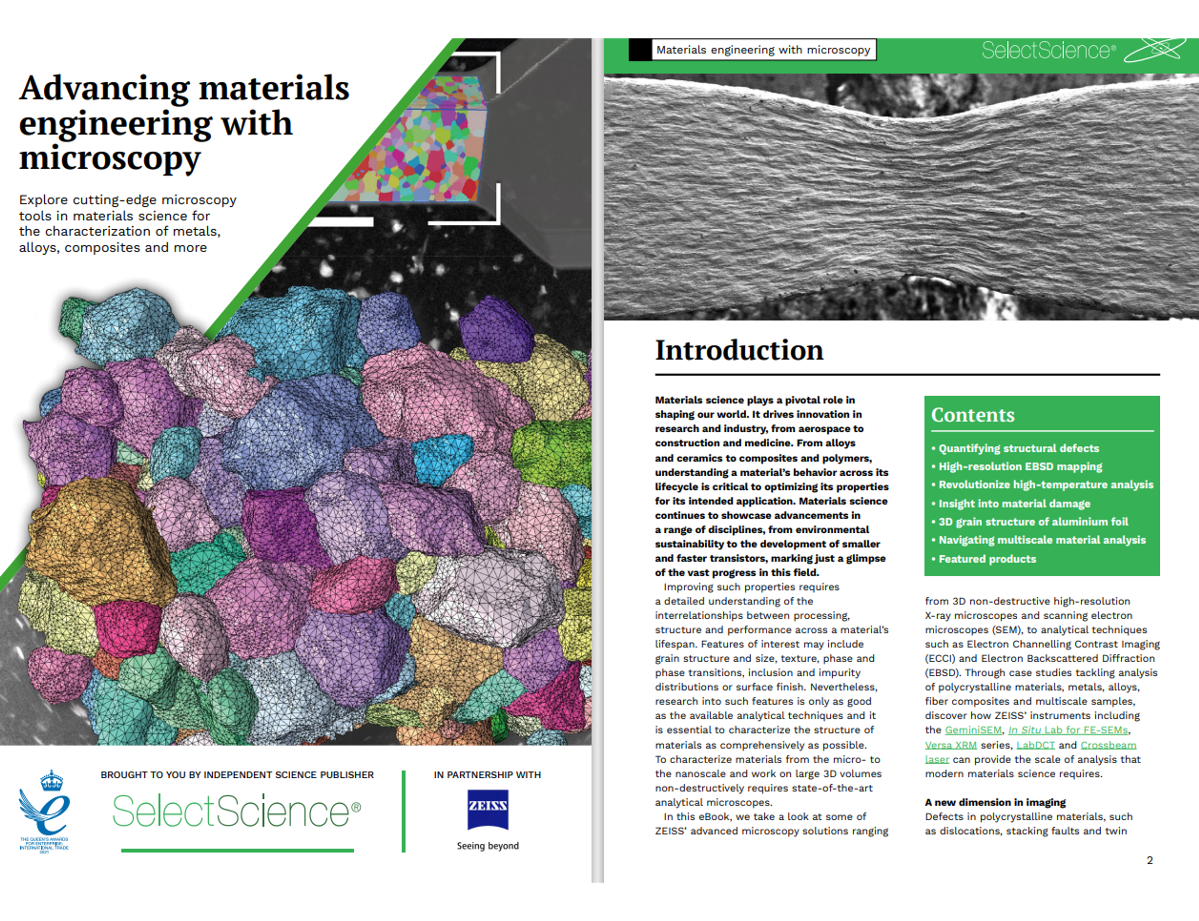
Advancing materials engineering with microscopy
Materials science is crucial in shaping our world and driving innovation across various fields like aerospace, construction, and medicine. It involves understanding the behavior of materials such as alloys, ceramics, composites, and polymers throughout their lifecycle. This knowledge is essential for optimizing their properties for specific applications.
Download this expert guide for free today and explore advanced microscopy solutions from ZEISS, including 3D non-destructive high-resolution X-ray microscopes and scanning electron microscopes (SEM). The eBook also covers analytical techniques such as Electron Channelling Contrast Imaging (ECCI) and Electron Backscattered Diffraction (EBSD).
Plus, find case studies on polycrystalline materials, metals, alloys, fiber composites, and multiscale samples, and learn about the tools essential for modern materials science analysis.

Investigating structure-property relationships in a carbon-fiber composite
Characterizing composite materials is a challenging task. Understanding the nucleation processes is critical toward engineering against failure, but traditional bulk testing methods are insufficient to describe this process. ZEISS presents how correlative microscopy is a viable conduit into the digital material testing approach. A carbon fiber reinforced composite hockey stick was used as the subject of the characterization study, though this same technique can apply to any variety of materials, from glass composites to metal matrix composites, as well as to monolithic materials. Correlative microscopy enabled a robust imaging-to-simulation workflow, producing a model that is available for further digital modification and analysis. Through implementation of this procedure in a regular basis, material development efficiencies may be enhanced, leading to high-performance products in a reduced amount of time.
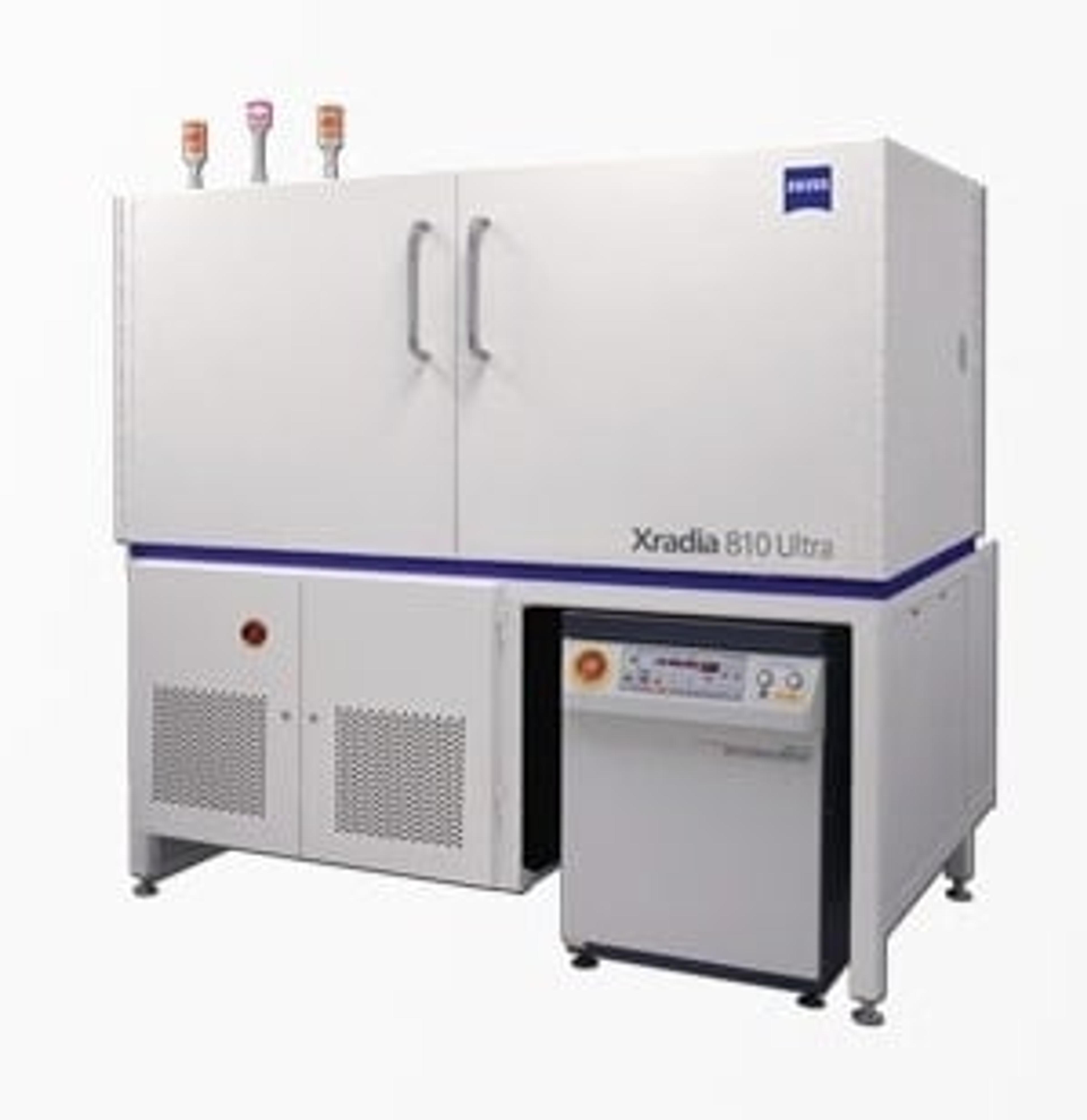
High-resolution characterization of solid foams
Characterization of the 3D morphology of solid foams is extremely important but has been limited due to the shortcomings of conventional techniques. High-resolution techniques such as physical sectioning coupled with optical or electron microscopy are not only destructive and time-intensive, but can also introduce physical artifacts. ZEISS highlights how the ZEISS Xradia Versa and the ZEISS Xradia Ultra series of 3D X-ray microscopes provide a unique solution for non-destructive submicron resolution and the highest contrast in foam imaging that can be achieved. Foam structures can be imaged to submicron spatial resolutions: down to 500 nm on ZEISS Xradia Versa and 50 nanometers on ZEISS Xradia Ultra.
X-ray microscopy of unstained biomaterials with propagation-based phase-contrast for enhanced full-field strain measurement
In this application note, ZEISS highlights the challenges in imaging soft tissues like cartilage, which require contrast enhancement often achieved through staining techniques that can alter tissue properties. To address this, propagation-based phase-contrast imaging is employed in the lab-based XRM ZEISS Xradia 510 Versa. This method enhances contrast in both articular cartilage and mineralized tissue without inducing tissue alterations, enabling digital volume correlation (DVC) to measure full-field strain at the cartilage-bone interface.
An overview of 3D X-ray microscopy
This publication offers an overview of the latest XRM technology and its unique advantages.

Quantitative characterization of asthma inhalation blends using correlative, AI powered X-ray microscopy
Discover the interdependent relationship between the structure, properties, and performance of drugs in pharmaceutical material science.
In this webinar, Dr. Ben Tordoff, Head of Materials Science Business Sector at ZEISS Research Microscopy Solutions, Parmesh Gajar, principal scientist at the University of Manchester and Darragh Murnane, Professor of Pharmaceutics at the University of Hertfordshire will discuss the importance of the microstructure for producing respirable particles for effective therapeutic benefits of a typical dry powder inhaler (DPI) formulation.
Additionally, they will describe how X-ray microscopy (XRM) can be combined with molecular modeling to calculate inter-particulate forces that underpin powder cohesion. They will also explain how the LabDCT technique can be utilized to gather 3D crystallographic information on crystalline bulk powders of lactose particles.
Finally, they will illustrate how artificial intelligence (AI) image analysis is used to visualize the powder microstructure and simulate the drug release performance.
Key learning objectives
- Learn how to evaluate heterogeneous microstructures
- Discover how to combine crystallographic information and micro-CT imaging
- Understand why drug delivery is dependent on the microstructure and how it can be simulated
Who should attend?
- Materials researchers from academia and industry, X-ray microscopy users, drug designers, pharmaceuticals material researchers, PhD students, principal investigators, and lab managers
Certificate of attendance
All webinar participants can request a certificate of attendance, including a learning outcomes summary, for continuing education purposes.

Characterize your Biologic like a Boss with Stunner
The Problem
Quantification and sizing are essential parts of protein characterization. Old school ways are one-by-one and only provide answers on one or the other, resulting in more precious sample and hands-on time required to get all the data you need. Not to mention the extra time it takes to combine and analyze results from multiple instruments.
The Solution
Stunner is the only platform that pairs up intelligent, short-length UV/Vis with rotating angle DLS (RADLS) to give sizing, aggregation detection and protein quantification in under two minutes from only 2 µL.
The Proof
In this webinar, Nelis Denys, Market Manager at Unchained Labs will explain how Stunner measures proteins and ADCs, detects aggregates anytime you want and gives you information on molecular weight and colloidal stability. Nelis will also cover some real-life results of how RADLS on Stunner provides protein quant, size, PDI, aggregation and molecular weight. See how Stunner provides quick QC checks to make sure every sample is good to go.
Key learning objectives
- Learn how to measure protein quant and sizing, fast, with as little as 2 µL
- Explore how to gain information on molecular weight and determine aggregation early with advanced light scattering
- Understand how to quantitate labelling efficiency of an ADC
- Discover how to assess colloidal stability through B22 kD
Who should attend?
- Biology researchers working on protein therapeutics, industrial downstream biotech – CMO, scientists, senior scientists, directors of assay/analytical development, VPs of protein/biologics products, directors of QC
Certificate of attendance
All webinar participants can request a certificate of attendance, including a learning outcomes summary, for continuing education purposes.
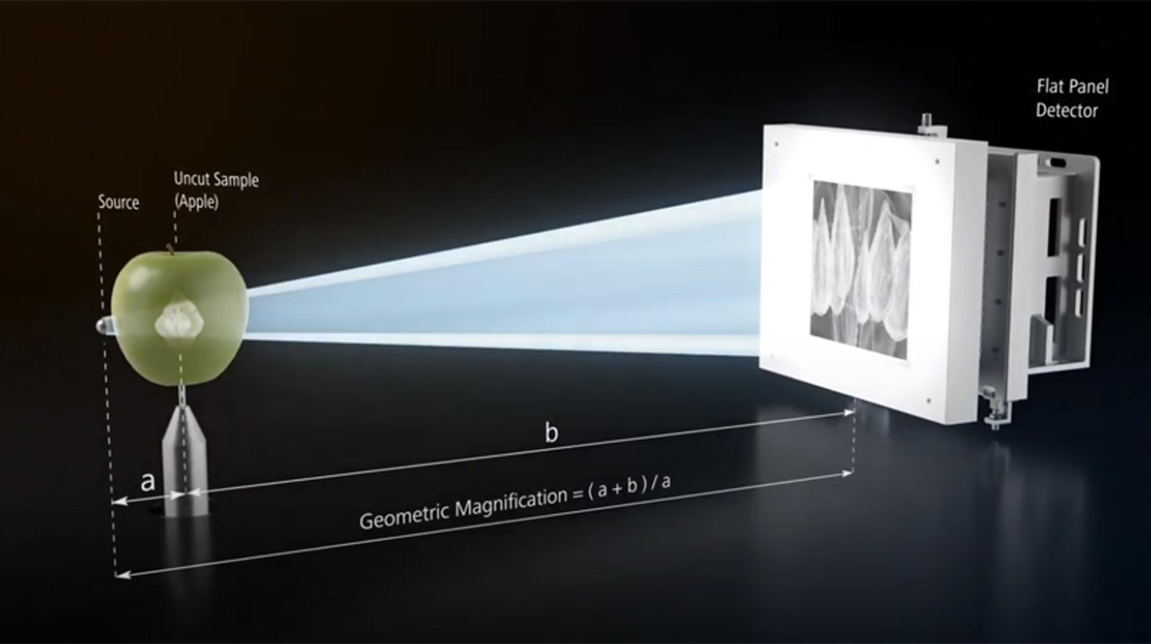
Resolution at a Distance: High resolution images, without destroying your sample
Do you want to look at the interiors of a sample at highest resolution without destroying it? Do you have to make a tradeoff between sample size and resolution? “Resolution at a Distance (RaaD)” technology on ZEISS Xradia Versa 3D X-ray microscopes overcome these limitations of Micro-CT systems.

Biomaterials: Shining a new light on bone disease
Prof. Silke Christiansen describes how she explores the mechanical properties of bone to help better treat osteoporosis and highlights the technological advances enabling new insights