Webinar Highlights: New Acquisition and Detection Modes with ZEISS Airyscan
13 Oct 2015
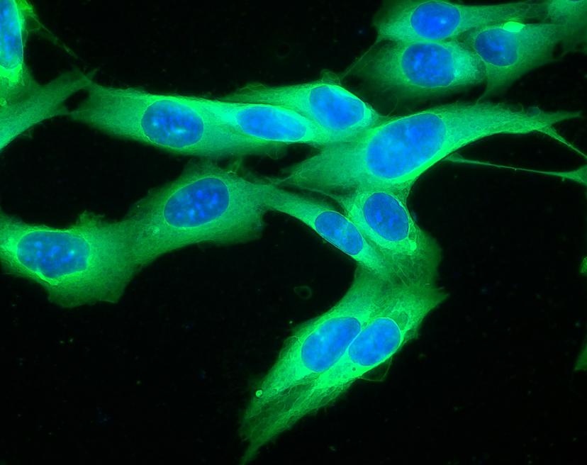
Learn more about Airyscan with our exclusive webinar, or read the Q&A highlights below
ZEISS Airyscan, a new detector concept designed for improved laser scanning confocal microscopy, enables the simultaneous increase of both resolution and signal-to-noise ratio over traditional confocal imaging. Learn about two new detection and acquisition strategies for the ZEISS Airyscan detection module for laser scanning microscopy. Both modes – sensitivity and two-photon – extend the Airyscan benefits of resolution and signal-to-noise ratio to address more sample types.
In this webinar, Joseph Huff, Product Marketing Manager for Laser Scanning and Superresolution Microscopy for ZEISS Microscopy, discussed the use of use of Airyscan to increase performance of laser scanning microscopy systems on samples including cell spheroids, Drosophila brain and live cells. Read on for a summary of the Q&A session, or watch the webinar on-demand here. Learn more about the ZEISS LSM family in our product directory.
What depth can the Airyscan image with both one photon and two photon excitation?
Airyscan will perform at whatever depth you are imaging with your confocal microscope today, which is typically up to ~100microns. Once Airyscan is combined with two-photon excitation, this depth can be extended beyond 100 microns to a few hundred microns or even further, depending on the nature of the sample.
What is the maximum scan rate of Airyscan?
This depends on sampling. In superresolution mode, the Airyscan can achieve five frames per second at 512x512 pixels. If the sensitivity mode is used, the same field of view can be imaged at around four times that speed while maintaining the same signal-to-noise increase.
To compare with a confocal image, do you need to use higher laser power for an Airyscan image? If you do, do you have quantitative comparison?
Quite the opposite. In fact, due to the detector collecting more light, and the utilization of the pixel reassignment and deconvolution, you can actually use less laser power with the Airyscan and still image with a higher signal-to-noise ratio.
If you use the deconvolution on a confocal image, by how much can the resolution be improved?
In theory, deconvolution can improve a standard confocal image resolution by ~1.4x. However (and this is a big however), in order to achieve this resolution increase, either more laser power or more averaging will be required in order to have enough image signal-to-noise to have reliable deconvolution. This will impact using LSM+DCV for live cell imaging or any other sample where there can be photo damage or toxicity. Download a white paper on this subject here.
Are all 32 signals acquired and digitized really simultaneously?
Yes, all 32 channels are acquired and digitized simultaneously. Because this is possible, we can create a pinhole plane image at every pixel.
Can you specify the objective used for the acquisitions?
Most of the samples were acquired with the one of the following objectives listed below. The LSM 880 version of the Airyscan has adjustable zoom optics that allow different objectives to be used with the detector. The LSM 800 Airyscan can be used with either a 63x or 40x objective.
- Plan-Apochromat 63x/1.4
- LD LCI Plan-Apochromat 25x/0.8
- LD C-Apochromat 40x/1.1
If you missed this interesting webinar, you can watch it on-demand here.
Related products
Request Quote for All Products
ZEISS LSM 880 with Airyscan
ZEISS Research Microscopy SolutionsYour New Standard for Fast and Gentle Confocal Imaging