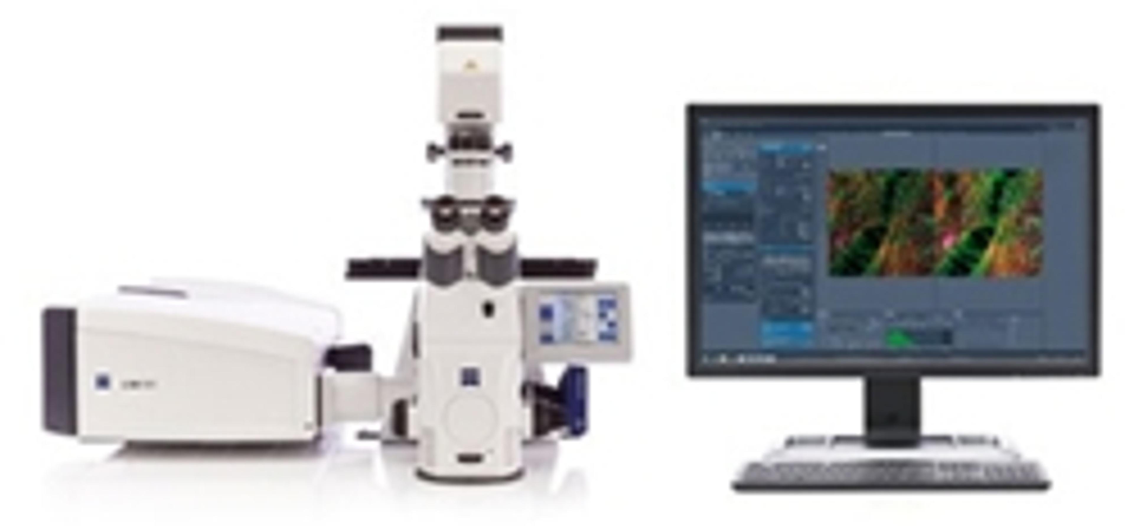
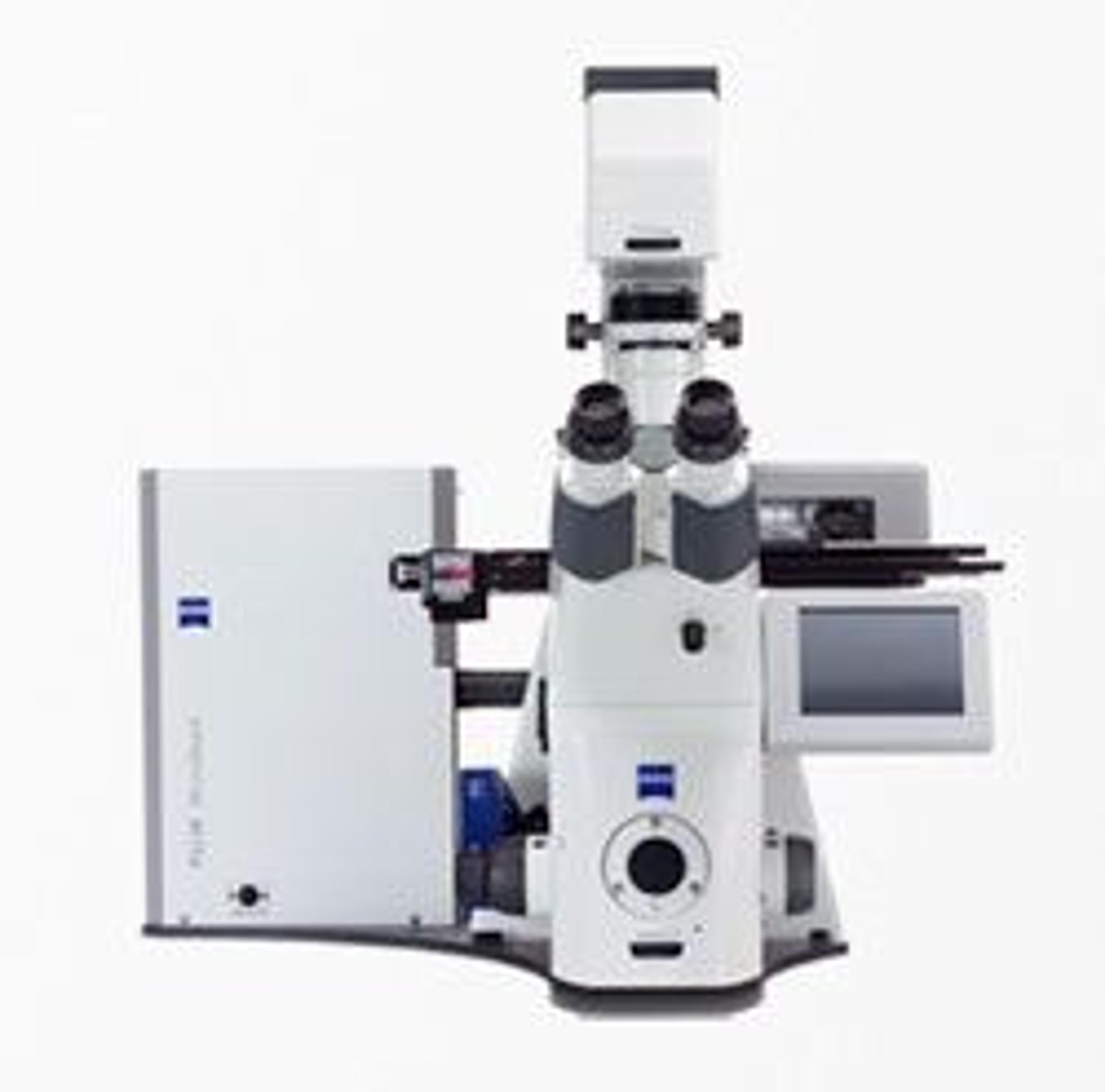
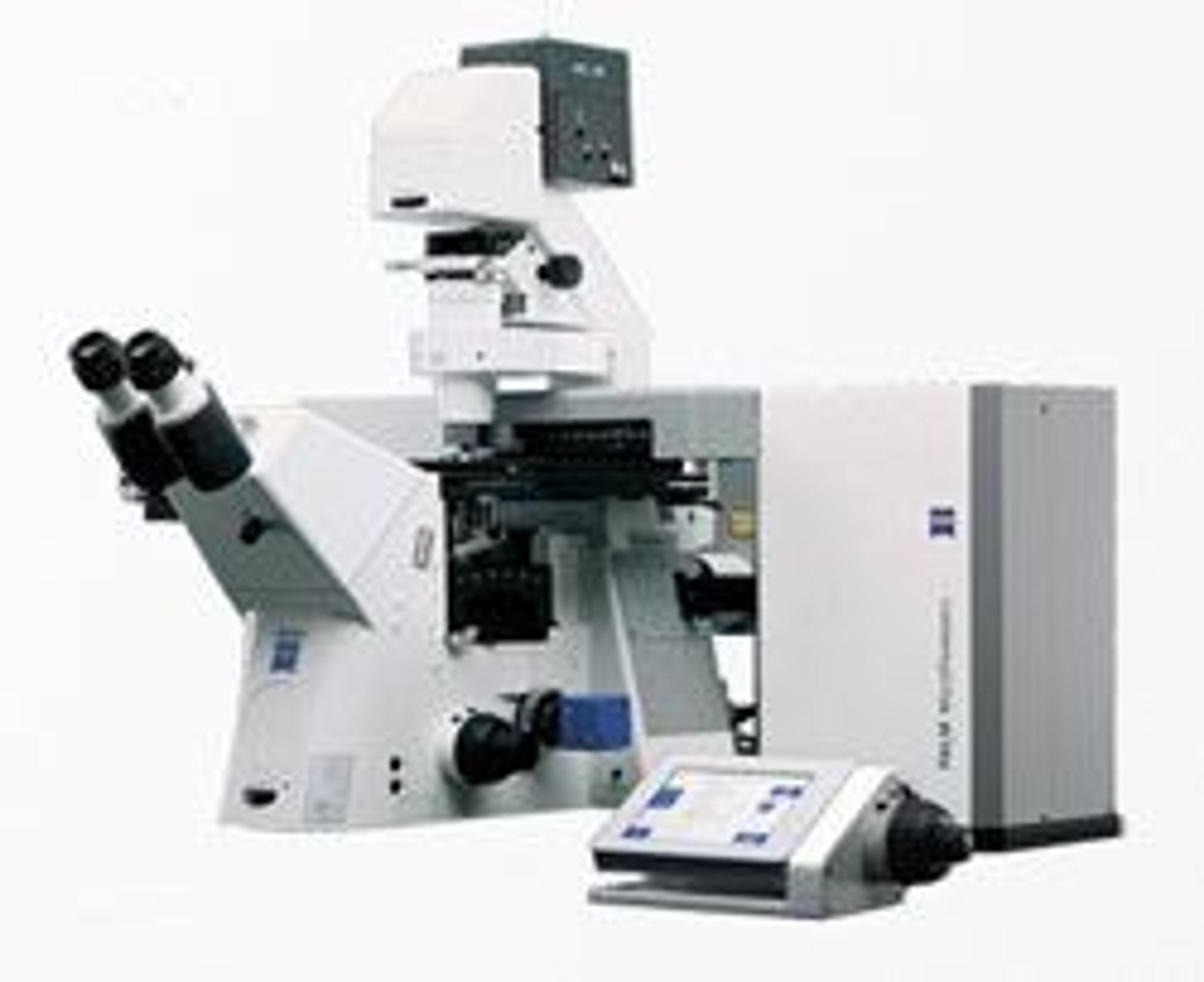
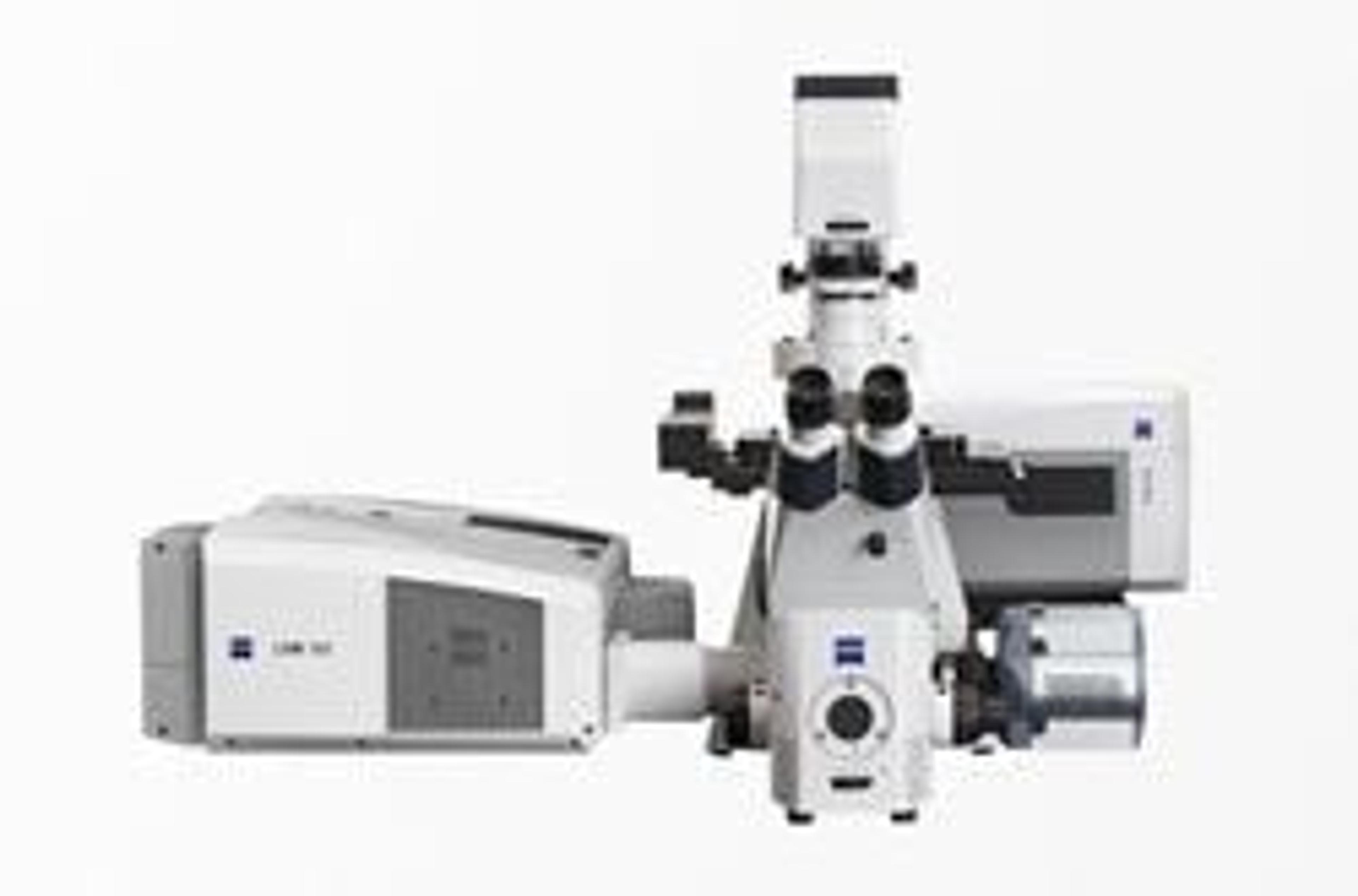
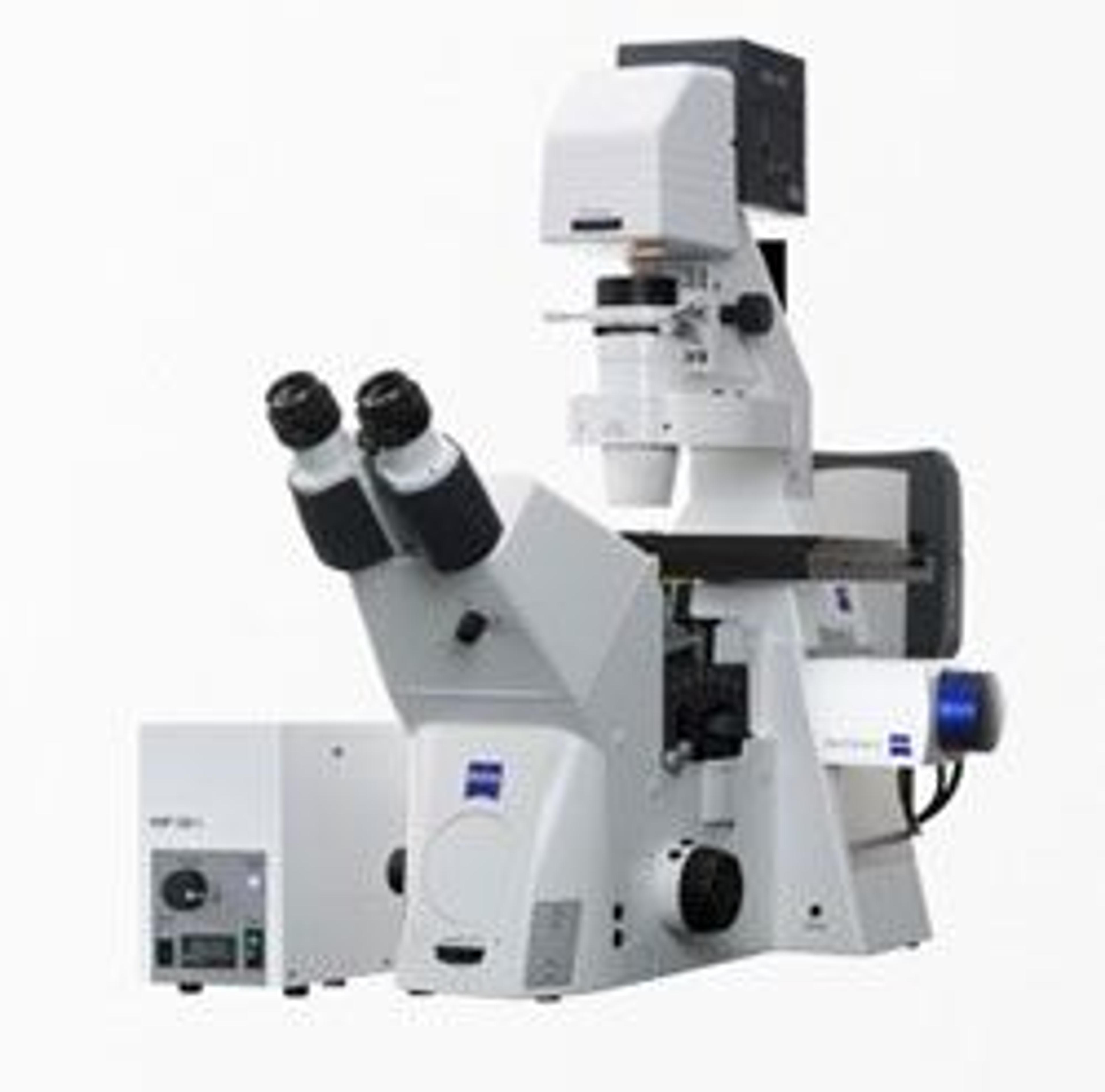
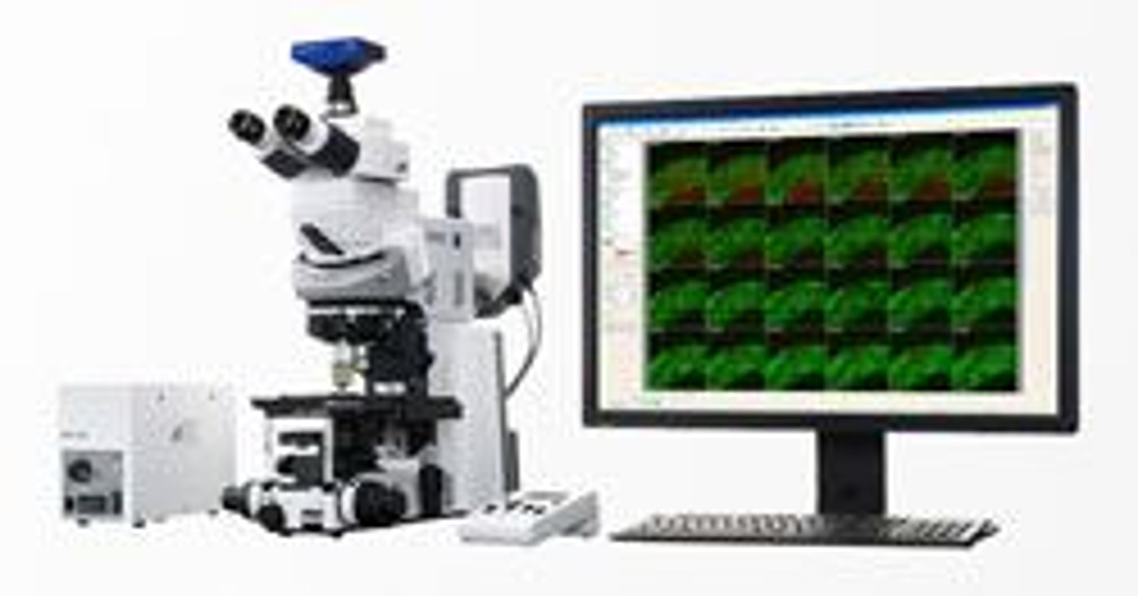
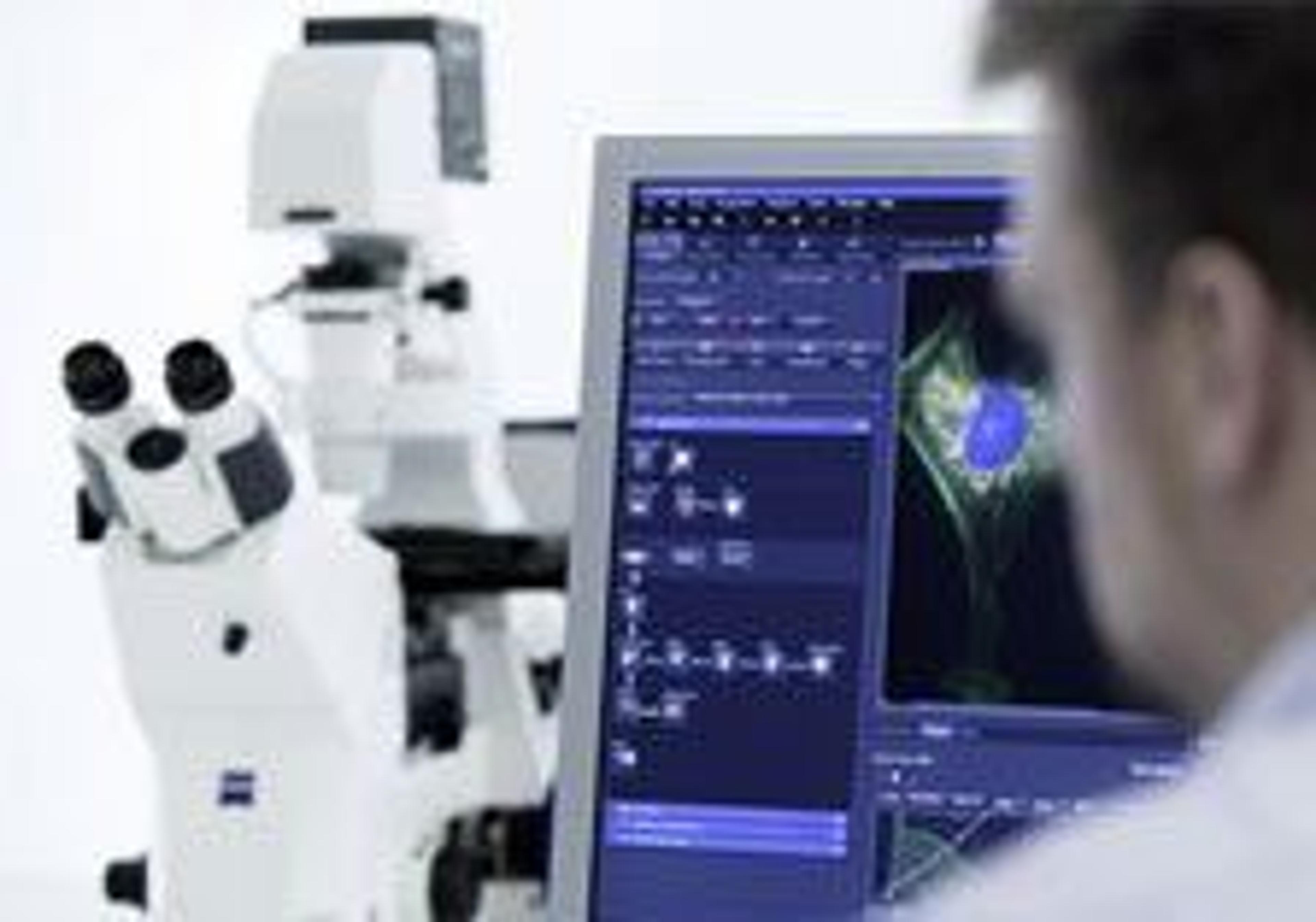
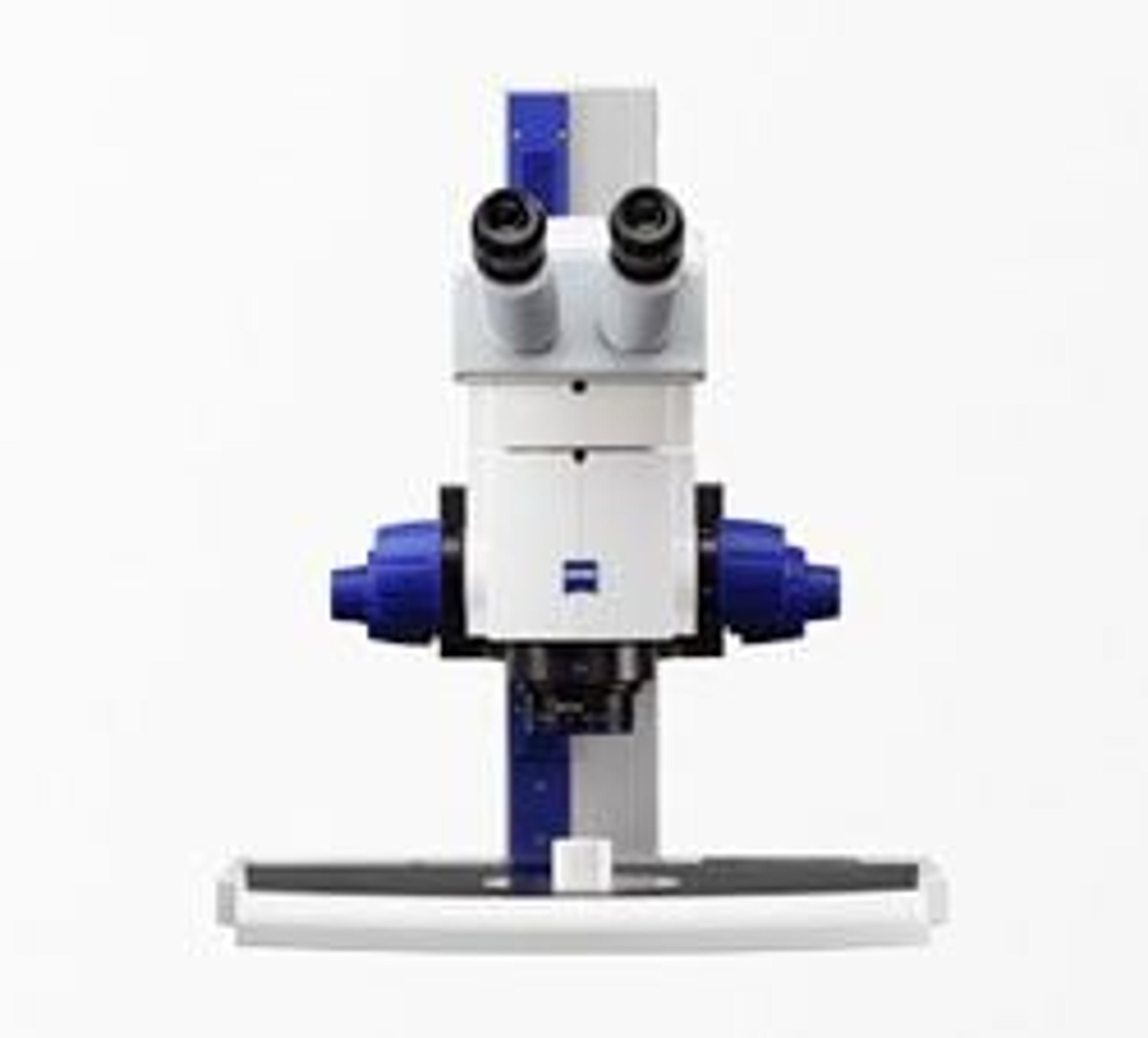
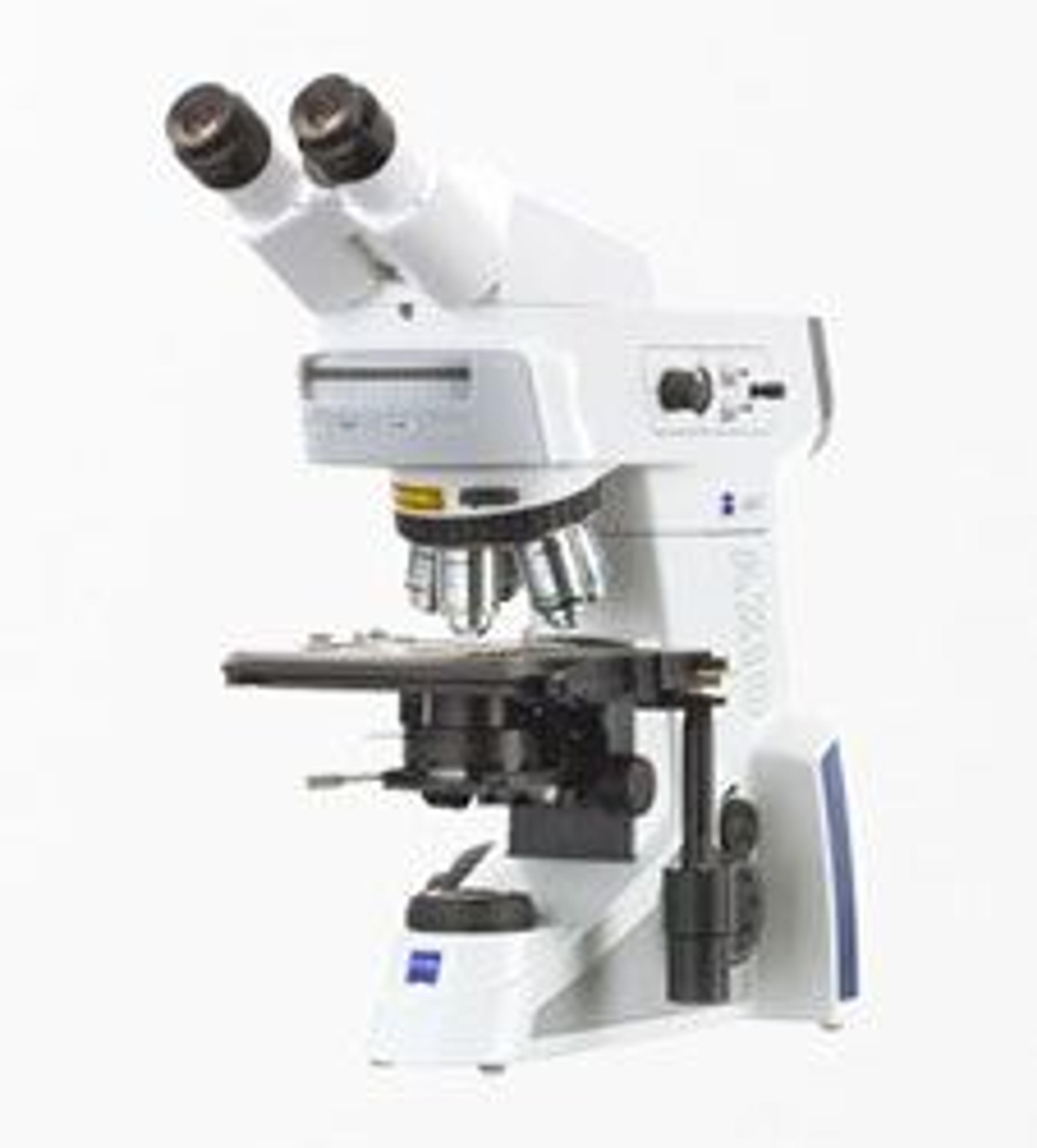
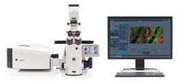
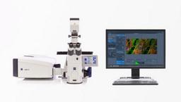
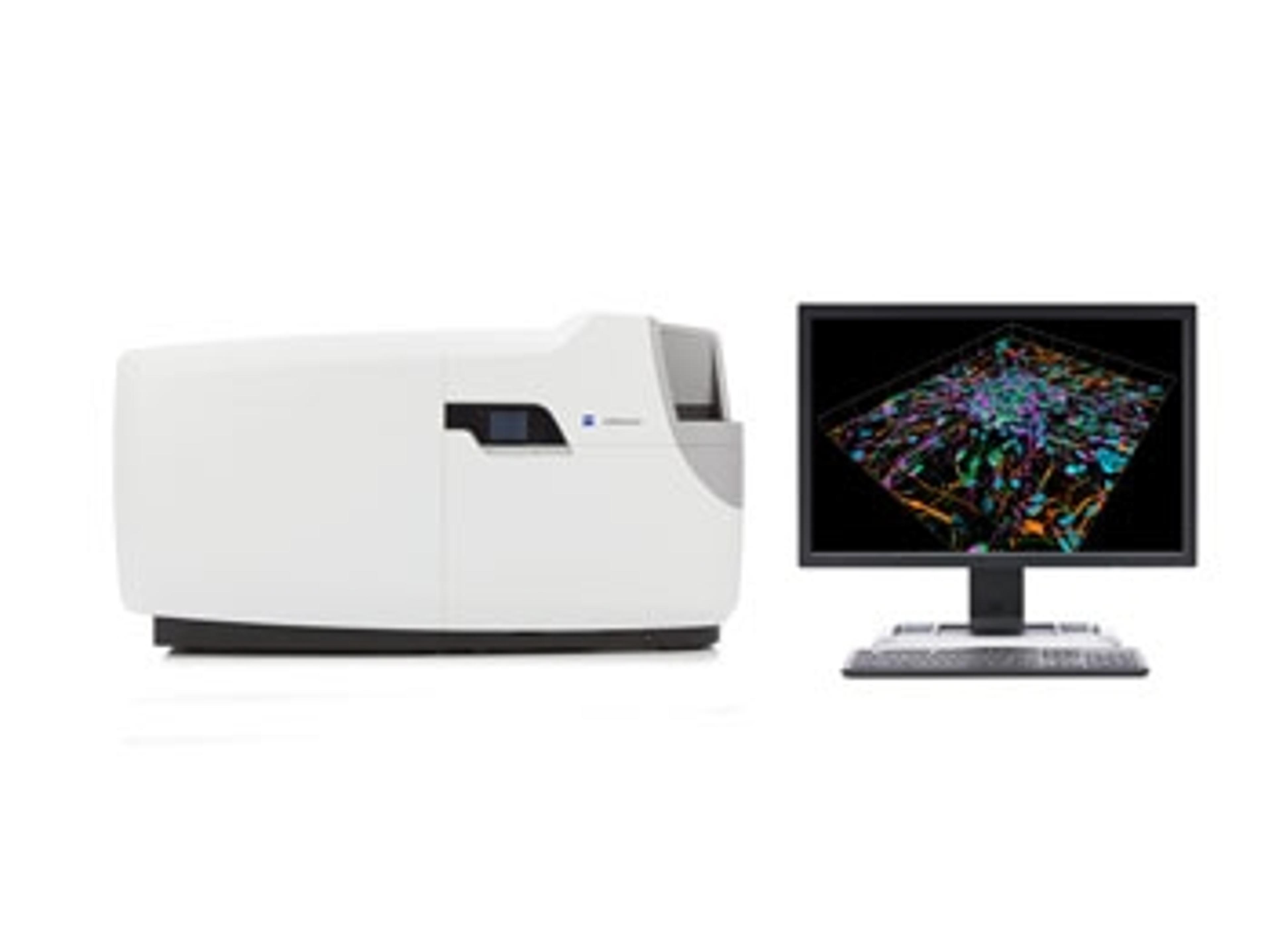
ZEISS LSM 880 with Airyscan
Your New Standard for Fast and Gentle Confocal Imaging


Receive your quote directly from the manufacturer.
Airyscan is great.
Neuroscience
Zeiss Airyscan is a very good tool for neuroscience research. When I really need to capture a little puncta significantly, the airyscan saves me.
Review Date: 16 May 2021 | ZEISS Research Microscopy Solutions
Fantastic instrument which is the backbone for much of our research.
Tissue and cell targeting of biotherapeutic compounds
We use the LSM 880 on a daily basis and it is fantastic for our purposes. It is used to examine tissue for cellular and subcellular targeting of biotherapeutic compounds. It offers a very flexible and capable microscopy platform. The learning curve may be a bit steep for new users, but in the hands of experienced personnel, it is a very powerful tool.
Review Date: 8 May 2019 | ZEISS Research Microscopy Solutions
It is an amazing microscope, it is our everyday workhorse.
Biomolecular imaging, genetic tagging and immunohistochemistry
This product is reliable, easy to operate and it is value for money. The results obtained are of high quality in terms of resolution so it is an important tool to have in every lab in order to bridge the gap between light and electron microscopy techniques.
Review Date: 8 May 2019 | ZEISS Research Microscopy Solutions
Great system for high-end results and ergonomy.
Microbiology
The LSM 880 is a very robust laser scanning microscope with liquid cooling (no fans, no vibrations) attached to an AxioObserver 7 with ergotube and side buttons also buttons on the Z focus knobs sample loading very easy along with light control. The system is very easy to use, most of the components are motorized and can be controlled by a touch screen or by ZEN software, high-quality reproducible images with minimum phototoxicity. The support team is very friendly and responsive.
Review Date: 22 Mar 2019 | ZEISS Research Microscopy Solutions
Great instrument.
Imaging at high resolution
The Airyscan function of LSM 880 is very useful for high resolution imaging.
Review Date: 18 Feb 2019 | ZEISS Research Microscopy Solutions
Good product
Microscopy
Perfect resolution, high quality, correct price and good sales care
Review Date: 8 Aug 2016 | ZEISS Research Microscopy Solutions
Imaging - core facility
Extremely sensitive. Very well resolved images. Fast and reliable. User friendly software.
Review Date: 13 Aug 2015 | ZEISS Research Microscopy Solutions
Microscopy of RPE tissue
A good combination of quality and speed of acquisition.
Review Date: 27 Apr 2015 | ZEISS Research Microscopy Solutions
Imaging
Excellent quality with improved imaging over regular confocals.
Review Date: 27 Apr 2015 | ZEISS Research Microscopy Solutions
Researching cereals and animal by-product meals
It is an excellent microscope which saves me time during my research. It has excellent resolution and by using multiple challenges at once it allows exploration of more complex cells.
Review Date: 27 Apr 2015 | ZEISS Research Microscopy Solutions
To get ahead in your research you may want to image the smallest structures, catch the faintest signal or track the fastest processes – or do all of that at once.
When it comes to getting accurate data from live cells or other weakly-labeled samples, there is no such thing as too much sensitivity, resolution or speed.
Each photon of emission light is precious. With Airyscan you have the unrivaled combination of fast superresolution and sensitive confocal image acquisition at hand.
Use multicolor samples with any label and get image quality like you’ve never seen before. Decide for this novel detector design and get a 4-8 × improvement in signal-to-noise ratio (SNR) as compared to imaging with conventional confocal GaAsP detectors. And 1.7 × higher resolution for your single photon or multi-photon experiments. The choice is yours.

Confocal Imaging with Improved Signal-to-Noise Ratio and Superresolution
Confocal imaging has grown to become the standard choice for most fluorescence microscopy applications. The optical sectioning ability of a confocal imaging system has been brought about by placing a field stop, the pinhole. This application note looks into the details of the recent development and market introduction of the Airyscan detector from ZEISS. Where traditional pinhole and detector design have been reworked to offer greatly improved resolution and signal-to-noise ratio (SNR).
Cryo-Confocal Imaging with Airyscan Improving Resolution and Signal-to-Noise in Cryo-Fluorescence Microscopy
This application note describes how the ZEISS Airyscan can improve resolution and Signal-to-Noise in cryo-fluorescence microscopy, it allows the user to record high-quality cryo- fluorescence data even without immersion optics, thanks to its novel confocal detection scheme.
ZEISS LSM 880 with Airyscan: Introducing the Fast Acquisition Mode
In this application note ZEISS introduce the features and benefits of the Airyscan detector concept for confocal laser scanning microscopy (LSM). With this feature additional light and spatial information can be collected, improving the spatial resolution and signal to noise ratio.
Confocal Imaging with Improved Signal-to-Noise Ratio and Superresolution
This application note explains how the Airyscan detector hardware is implemented and describe how the resolution and SNR increase is achieved and how it compares to traditional laser scanning microscopy imaging. Over the last 25 years the technique of confocal imaging has grown to become the standard choice for most fluorescence microscopy applications. The increase in utilization of confocal imaging systems in basic biomedical research can be attributed to the ability of a confocal imaging system to produce optically sectioned images with high contrast while providing acquisition versatility to address many sample and application demands. Over time, novel approaches and options to increase image contrast and instrument versatility have been developed but creating the optical section has not changed.
Cryo-Confocal Imaging with Airyscan Improving Resolution and Signal-to-Noise in Cryo-Fluorescence Microscopy
A major limitation for correlative cryo-fluorescence microscopy is the current unavailability of cryo-immersion optics that could yield a higher numerical aperture. In this application note, combining ZEISS LSM 880 with Airyscan and cryo-fluorescent imaging, allowed data to be obtained with a significant increase in resolution and signal-to-noise compared to standard confocal images under cryoconditions.
The Basic Principle of Airyscanning
Airyscanning is a technique based on confocal laser scanning microscopy. This white paper introduces a detector concept that drastically improves signal by utilizing light that otherwise is rejected by the confocal pinhole. Learn how the Airyscanning principle is put to work technically as an add-on to ZEISS LSM 880 and how Airyscan technology compares to structured illumination microscopy.
Airyscanning: A Novel Approach to Confocal Imaging
Confocal laser scanning microscopy is the recognized standard for 3D fluorescence microscopy. This application note describes how Airyscanning releases the full potential of a confocal microscope, achieving resolutions comparable to an extremely small pinhole but with a much better signal-to-noise ratio. It combines excellent optical sectioning performance with flexible scanning strategies for imaging and photomanipulation, making it the method of choice for a vast range of applications.
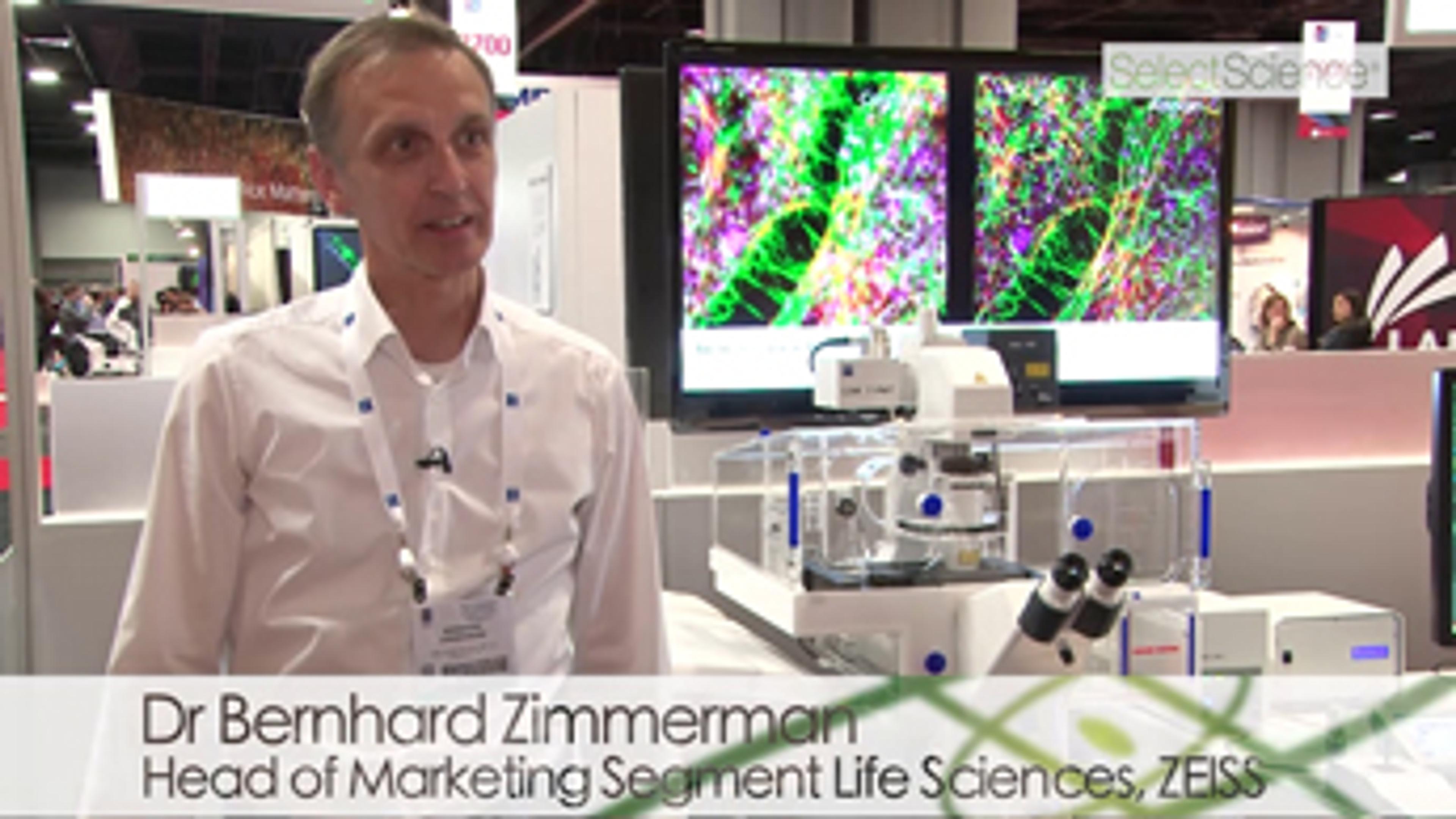
Learn How ZEISS LSM 880 with Airyscan Can Enhance Your Confocal Imaging
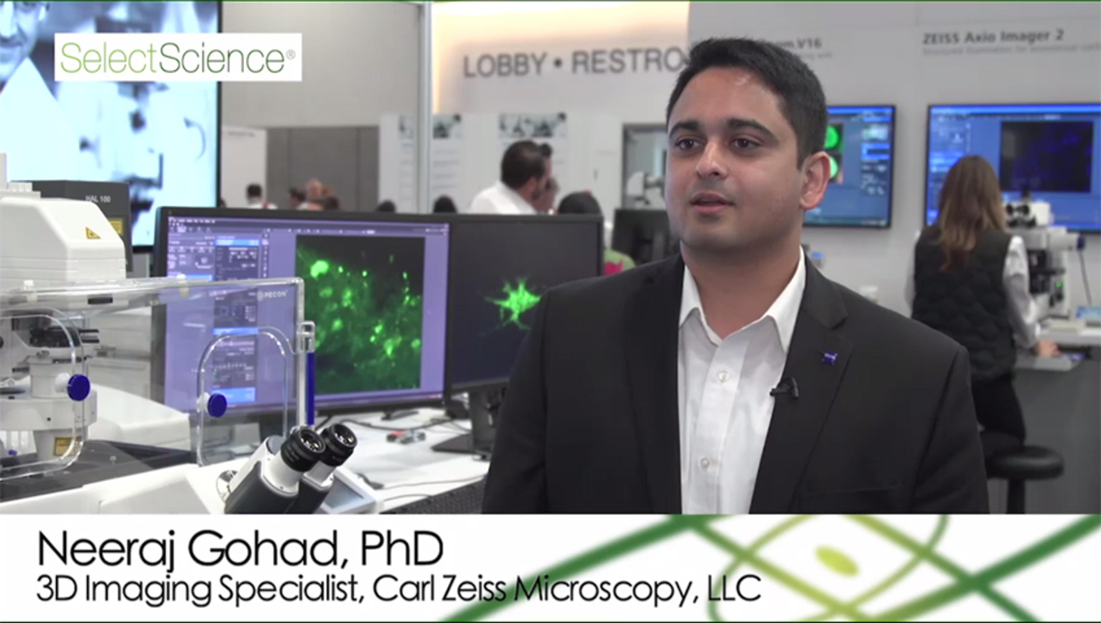
In this video, Neeraj Gohad, Carl Zeiss Microscopy, discusses the benefits of Zeiss LSM 880 with Airyscan, which can be utilized for live cell imaging. These include 4x faster imaging speed, as well as the production dynamic and super resolution images.
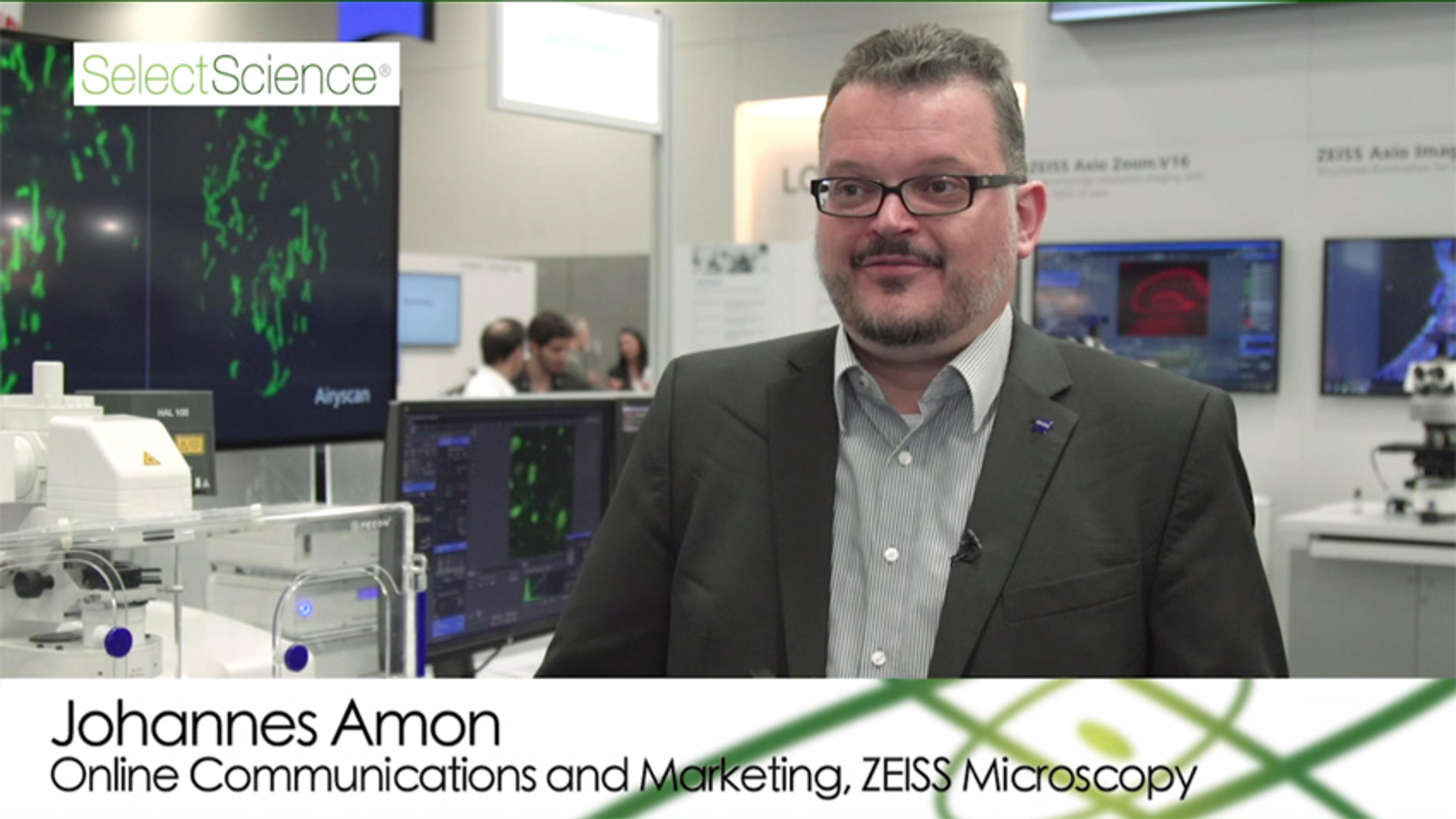
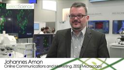
ZEISS Winner of #MyNeuroVote 2016
Hear Johannes Amon accept the #MyNeuroVote 2016 award on behalf of ZEISS Microscopy for the ZEISS Fast module for LSM 880 with Airyscan. Over 500 neuroscientists voted from a shortlist of nominated research products at the 46th Society for Neuroscience annual meeting, in San Diego, USA.
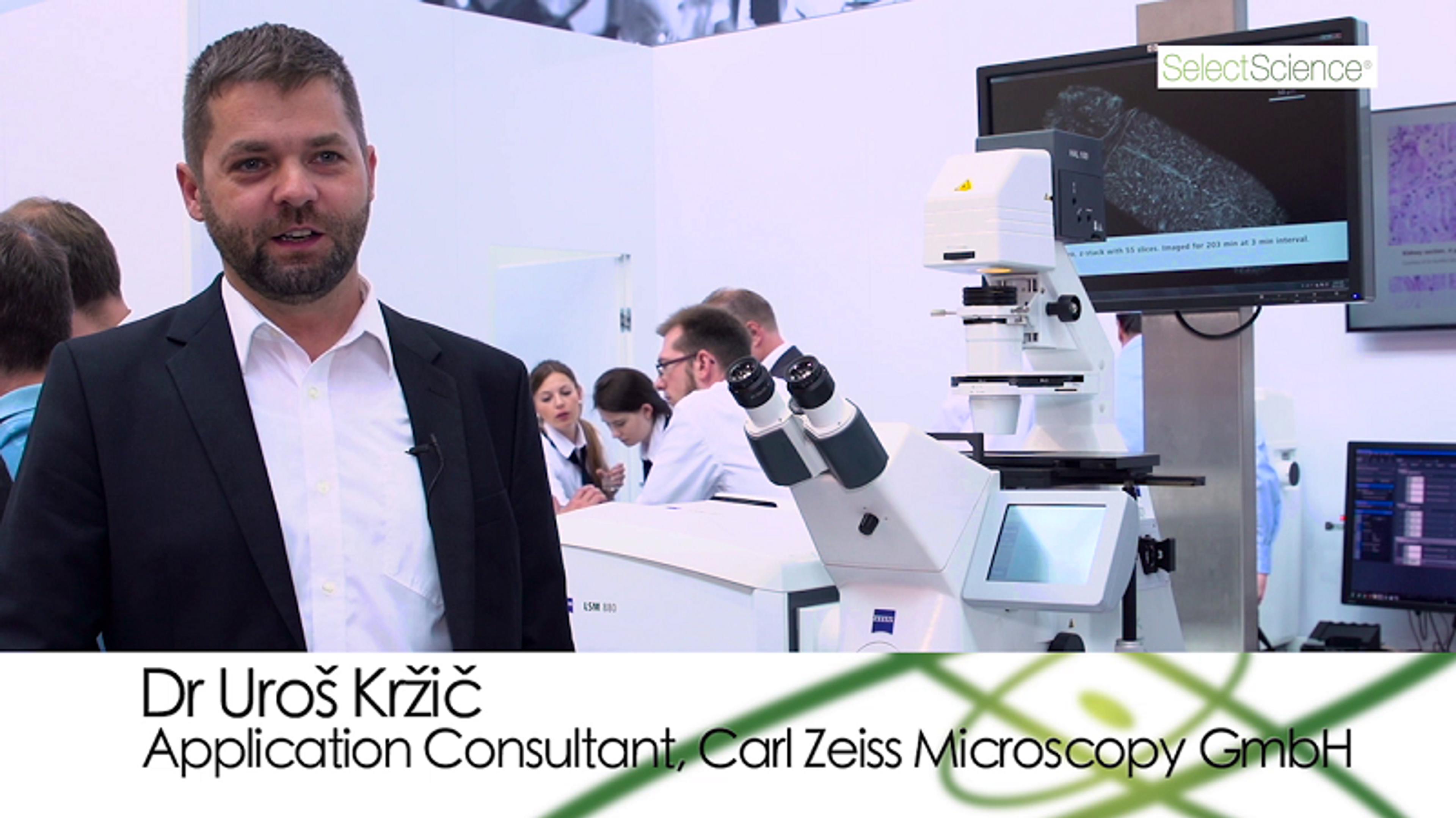
Enhanced Speed for Sensitive, High Resolution Confocal Microscopy
Dr Uros Krzic, application consultant at Carl Zeiss Microscopy GmbH, describes how the ZEISS Airyscan with fast acquisition mode facilitates sensitive, high resolution confocal microscopy that is ideal for imaging transient events.
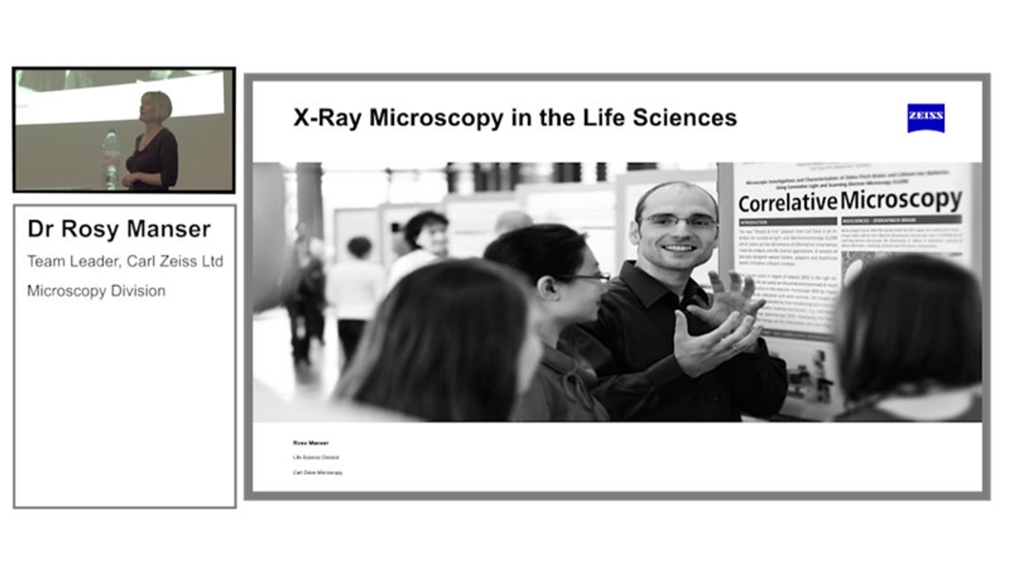
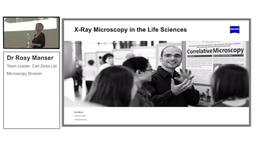
Presentation: X-Ray Microscopy in the Life Science
Dr Rosy Manser, Carl Zeiss Microscopy GmbH, Germany, discusses new X-Ray Microscopy applications enabled by the latest ZEISS technology, including the imaging of unstained tissue using the ZEISS VERSA XRM. Also, learn how the technology can be used as part of a correlative microscopy workflow.
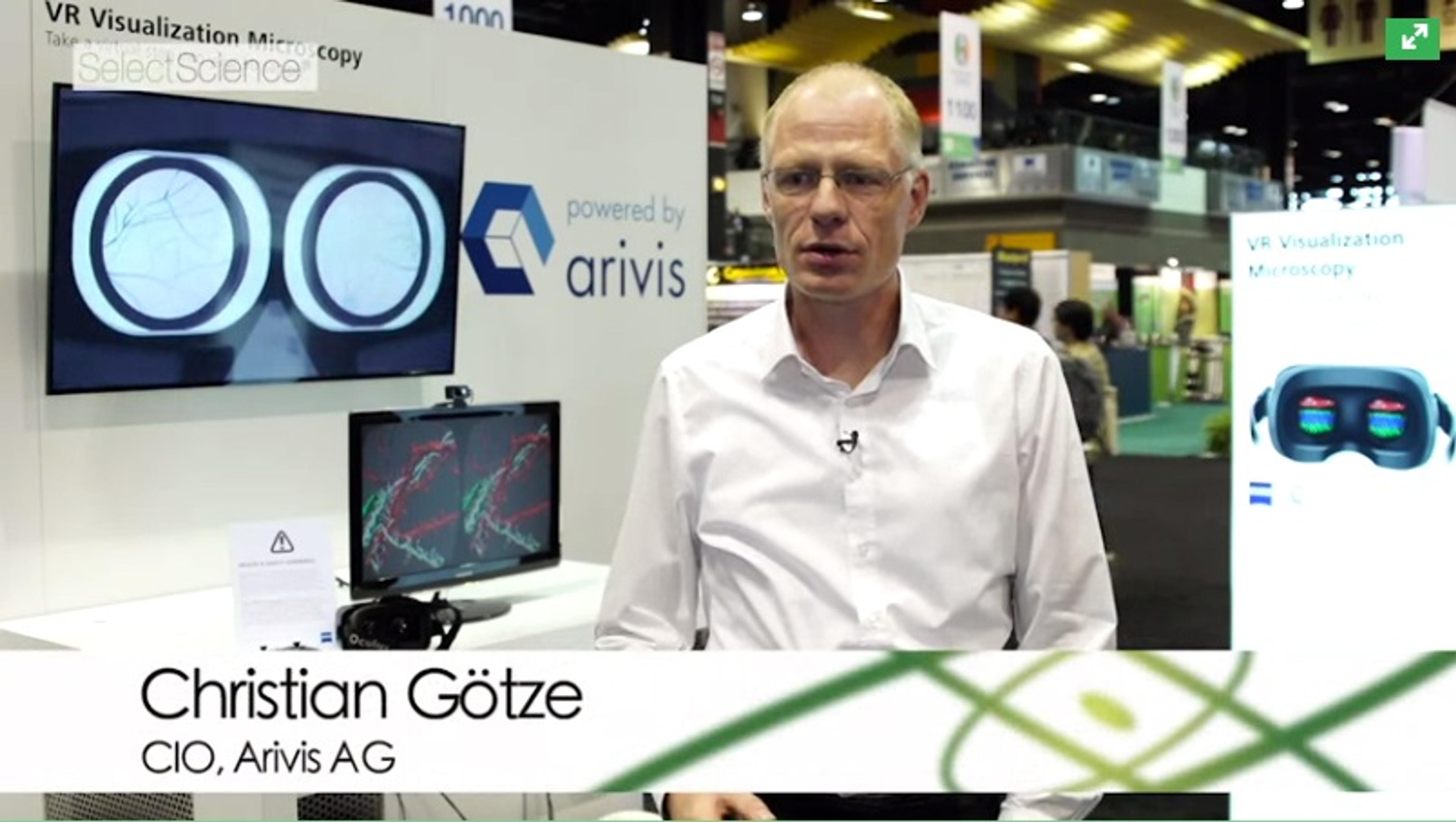
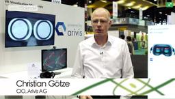
Virtual Reality Visualization of Complex Data Sets Acquired on ZEISS Microscopes
In this video, Christian Götze, Head of Development at arivis AG, explains a new approach to look at the large data acquired with microscopes. ZEISS and arivis partnered to create a prototype which allows you to render and visualize terabytes of volume data with the Oculus Rift virtual reality headset. You get a deeper insight into very complex brain structures and intuitively understand your data. You can virtually step into your sample, fly through a brain, past brain cells and analyze the image from every angle, and from inside.
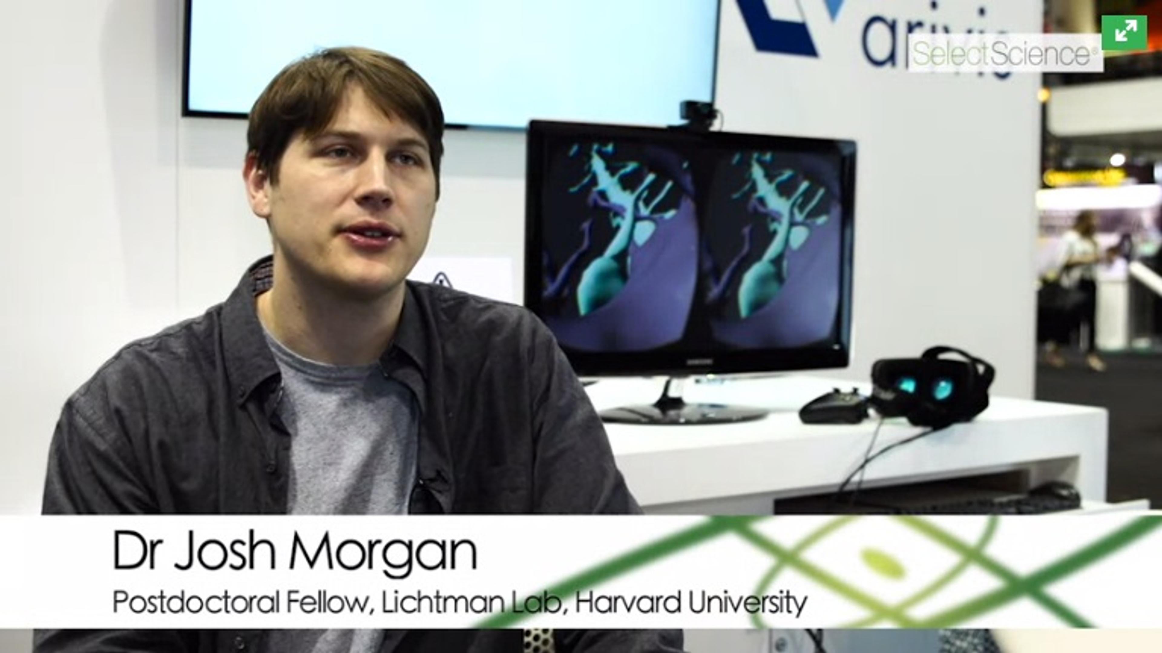
How Large Scale Electron Microscopy is Revealing Animal Behavior at Harvard University
Discover how Dr Josh Morgan, Postdoctoral Fellow in the Lichtman Lab at Harvard University, is trying to understand how the nervous system works by looking at how neurons organize their synapses with one another. All behavior in animals depends on cells talking to each other via specific connections, sending out long processes and forming synapses with each other. Understanding the pattern of those connections is one way of understanding animal behavior. Using large scale electron microscopy to reconstruct neural circuits allows you to see every cell and synapse in a circuit and find how they’re wired to one another.
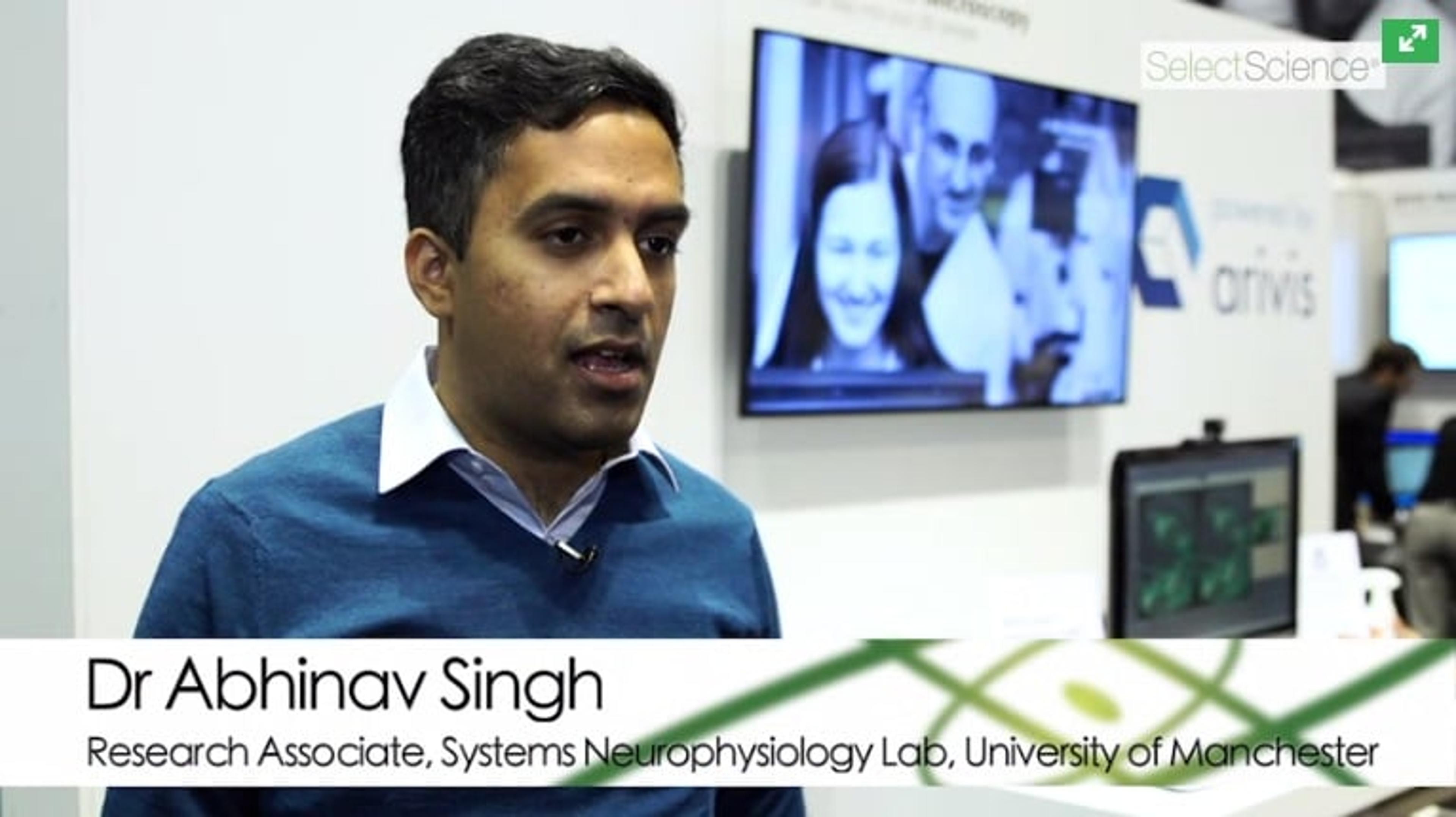
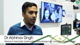
Solving the Problem of Neural Population Coding at Manchester University
Watch this video to hear Dr Abhinav Singh, Research Associate in Systems Neurophysiology Lab at University of Manchester explain the problem of population coding. When information about the external world is received via sensory signalling, it is transformed by our brain. Exactly how this transformation happens is unknown. Dr Singh is looking into how spikes in the pre-frontal cortex encode information about rule learning, and also discusses the fantastic viewpoint virtual reality offers in data analysis.
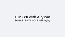
Revolutionize Your Confocal Imaging with LSM 880 with Airyscan
Watch this video to discover how Airyscan technology from ZEISS allows better signal-to-noise, resolution and image collection speed. Image areas lost in traditional confocal microscopy with super resolution - even single molecules!
Extend Your System Further with Airyscan Technology from Zeiss
Watch this video to hear Dr. Ralf Engelmann, Product Manager for LSM880 and Airyscan at ZEISS, explain how the LSM880 allows you to extract more information from your samples, allowing higher throughput and in turn, allowed you to image more samples. Extend the sensitivity of your instrument further with Airyscan technology, which allows imaging of areas lost in traditional confocal microscopy with super resolution - you can even image single molecules!
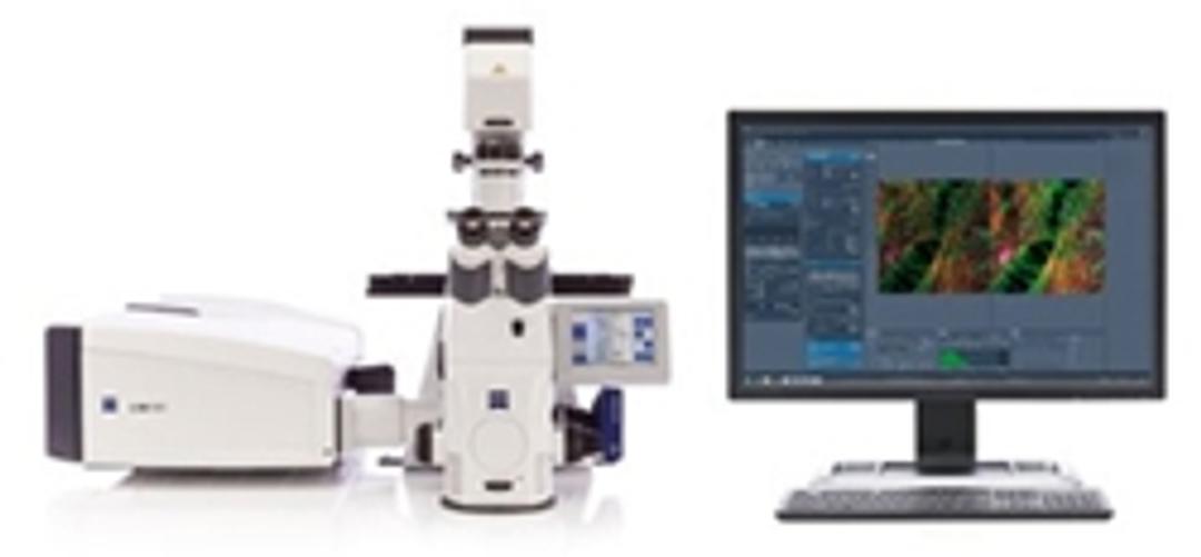
ZEISS LSM 880 Confocal Laser Scanning Microscope with Revolutionary Airscan Technology
Learn how Airyscan technology, incorporated in the ZEISS LSM 880 Confocal Laser Scanning Microscope, can increase the resolution, sensitivity and speed of your imaging. The multifunctional Airyscan detector enables detection of 5x smaller confocal volume compared to traditional confocal microscopy, making it ideal for a broad range of applications, including live cell and tissue imaging.

Biomaterials: Shining a new light on bone disease
Prof. Silke Christiansen describes how she explores the mechanical properties of bone to help better treat osteoporosis and highlights the technological advances enabling new insights
Automated Microscope for Gentle and Fast Confocal 4D Imaging
Enhancing ZEISS Celldiscoverer 7 with ZEISS LSM 900 for optical sectioning

Leveraging ZEISS Airyscan to Integrate Subcellular and Structural Detail with Inscopix Functional Neural Network Data
Expanding freely behaving microscopy by delivering increased resolution, signal-to-noise and optical sectioning through data correlation

ZEISS Opens New Microscopy Customer Center
From 3D Light to 3D Electron Microscopy: Highlights from the EMBL Workshop
Discover the latest news from this conference on correlative microscopy

How Advanced Confocal Imaging Revealed a Key Role for the Cytoskeleton in Pathology of Heart Disease
Dr Ben Prosser discusses his mechanobiology research, recently published in Science

Latest Clinical Downloads Available from the SelectScience® Library
A selection of the latest clinically relevant application notes, infographics and case studies

Exclusive Videos from analytica 2016
Watch exclusive interviews filmed at the 25th analytica international trade fair for laboratory technology, analysis and biotechnology
Top 5 Webinars to Improve Your Imaging Techniques
Learn about the latest imaging technologies and innovative applications